A/1 Causes, morphology and mechanisms of cell necrosis.
Necrosis is a form of cell death in which cellular membranes fall apart, and cellular enzymes leak out and ultimately digest the cell.
Causes:
- Oxigen deprivation: Hypoxia, ischemia
- Infectious agents
- Physical agents: radiation, temperature
- Immunologic ractiton
- Aging: telomeres
Morphology:
- Eosinophilia
- Glassy appearance
- Vaculated appearance
- Myelin figures
- Membrane fragmentation
- Nuclear changes: Karyolysis, karyrorrhexis, pyknosis
Mechanisms:
- ATP depletion
- Mitochondrial damage
- Ca++ influx
- ROS: DNA, lipids, proteins
- Membrane damage
- DNA damage
Types:
- Coagulative
- Liquifactive
- Gangrenous: Sicca, Humida
- Caseous: Coagulative + liquifactive
- Fat: pancrease, enzymes leak out -> Fat released (chalky white appearance)
- Fibrinoid: Endothil damage -> fibrin leaks out -> Ag-Ab complexes combine with it.
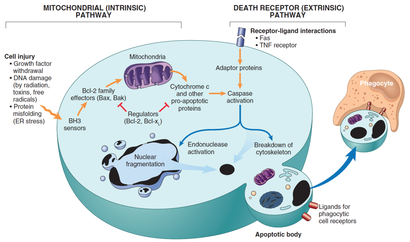
A/2 The causes and mechanisms of apoptosis. Apoptosis in pathologic conditions.
Pathway of cell death in which cells activate enzymes that degrade the cells’ own nuclear DNA and nuclear and cytoplasmic proteins.
Causes:
- Physiologic:
- Embryogenesis
- Hormone dependent
- Death of inflammatory cells
- Death of proliferating cells
- Pathologic:
- DNA damage
- Mis-folded proteins
- Viral infection: viral (cell cycler arrest)
- Atrophy
Mechanisms:
- Intrinsic:
- Cell injury -> BH3 receptors -> Bcl2 effectors (Bak-Bak, Bax-Bax channels) -> Cytochrome C leaks out -> Pro-caspase -> Caspase -> activation of effector caspase 3,6,7.
- Regulated by Bcl-2
- Extrinsic:
- Fas/TNF -> FaDD-> Pro-caspase -> Caspase -> activation of effector caspase 3,6,7.
Morphology:
- Apoptotic bodies (Contain organels) -> undergo phagocytosis
- Eosinophilic cytoplasm
- Nuclei changes: Karyorrhexis
4.
A/3 Coagulative necrosis, organ manifestations
architecture is preserved. firm. denaturation of the proteins and enzymes -> coagulated. inflammatory cells migrate and digest.
Infarct: ischemic necrosis caused by occlusion of the arterial supply or the venous drainage.
- white (anemic) infarct:
- Solid organs
- red (hemorrhagic) infarct: occurs in:-
- Venous occlusion
- Loose tissues
- Dual circulation
- Reperfusion
Organ manifestation:
Anemic:
- Kidney + spleen:
- obstraction of an end artery
- thromboemboli (usually from the heart - aneurysme/fibrilation)
- Wedge shaped, pale, necrotic area.
- Inflammatory response -> fibrotic scar
- In kidney: Might lead to hypertension (renin-angiotensin-aldosteron).
- Gangrena sicca:
- Atherosclerosis in extrimities
- Mummification (might fall off)
- Claudicartion intermittans
- AMI:
- .
Hemorrhagic:
- AMI:
- .
- Pulmonary:
- Usually from DVT
- Morphology depends on size:
- Large -> hypoxia -> R sided HF -> death
- Small -> damage to endothelial + pulmonary hemorrhage
- Bronchial circulation can prevent.
- Affects lower lobes
- Wedge shaped, Red-blue -> 48 Hr -> red-brown (hemosiderin)
- Final stage: fibrous replacment -> whitish scar.
- Intestinal:
- Superior mesenteric-> duodenum + jejunum
- Inferior mesenteric -> no infarct
- Portal (hypercoagulability) -> entire intestine
- Transmural -> 24 hr -> Bacteria -> gangrene -> perforation
- Mural (mucosa + sub-mucosa) - > Multi focal lesions -> Edematorious thickening of the mucosa + Inflammation due to bacterial superinfection
- Mucosal -> bacterial superinfection
- Abdominal pain + bloody diarrhea
A/4 Liquefactive necrosis, organ manifestations
enzymes are active and digest the tissue. seen in bacterial and fungal infection. heterolysis. autolysis. the tissue transforms into a liquid viscous mass. if initiated by an acute inflammation -> pus.
Anemic
Gangrene humida:
- infection superimposed the necrotic tissue.
- No immune.
- Microbe’s enzymes digest the tissue
Cerebral infarct:
- Thrombosis / Embolism (carotid atheroscelrosis / heart)
- Morphology:
- Encephalomalacia alba: anemic progress to:
- Encephalomalacia flava: liquifactive necrosis
- Cysta post encephalomalacia: astrocytes around the cyst
Pulmonary abscesses:
- suppurative necrosis -> formation of cavities with pus surrounded by inflammatory cells.
- Morphology:
- Aspiration -> Right: Upper lobe/upper part of lower lobe
- Pneumonia / Bronchial obstraction -> Multiple, basal and diffusely scattered.
- Septic / hematogenous spread -> multiple and everywhere
- Bloody and purulent sputum
- Consequences:
- Rapture to the A.W
- Bronchopleural fistula -> pneumotorax + empyema
Hemorrhagic:
- Encephalomalacia Rubra due to:
- Reperfusion
- thromb us in a vein
- border line necrosis
A/5 Acute myocardial infarction
Pathgenesis:
- Fixed coronaries + dynamic changes
- Fixed coronaries - 70-75% due to atherosclerosis
- Dynamic changes - disruption of the plaque (intra plaqueal hemorrhage, erosion, rapture) -> Platelet AAA -> ADP, TXA2, serotonin -> Vasospasem -> occlusion => ischemia
- ischemia -> ATP decrease -> pH decrease -> loss of contractility + reversible changes. (after 30min -> irreversible)
- “Wavefront theory”: subendocardial affeceted first (Higher pressure & contrasction compresses capillaries) => transmural in 3-6 hours (theraputic window)
Morphology:
- According to the
- 0-0.5:
- EM - swollen myofibers and swollen mitochondria
- 0.5-4:
- EM - swollen myofibers and swollen mitochondria
- Diaphorase reaction (DH)
- 4-12:
- Coagulative necrosis starts -> Eosinophilia
- Pale myocardium
- 12-24:
- Nuclear changes
- Dissociation of the organelles
- 2-3 day:
- Reddish border line -> migration of PMNI
- Macrophage system -> starts to dissolve the tissue
- 3-7 days:
- Yellow myocardium (lipid deliberation)
- Myomalacia (weakening of the texture of the myocardium)
- 7-30 days:
- Regeneration, capillary proliferation.
- fibroblasts ingrow from the capillaries and collagen deposits
- Scar formation (firm whitish tissue)
Reperfusion methoods: ballon (w/wo stent), thrombolysis (tPA, streptokinase), bypass
- Reperfusion injuries: ROS, Ca++ influx, Hemorrhage.
Clinical features:
- Substernal crushing pain + radiation - not relived by nitroglycerin. dyspnea, diaphoretic.
- ECG - ST elevation, Q-wave
- Serum levels of: CK-MB, Troponin T, Troponin I.
Consequnces: (CAMP CART)
- Cardiogenic shock
- Arrythmias
- Myocardial apture
- Pericarditis
- CIHD
- Aneurysm
- Reinfarction
- Thrombos formation
A/6 The morphology and mechanisms of reversible cell injury, examples
Morphology and Mechanisms:
- Cellular swelling:
- ATP decrease -> pump function decrease -> swelling
- Vacuoles of pinched-off segments of ER.
- Fatty changes:
- Accumulation of TAG
- Fat vacuoles appear around the nucleus, pushon it aside
- Reasons:
- Starvation: increase fat mobilization
- Hypoxia: Decrease oxidation
- Alcohol: Decrease NAD+ (decreased oxidation)
- Obesity
- Microscopic changes:
- Plasma membrane alterations
- Mitochondrial swelling (phospholipid-rich amorphous densities)
- Dilation of ER + ditachment of ribosomes
- Clumping of chromatin
Examples:
- Hepatic steatosis:
- Diffused
- Reduced NAD; imparied lipoproteins.
- Alcohol is the main reasone; DM (insulin resistance -> Fat intake)
- Morphology: Vacules, displace the nucleus; enlarged liver (4-6kg), soft, yellowish, tense capsule.
- Revarsible until fibrosis developes, start, around the central vein -> cirrhosis (oblitiration of the nodules creats scar tissue)
- Nutmeg liver
- Spotty, not diffused
- Tiger heart
- Hypoxia
- Due to anemia, CO, COPD
- Fatty heart
- Fat arround the cells (in the Right side)
A/7 Pathomorphology and complications of atherosclerosis
Chronic inflammatory response due to non denuding endothelial injury.
Risk factors
- Environmental than genetic
- Multiplication effect - exponential increse
- Major constitutional (non modifiable)
- Age (40-60)
- Sex (female premenopausal)
- Genetics
- Major Modifiable
- Hyperlipidemia
- Smoking 1pack/day * 200
- Hypertension
- Diabetes
- Additional
- Obesity
- Lack of exercise
- Stressful life
Pathogenesis
Non denuding EC injury -> ROS -> LDL oxidation -> Oxidzed LDL-scavenger receptor -> Macrophage become foam cells -> recruit more monocytes + ROS + release PDGF -> SMC (plaque stabilization) -> Fatty streak
Morphology
- Fatty streak - Lipid filled foam cells
- Primary plaque -
- Lipid core - Cholesterol chrystals, foam cells, EMC, Cell debris, Calcium
- Fibrous cap - SMC, EC, Collagen, neovascularization.
- 2 shoulders - vulnerable areas of the plaque
- Secondary (complicated) plaque - THE CAR
- Thrombus
- Hemorrhage
- Embolism
- Calcification
- Anurysem
- Rupture
Prevention
- Primary prevention - exercise, hypertension control, diet, stop smoking
- Secondary prevention - Statins, aspirin, Beta blockers, Bypass.
A/8 Amyloidosis
Extracellular deposits of fibrillar proteins responsible for the tissue damage (aggregation of misfolded proteins)
Proteins can be accumulated in the following way:
- Excess protein is presented to the cell (proteinuria)
- Increased protein synthesis (plasma cells)
- Cell injury. Alcohol (Mallory bodies)
- Abnormal folding of proteins (problems with chaperons)
Pathogenesis
- Misfolded proteins -> fibrilis -> 4-6 fibrils in β pleated configuration form an amyloid (resembles starch)
Important amyloid types:
- AL - precursor is immunoglobulin light chains, monoclonal B cell proliferation.
- AA - precursor is SAA normally synthesized in the liver
- Aβ - Cerebral lesions of Alzheimer disease (APP is the precursor)
- Transthyretin - binds thyroxine and retinol, deposits in the heart (Senile systemic amyloidosis)
Clasification of amyloidosis: RELIFA
- Reactive: AA type, systemic
- Endocrine: APUD-OMAs
- Localized: plasma cytoma
- Immunologic: Multiple myloma, AL type, systemic
- Falmilial: Familiar mediterinian fever, AA type, systemic
- Aging: TTR, Senile, systemic
Morphology
- Can be seen macroscopically when painted with iodine and sulfuroc acid
- Microsocpic staining - congo red
- Kidney - deposits in the peritubular area
- Spleen - Tubular or nodular
- Liver - Disses space, sinuses
- Heart - subendocardial elevations
- GI - is good for biopsy (Rectum, oral)
Clinical correlation
- Difficult to recognize (non-specific symptoms)
- Kidney failure
- Conduction disturbances
- 1 - 3 yr after diagnosis
- Amyloid can not be removed
- primary is the most common
A/9 Exogenous and endogenous pigments
Exogenous: Tattoo, Carbon, Silicium
- Carbon:
- Urbanic life
- antraxosis
- Silicium -> pneumoconiosis
- Phagocytosed by macrophages -> kill the macrophages -> leakage of lysosomal enzymes -> necrosis -> fibrosis -> increased lung resistance -> cor pulmonale -> Right sided heart failure
- Types:
- asymptomatic anthracosis
- sCWP - little to no pulmonary disfunction (coal macules and nodules; upper lobe and upper of lower lobe)
- cCWP - excessive fibrosis, lung function is compromised (Coalescence of the coal nodules)
Endogenous: Lipofuscin, hemosiderin, melanin
- Lipofuscin
- Wear an tear, Brownish color perinuclear
- Brown atrophy of the heart:
- Heart is small, sharp edge, pointing downward; apical shortening, coronary are curly; brown color is seen. (this is a side affect)
- Hemosiderin
- Hemoglobin derived granular pigment, yellow-brown, accumulates when there is a systemic/local excess of iron.
- apoferrtin
- stored in macrophages in the bone marrow, spleen and liver.
- Prussian blue -> blue color
- Local:
- פנס בעין
- Heart failure cells
- Systemic:
- general hemolysis
- autoimmune hemolytic anemia
- Toxic
- All the organs become brown
- general hemolysis
- free iron has a toxic effect (produce free radicals)
- Hemochromatosis
- Autosomal recessive (HFE gene on chromoosome 6)
- lipid peroxidation, collagen formation, DNA damage
- Morphology:
- Liver -> cirhosis
- Pancrease fibrosis -> DM
- Heart -> cardiac dysfunction
- joints -> atypical arthritis
- Skin -> pigmentation
- Melanin:
- Brown-black
- thyrosinase enzyme
- Alterations:
- Frackels
- Melasma: pregnant woman in the sun
- Vetiligo
- hereditary: thyrosinase enzyme
- Autoimmune: Autoantibodies against melanocytes
- Melanocyte nevus:
- Benign congenital / acquired neoplasm
- Junctional -> compound -> Dermal.
- transformation leads to melanoma.
A/10 Pathologic calcifications
Abnormal deposition of calcium salts + iron / magnesium
- Dystorphic calcification
- associated with necrosis + aging
- Pathogenesis:
- Extracellular:
- Membrane bound vesicles with Ca++ inside -> membrane bound phosphatase -> phosphatase ions generation -> phosphate ions bind Ca++ -> Crystal formation -> more and more (propagation)
- Intracellular:
- initiated in the mitochondria of dead/dying cells
- Morphology:
- grossly - white granules or clumps
- Somtimes tuberculous lymph -> radio-opaque stone.
- Extracellular:
- Diseases:
- Calcifing aortic stenosis
- arteficial valves
- Atherosclerosis
- Neoplasm
- Infectious disease (tuberculosis)
- Metastatic calcificartion
- Increased Ca++ serum levels due to:
- Neoplastic disease (multiple myloma osteoclast activation)
- Increased PTH
- VItamin D related
- Renal faliure (increased phosphate retention)
- Morphology:
- Everywhere but principally affects the lung, vasculature, kidney, gastric mucosa.
- Increased Ca++ serum levels due to:
A/11 Stone formation
Gallstone (cholelithiasis)
- Risk factors:
- Age, Gander (Women), Ethnic, heredity, decreased motility
Cholesterol stones
- Crystalline choloesterol monohydrate crystal
- Conditions for stone formation:
- supersaturation
- nucleation
- hypomobility
- Mucus hypersecretion
- Risk factors (more specific):
- Demogrophy
- Females (estrogen)
- Obesity (weight reduction)
- Hyperlipidemia
- Morphology:
- Exclusively in the gallbladder (50-100% cholesterol):
- Pure: yellow
- Mixed with Ca++-carbonate, phosphate, bilirubin: gray-white-blck
- Firm. 80% radiolucent (Ca++-carbonate are radio opaque)
- Exclusively in the gallbladder (50-100% cholesterol):
Pigment stones:
- uCB
- Risk factors (more specific):
- Asian
- Chronic hemolytic syndrome
- Biliary infection
- GI disorders (chron, CF …)
- Morphology:
- Black: sterile gallbladder, small in large quantities, crumble easily, 50-75% radiopaque.
- Brown: Infected ducts, singly/small quantitie, soft, radiolucent
Clinical features:
- 70-80% asymptomatic throughout life
- Symptoms: pain, GB inflammation, empyema obstructive cholestasis, pancreatities.
Renal stone:
- Urolithiasis
- Types:
- 80% - Calcium oxalate
- 50% - Idiopathic hyperclciuria
- 10% - Hypercalcemia -> hypercalcuria
- 20% - Hyperuricosuria
- High pH
- 10% struvite (Mg, NH3, PO4-3)
- UTIs
- 6-7% uric acid
- Hyperuricemia (gout / leukemias)
- Low pH
- 1-2% cystine
- 80% - Calcium oxalate
- Increased urine concentration of the stone’s constituent
- Morphology:
- Unilateral in 80%
- Staghorn calculi: branching
- Clinical features:
- Large stone -> asymptomatic
- Small stone may pass the urether -> intense pain, hematouria
- If blocks urine flow -> ulceration and bleeding
- Patient is exposed to bacterial infection
- Diagnosed radiologically
-
A/6-11 CELL INJURY, ACCUMULATIONS, PIGMENTS, CALCIFICATION (Leiel)62
-
A/1-5 NECROSIS, APOPTOSIS (Tamer)25
-
A/12-15 CELLULAR ADAPTATION TO STRESS, REGENERATION, WOUND HEALING (Leiel)48
-
A/16-21 HEMODYNAMIC DISORDERS (Leiel)53
-
A/22-25 INFLAMMATION (Tamer)37
-
A/26-33 IMMUNPATHOLOGY (Tamer)104
-
A/34-38 GENETIC DISORDERS AND DEVELOPMENTAL ABNORMALITIES (Leiel)48
-
A/39-44 PEDIATRIC DISEASES (Leiel)51
-
A/45-49 GENERAL PATHOLOGY OF INFECTIOUS DISEASES (Tamer)59
-
A/50-52 ENVIRONMENTAL CAUSES OF DISEASE (Leiel)28
-
A/53-71 NEOPLASIA78
-
B/1-15 PATHOLOGY OF CARDIOVASCULAR SYSTEM21
-
A/26-33 IMMUNPATHOLOGY (Tamer) COPY104
-
HistoPATHOLOGY54
-
111
-
B1
-
Heart95
-
heart 229
-
Tissue repair14
-
types of exudate7
