1-Carious Lesion Detection
2-Carious Lesion Assessment
3-Caries Disease Diagnosis
1-recognition of changes in enamel/dentin/cementum that are consistent w/ having been caused by caries process
2-severity/extent—evaulation of characteristics of caries lesion once its been detected. Characteristics= optical, physical, chemical, or bio, color, size
3-caries disease diagnosis= professional summation of all signs/symptoms of disease to arrive at ID
1-Lesion Detection Assessment
2- Diagnostic Tests
1-establish level of destruction present, aid in caries diagnosis, determine treatment decisions
2- Valid-test measures what is intended to measure= white spot lesion
Reliability- test can be repeated w/ same result—same lesion
1- true +
2- false +
3- true -
4- false -
1-caries= present and test identifies it
2- diagnostic test incorrectly IDs when caries is absent
3- test correctly identifies individual as caries free
4-has caries & test incorrectly= caries free
1-Sensitivity
2-Specificity
1-proportion of true positive that are correctly identified
2-proportion of true negatives that are correctly identified
Detection Methods—Ideal Method
- reproducible, accurate
- easy to use/learn
- useful on surfaces
- influence on treatment
Visual Examination of Caries
- widely used, quick, cheap, easy
- dry, clean tooth w/ good light, and mirror
- all surfaces
- occlusal, smooth surface (proximal), root caries
- dichotomous decisions—presence / absence
- Can’t see interproximal so it is absent upon examination
Explorer
- explorer can break off part of the tooth because it is fragily
- –doesnt add anything to detection yield
- use it to feel margins/defects
- clean debris from fissures/interproximal spaces and confirm/assess cavitations
- hardness of root/dentin
- texture of white spot
Occlusal Surfaces
-low sensitivity= 0.30 and high spec.
ICDAS
0- sound surface
1-first visual change in enamel
2-distinct visual change in enamel
3-localized enamel breakdown bc of caries w/ no visible dentin
4-non cavitated surface w/ underlying dark shadow from dentin
5-distinct cavity w/ visible dentin
6-extensive distinct cavity w/ visible dentin
Interproximal Detection
- visual inspection via bitewing radiographs
- but doesnt detect early subsurface demineralization or lesion activity
Caries—
- Biofilm
- Pellicle
- Enamel
- Dentin
1-E Classification
2-D1 Classification
3- D2 Classification
1-lesion penetrates through part of the enamel
-radiolucent triangle w/ base at enamel and point to DEJ
2-lesion penetrates into dentin but is less than 1/2 through dentin toward pulp…radiolucent triangular lesion in enamel
3- lesion extends= more than 1/2 but less toward pulp, deeeeep appearance
1-Transillumination
2-DIAGNOdent
1-intense beam of white light, tip on facial surface, caries has lower index of transmitted light, detection of proximal lesions, inexpensive (light through tooth)
2-detection of early occlusal lesions, fluorescence from lesion=produced from bacterial porphyrins, tip on tooth, normal enamel exhibits, intensity=size…drawbacks= heavely stained fissues and false positives
Caries Disclosers
- colored dye stains organix matrix of less mineralized dentin
- drawback= over prep of pulp exposure due to natural differences in colalgen content in diff parts of dentin
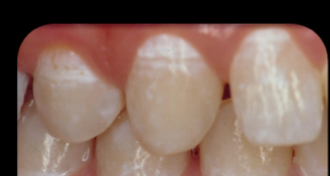
Active Lesion
- chalky opaque, white dull
- rought/soft
- plaque stagnation–covered by plaque
- close to gingiva
- high surface porosity
Inactive Lesions
- translucent/white-brown
- shiny surface
- smooth/hard
- non-plaque stagnation
- away from gingiva
- low surface porosity
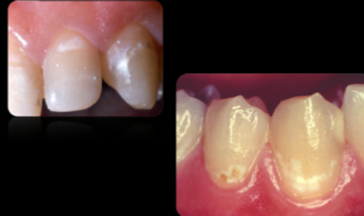
Active Lesion
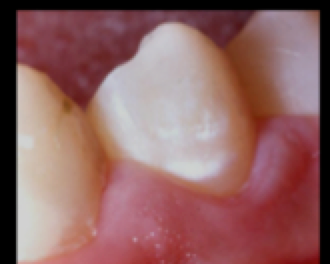
Inactive White Spot Lesion—–reversible & stable
Differential Diagnosis
—loss of tooth structure
- Erosion
- Attrition
- Abrasion
- —white spot differential
- hypoplasia
- fluorosis
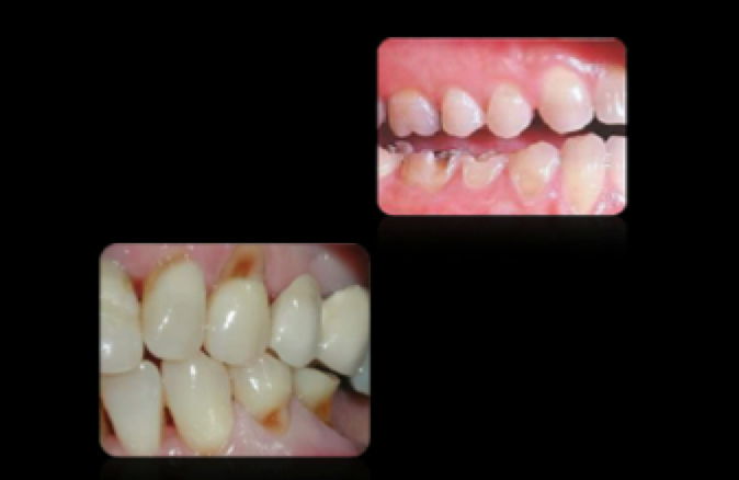
Decalcification
Fluorosis
1- alginate
2-gypsum
3- Dental impression media
1-impression making
2-dental casts
3- used to reproduce accurate replicas of intraoral and extraoral tissues
Impression Making Goals
1-obtain accurate negative images, all anatomic details from patients
2-transform images into positive physical or virtual casts= diagnostic, planning, and treatment
Digital Impression Media
- iTero (cadent)
- E4D (D4D)
- Lava (3M ESPE)
- —-we use—-CAD—CEREC (sirona)
-digitally imaged impressions to create virtual casts on restorations may be fabricated
