Atherosclerosis
It is the lipid laden deposits in the large and medium sized arteries in the tunica intima
Where do you see atherosclerosis
Thoracic and abdominal aorta, coronary arteries, peripheral arteries and carotid arteries
Interestingly internal mammary (thoracic) artery is protected from it
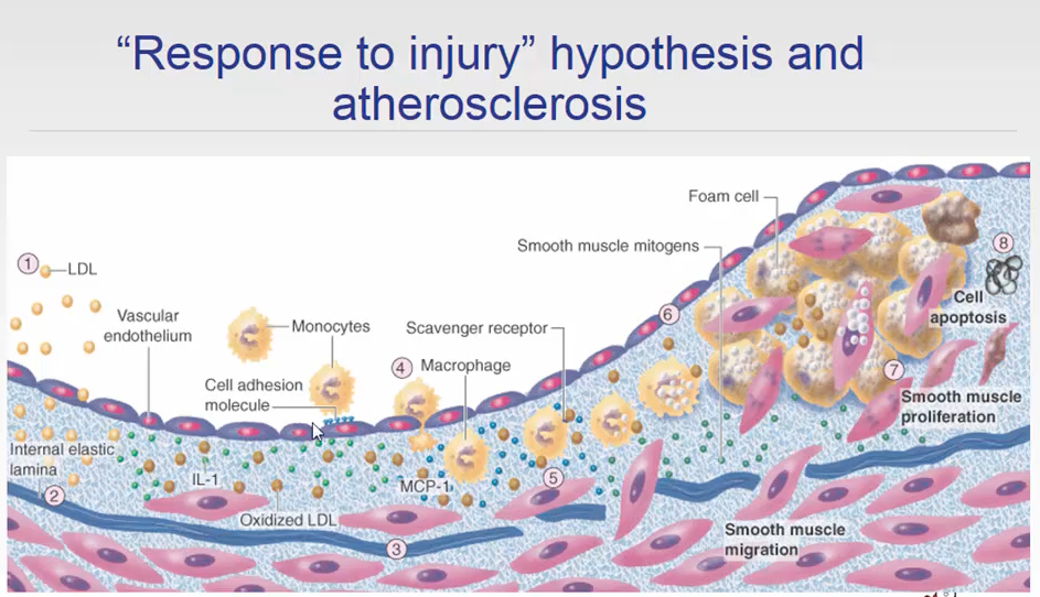
Pathogenesis
- Chronic endothelial cell injury
- Accumulation of LDLs
- Modification of lesional lipoproteins by oxidation
- Adhesion and migration of blood monocytes into the lesion, transformation into foam cells
- Adhesion of platelets
- Release of cytokines and growth factors, movement of smooth muscle into the intima
- Smooth muscle cells proliferate and produce extracellular matrix
- Enhanced accumulation of lipids
What is a C reactive protein a measure of in terms of atherosclerosis
It is a measure of vascular inflammation.
It is only a marker of vascular disease and does not play a direct role in vascular inflammation
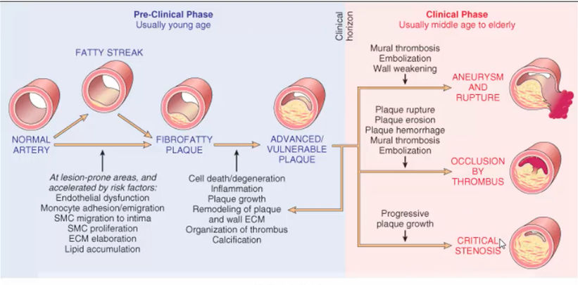
Natural history of atherosclerosis
It can be divided into 2 phases, pre clinical and clinical phase
- Preclinical starts with blood vessels that have lesion prone areas, accelerating risk factors can cause fibrofatty plaque deposition in these regions.
This leads to the formation of vulnerable plaque that mediates the clinical phase. It is important to know that the vulnerable plaque is still in the preclinical phase
- So 3 things can happen to a vulnerable plaque leasion: there can be progressive plaque formation which can lead to occlusion of the vessel and cause ischemia downstream, there can be ANEURYSM due to MURAL THROMBOSIS or EMBOLIZATION which can lead to rupture of the wall of the vessel and finally there can be OCCLUSION by THROMBOSIS
Details of the AHA classifications
- Early on there can be plaque formation which is asymptomatic
- Plaque formation can be occluded by thrombosis which can cause an acute MI, unstable Angina or sudden cardiac death
- If it doesnt get occluded by a thrombosis the plaque can continue to grow causing Angina pectroris
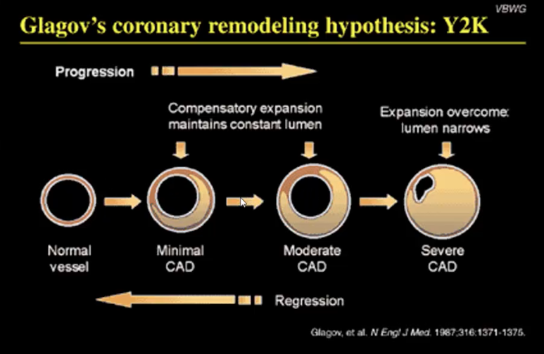
Risks of A by coronoary angiography
The progression and risks of A are often underestimated by coronary angiography.
It is important to know why since the lumen narrowing is a late phenomena and angiography can check for the thickening of the vessel
Where are occlusions and aneurysms common
In the coronory artery occlusions are common whereas in the aorta aneurysms are common. Usually aneurysms are not seen in the coronary artery
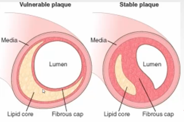
What is the difference between stable and vulnerable plaque
Stable plaque has a small lipid core with a thick fibrous core whereas a vulnerable plaque has a small fibrous layer and a thick lipid core.
The stable plaque has a narrow lumen but it has a better clinical prognosis as the thin fibrous cap on the vulnerable plaque is the one that gets damaged and causes rupture of the plaque
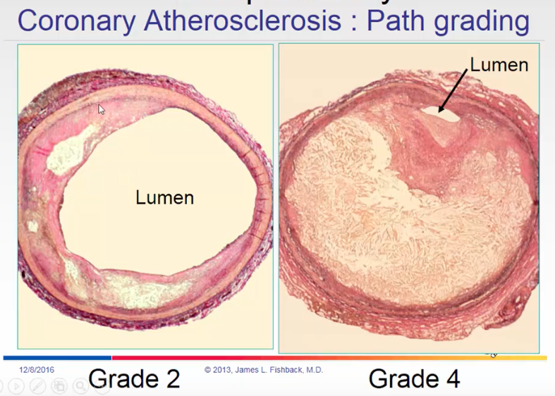
Grading of a plaque
Depends on the lumen
How do you image plaques
- Angioplasty can only assess the lumen of a vessel
- Ultrasound can be used to assess for the lipid content, data received from the ultrasound is used to develop a virtual histology of the wall of the vessel, it can also look for the calcification
- Optical Coherence Tomogrpahy is good for checking for the lipid content in a vessel, short is OTC
Patients with MI, how does thier vessels look like with angiogrpahy
Patients who have had acute MI, the plaques that rupture leading to atherosclerosis are not the ones that show stenosis in angiography
In other words, the tightest stenosis usually are less likely to cause MI
Mechanisms by which inflammation can lead to occlusion
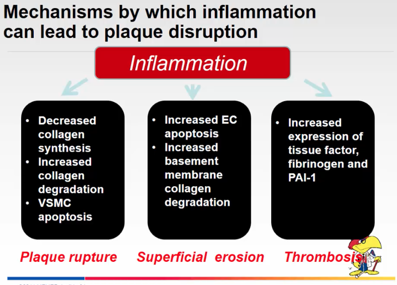
Pathogenesis of thrombosis
It is called the virchow’s triad:
- Increased coagubility of the blood
- Alteration in the blood flow, causing turbulent flow, it is called rheology
- There can be injury to endothelium, which can be chronic or acute
How does rheology explain the location of plaque formation in the peripheral areas such as the carotids
At the site of bifrication of the carotids (division) there are sites of lower shear stress due to turbulent flow which can cause increased endothelial injury at the sites and this is most often the place where plaques form.
This only explains some of the places where plaque formaiton occur most frequently, it doesnt explain why in some places the plaque doesnt form
What are some of the things that are common in aorta
- Occlusion is rare, due to its size
- Plaque formation is common, embolizations is common that can travel to the periphery and causes for example gangerene
These are usually silent manifestation
Lines of Zanes, thrombosis, healing, thrombosis, healing
How do you check for atherosclerotic embolism
If you see cholestrol crystals in the center of the lumen of the vessel then they probably came from upstream, cholestrol clefts dont form in the center of the lumen, they form at the edges but they can get dislodged and travel downstream
Aortic aneurysm
- These usually form at the abdomen and they do pulsate
- Risk of rupture is directly related to the size of the vessel, usually 5 cm of a diameter is a good cut off when intervention is required
- Operative risk is low if they dont rupture
Claudification
Plaque progression in the peripheral arteries can lead to cramping pain in the peripheral parts of the body during exertion due to occlusion of the vessel. This phenomena is called claudification
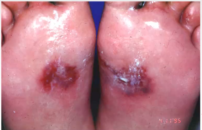
This is something that can be seen in the test and it is important to know that these kind of lesions are not a plaque progression problems but embolism problem and can be a marker for aortic embolism
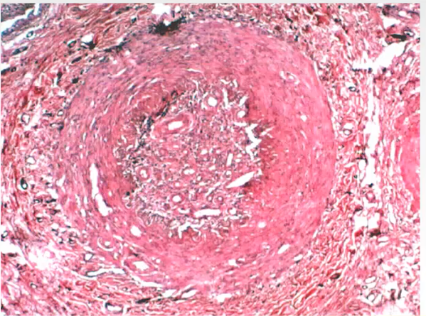
These are not cholestrol clefts but this is Recanalization where after oclusion of the vessel small vessels form within the artery to deliver blood, these vessels form by angiogenesis
-
Body Fluids and Electrolytes20
-
Cell Biology and Histology of Vascular System24
-
Heart Structures17
-
Lipoprotein structure10
-
Body Fluids Flipped Class4
-
Congenital Heart Defects32
-
Resting Potential Generation8
-
Cardiac Myocyte19
-
Histo Vascular12
-
Action Potential Generation29
-
Normal Cardiac Electrophysiology45
-
Normal Heart Exam20
-
Electrocardiography25
-
Abnormal Electrophysiology48
-
Autonomic Nervous System48
-
Cardiac Cycle27
-
Myocardial Performance41
-
Treatment of Cardiovascular Disorders27
-
Hemodynamics33
-
Infectious and Valve Disease26
-
Sepsis9
-
Vasculitis Disorder24
-
Microcirculation22
-
Hypotension19
-
Cardiac Valve Dysfunction14
-
Cardiac Output and Shunts17
-
Cardiomyopathies24
-
Regional Circulations28
-
Hypertension25
-
Coagulation and Fibrinolysis25
-
Atherosclerosis22
-
Development of Heart47
-
Drugs29
-
Drugs - Randomized29
-
Treatment of Cardiovascular diseases drugs12
-
Lipoproteins24
-
Katie Diseases5
