3 Major Locations for Brain Herniation
1) Cingulate gyrus herniation: the cingulated gyrus herniates medially beneath the falx cerebri
2) Uncal herniation: the medial temporal lobe herniates below the free edge of the tentorium
*can compress the midbrain and CN III resulting in a dilated pupil (ipsilateral)
*can compress the posterior cerebral artery which leads to infarction of that territory
*downward displacement of the midbrain and pons can lead to tearing of penetrating arteries and veins leading to Duret hemorrhages, flame shaped hemorrhages in the gray matter of the pons
3) Cerebellar tonsils: herniation of cerebellar tonsils into the foramen magnum, may be fatal when respiratory centers in the medulla are compressed
Amygdala
•nucleus at the anterior end of hippocampus
Arachnoid mater
- middle meninges
- neural crest mesoectoderm
- linked by tight junctions, forming a blood-CSF barrier
Arachnoid Villi
- protrusions of the arachnoid through the dura and into the sinuses
- enables CSF to drain from the subarachnoid space into the venous system
- bulk flow process - when the pressure in the subarachnoid space is greater than venous pressure (as is normally the case) CSF moves across the villi into the sinus
Basal ganglia
•masses of gray matter buried inside the cerebral hemispheres •lenticular and caudate nuclei (also some diencephalic and brainstem structures) •nuclear accumbens + olfactory tubercle = ventral striatum •caudate nucleus + putamen = dorsal striatum •dorsal striatum + ventral striatum = striatum •pallidum •motor functions - muscle tone, involuntary movements, initiating and stopping movement
Blood Brain Barrier
- tight junctions of arachnoid membrane
- choroid epithelial cells
- endothelial cells of blood vessels
Brainstem
•midbrain, pons, medulla •CN III-XII attach here (XI exits the cervical spinal cord) - brainstem processes their incoming info and sends it on to the thalamus, cranial nerve reflexes, motor commands out through CN •spinothalmic and corticospinal tracts traverse the brainstem - long tract functions of the brainstem •multiple collections of brainstem neutrons with widespread, diffuse connections (Ascending Reticular Activating System) - regulate our state of consciousness and are central to the sleep wake cycle
Calcarine Sulcus
- runs roughly horizontally through the occipital lobe
- primary visual cortex lives here
Caudate Nucleus
•The caudate nucleus is one of the structures that make up the corpus striatum, which is a component of the basal ganglia. While the caudate nucleus has long been associated with motor processes due to its role in Parkinson’s disease, it plays important roles in various other nonmotor functions as well, including procedural learning,associative learning[5] and inhibitory control of action,among other functions. The caudate is also one of the brain structures which compose the reward system and functions as part of the cortico–basal ganglia–thalamic loop.[1]
Central sulcus
•divides parietal and frontal lobes
Centrum Semiovale
- The centrum semiovale, semioval center or centrum ovale is the central area of white matter found underneath the cerebral cortex. The white matter, located in each hemisphere between the cerebral cortex and nuclei, as a whole has a semioval shape.
- The white matter, located in each hemisphere between the cerebral cortex and nuclei, as a whole has a semioval shape. It consists of cortical projection fibers, association fibers and cortical fibers. It continues ventrally as the corona radiata.
Cephalic flexure
•80º bend between the brainstem and the diencephalon
Cerebellum
- inferior and posterior to the forebrain
- receives huge amounts of sensory information, but is NOT part of the sensory system - lesions do not cause sensory deficits
- designs movements and adjusts movements once they have begun - lesions cause movement disorders (rate, range, force of motion - ataxic)
- tethered to the back of the pons by three cerbellar peduncles
- two large hemispheres, continuous across the midline through a narrow strip called the vermis
Cerebral Aqueduct
- connects 3rd ventricle and 4th ventricle
- passes through midbrain
Cerebral cortex
•covers the surface of the cerebral hemispheres •involved in perception, initiation of voluntary movements and in everything we think of as “higher function”
Cerebral hemispheres
•cerebral cortex, basal ganglia, subcortical limbic system
Choroid Plexus
- produces CSF
- specialized structure derived from pial capillaries and modified ependymal cells
- responsbile for secretion of CSF through active transport across the choroid epithelium
- choroid plexus is composed of thin walled, leaky capillaries, a thin CT layer, and a single layer of simple cuboidal cells (modified ependymal cells) connected by tight junctions and modified for secretion - the blood brain barrier in the choroid plexus is at the level of the epithelium
Communicating Hydrocephalus
•obstruction after CSF exits the ventricles (such as blockade of the arachnoid villi)
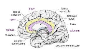
Corpus Callosum
- along with the anterior commissure interconnect most areas of the cerebral cortex
- splenium, body, rostrum
CSF
- cerebrospinal fluid, much like plasma but with less protein
- produced at a steady rate by the choroid plexus - because of this, anything that blocks the pathway resultsin hydrocephalus
- fill ventricles and percolate through brain parenchyma - pours out of three holes in the ventricular walls of the brainstem filling the spaces outside the CNS as well
- choroid plexus —> ventricular system —> out the brainstem apertures —> down around the spinal cord as well as up towards the arachnoid granulation and out into the superior sinus systems and eventually out the venous system
- partial flotation of the brain, indrectly regulates the brain’s extracellular fluid, route of distribution of neuroactive substances produced by nerve cells, and a spatial buffer - moving in and out of the cranial cavity to make room for arterial pulses
Diencephalon
•thalamus - major relay station through which nearly all specific information reaches the cerebral cortex: sensory, outputs from basal ganglia (not olfactory system - bulb reaches cortex directly) •hypothalamus - major control center for ANS, drive related behaviour (hunger, thirst, temperature regulation, neuroendocrine control etc…)
Dura mater
•outermost layer of meninges
Dural Sinuses
- where the dua doubles upon itself, dural sinuses are formed
- thin walled, endothelium lined venous channels within the dura that can be found at several locations within the CNS
- cerebral veins empty into the dural sinuses
- superior saggital sinus - along the attached edge of the falx cerebri
- straight sinus - along the falx/tentorium line of attachment
- transverse sinuses - (left and right) along the attached edge of the tentorium cerebelli
- sigmoid sinuses - (left and right) continuation of each transverse sinus after it leaves the tentorium
- confluence of the sinuses - junction of the superior, saggital, staright and both transverse sinuses
Falx Cerebri
•dural reflection located between the cerebral hemispheres
