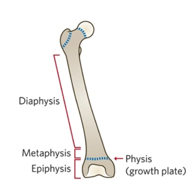
How can you tell a paediatric CXR?
The growth plate (physis) will be seen on CXR in a child
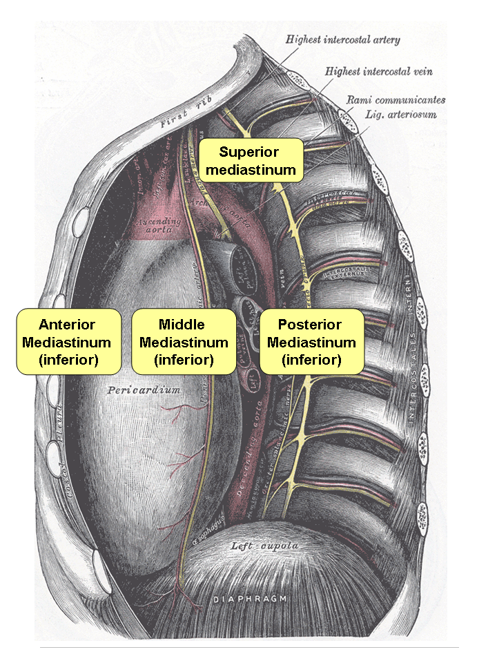
How can you tell on a CXR what area of the mediastinum an opacity etc is in?
- If Trachea is deviated on CXR
- = Anterior mediastinum
- If hyelum if affected
- = middle mediastinum
- if aorta is affected (lower down)
- = posterior mediastinum
What is the appropriate method for systematically checking a CXR?
- pt details
- RIPE
- ABCDE
What patient details should be checked when/before analysing a CXR?
- pt details
- name
- DOB
- image type -
- normally the image is PA
- if it is AP then normally will be written
- imaging indications for CXR
How do you present RIPE?
- R = rotation
- I = inspiration
- P = Penetration
- E = exposure
so ask/state:
- (R) are the clavicular heads and spinous processes equidistant? Spinous processes and clavicles level?
- (I) are there 5-6 anterior ribs / 8-10 posterior ribs?
- (P) Can you see the vertebral bodies in cardiac shadow? ~should be barely visible otherwise it is over or under penetrated
- What penetration is this PA or AP?
- NB: AP cant comment on cardiomeg
- PA cant see scapula
- (E) can you see the glenohumeral joints, upper lobes/apices? & can you see the costophrenic and cardiophrenic angles?
What is an air bronchogram?
an air bronchogram is a tubular outline of an airway made visible by filling of the surrounding alveoli by fluid or inflammatory exudates. Six causes of air bronchograms are; lung consolidation, pulmonary edema, nonobstructive pulmonary atelectasis, severe interstitial disease, neoplasm, and normal expiration.
- tubular outline because around is gunk
- pulmonary vascularture is more prominent
- boarder block = left lingular, (inferior protion of left upper lobe) or right middle
What is reticular opacity?
- meshwork/spidery
- e.g. fibrosis, oedema and some pneumonias
What is patchy (fluffy) opacity?
- palignancy
- pneumonia
- oedema
- haemathorax
e.g. takes a lobe
What is ABCDE in CXR interpretation?
- Airways
- Breathing
- Circulation
- Diaphragn
- Everything else
- THEN ‘in summary’ [ddx, conclusions –> investigations]
What should you be thinking when looking at the airways (A) on x-ray?
- think: trachea, carina and bronchi
- is trachea deviated?
- is the carina narrowed?
- are the bronchi blocked?
- hilum
- are the hilar asymmetrical / symmetrical?
- symmetrical hilar bulking = HTN / sarcoidosis ~more systemic
- asymmetrical hilar bulking = malignancy or TB
- are the hilar asymmetrical / symmetrical?
What should you be thinking when looking at the breathing (B) on x-ray?
- Lung fields
- Pleura
- There are 3x zones in the lung fields to check
- Upper zone / Apex; middle zone: lower zone
- (as cba for 3xRHS and 2xLHS)
- ?consolidation - pneumonia
- ?solid mass - tumour/abcess
- ?boule - emphsema
- widespread bilateral shadowing e.g. pulm oedema?
- there are 2 main boarders to check
- cardiophrenic
- costophrenic
- are the pleura extending to the edges?
- pneumothorax = gap of air on top of lungs as lungs are heavier
- There are 3x zones in the lung fields to check
What should you be thinking when looking at the circulation (C) on x-ray?
- heart boarder
- heart size
- mediastinum
- heart boarder - LH = LV [e.g. cardiomegaly due to mitral regurg sloshing back in]; RH boarder mostly RA
- -silhouette sign –> consolidation, pneumonia
- can split into right upper, middle (cardiac boarder), lower (diaphragm blocked)
- -silhouette sign –> consolidation, pneumonia
- heart size- only commentable on PA (<50% heart:thorax ratio)
- as ap is bigger due to heart being closer to rays
- mediastinum - visible aortic knuckle/notch? - width ?coarctation of aorta & pulmonary trunks? & Hyelum - prominence?vasculature?
- heart boarder - LH = LV [e.g. cardiomegaly due to mitral regurg sloshing back in]; RH boarder mostly RA
What should you be thinking when looking at the diaphragm (D) on x-ray?
- diaphragm and hidden areas
- should see clear definition above (lung air)
- unclear definition below (abdo contents)
- RHS is normally slightly higher due to liver
- ? perforation
- free air under diaphragm
- or perforation of abdo viscous
What should you be thinking when looking at the everything else (E) on x-ray?
- everything else
- bones
- are bones intact?
- soft tissue
- any swellings?
- Devices / lines / artefact
- any lines? - NG tube, central lines
- any artefact?
- devices e.g. pacemakers?
- bones
after completed ABCDE do “in summary,” investigations: bedside, bloods scans
e. g. LFTs as derrangement can impact choice of Abx
e. g. urea to be done for CURB-65 + dehydration
how can you tell the difference between pleural effusion and consolidation?
effusion e.g. free fluid has a meniscus (waterline) and is thicker than consolidation
- the air and fluid is horizontal
- homogeneous density throughout
- pleural effusions may push mediastinum away
Consolidation = fluid within the alveoli
- patchy opacity through lobe - can see air patches (black)
NB: if stuck say increased density which means its whiter
- while less density = clear black
What are the signs of pulmonary oedema?
- horizontal fissre w/t lined in -
- kerley b lines
- hilar expansion (batwings)
What should you do if you see pneumothorax on a CXR?
- 1* pneumothorax
- spontaneous
- if only small rim then no tx needed
- 2* pneumothorax
- trauma / underlying disease
- get chest drain as unlikely to expand on own
- tension pneumothroax
- break in lung (flap of tissue) creates 1x way valve so air goes in that pushes mediastinum to the side
symptoms of tension pneumothorax:
deviated trachea, hyperesonance on pneumo side, ; unwell pt, use cannula in 2nd ICS
check scars e.g. lobectomy as rest of lung will fill the space
How do you tell the difference between a pacemater and an implantable cardioverter defib?
- Pacemaker
- single lead pacemaker
- smaller
- will have 1x area of increased density
- screws into myocardium
- implantable cardioverter defib (ICD)
- bigger - w/ability to shock
- 2x density leads as need to shock across heart
-
Psychiatry history18
-
ECGs to interpret19
-
Alcohol Hx condensed4
-
Systemic review10
-
Summarising and investigations4
-
General Hx3
-
CXR18
-
Information giving56
-
Neurological Exam21
-
Cardiorespiratory Histories20
-
Case Histories5
-
Neurological HISTORIES22
-
Renal medicine and urology Hx's14
-
MSK Hx's35
-
Thyroid Status and Exam19
-
Cardiovascular Examination6
-
MSK Examination38
-
ABGs Interpretation9
-
Intimate Examinations21
-
Breast Lump History6
-
Peripheral vascular examination and ABPI13
-
GI Differentials32
-
Cardioresp differentials81
-
Clin/comm & diversity and Homelessness12
-
ECGs-how-to46
-
AXR interpretation12
-
Motivational Interview8
-
Angry patient / strong emotions4
-
Common drugs to explain49
-
Constitutional DDx27
