What makes up the skeleton of the head?
Skull, mandible, hyoid apparatus, ossicles of the ear, cartilages of the ear and nose.
Define the face, cranium and the mandible.
Face - boney extension of the skull enclosing nasal cavity and forming the roof of the mouth. Cranium - boney box protecting the brain. Mandible - lower jaw bone
The skull has two materials of origin, name and describe them with an example of a structure from each.
Endochondrial bones - arise from unpaired (mainly) structure - eg Basisphenoid. Intramembranous bones - arise from foci developing in mesenchyme, paired - Frontal.
Name the unpaired bones of the skull.
Supraoccipital, Basioccipital, Basisphenoid, Presphenoid, Ethmoid, Vomer
Name four paired bones of the skull.
Temporal, Frontal, Parietal, Exoccipital, Nasal, Maxilla, Zygomatic, Palatine, Lacrimal, Pterygoid, Mandible, Dorsal, Ventral and ethmoid turbinates.
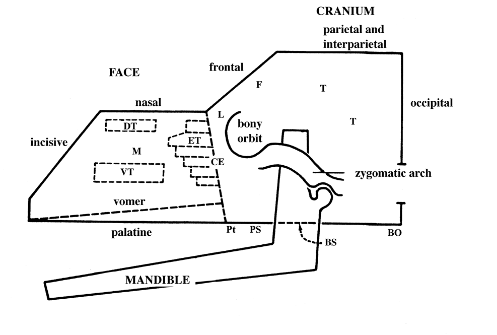
Label the bones of this laterally placed skull. (x7)
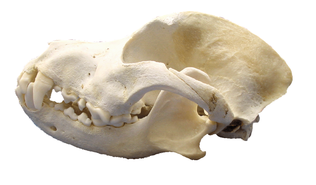
Incisive, nasal, maxilla, frontal, parietal, zygomatic, temporal
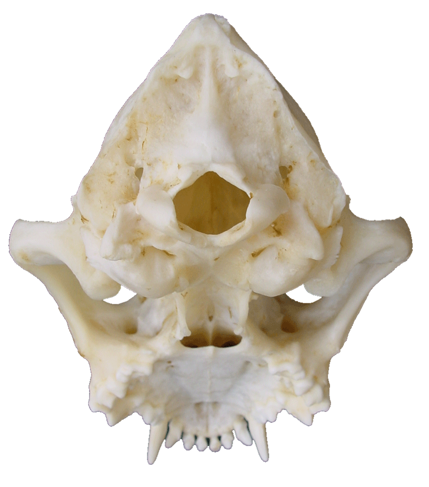
Label the bones of the ventral skull. (x9)
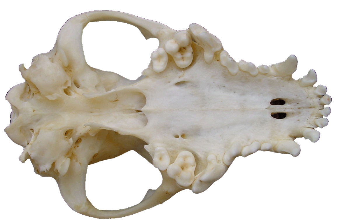
Occipital, bulla, pterygoid, vomer, maxilla, incisive, palatine, presphenoid, basisphenoid
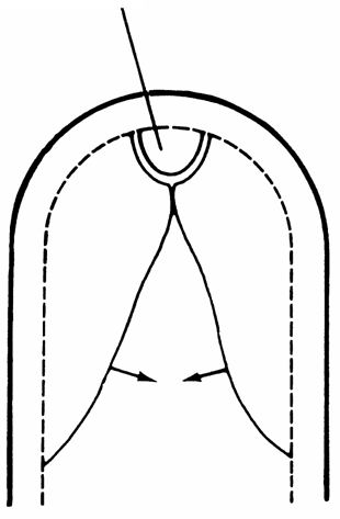
Draw the structure of the primary and secondary palate and describe their development. What definitive structures does each form?
What does failure do close of the primary and secondary palates result in?
Primary - unpaired semilune shape - lip and incisive bone,
Secondary - paired, move to midline from the side - hard and soft palate.
Failure to close the primary palate results in cleft lip, and failure to close the secondary palate results in cleft palate. AKA Congenital oronasal fistula – an abnormal communication between the oral and nasal cavity. Aspiration pneumonia
Describe the structure of the occipital bone.
What condition can be caused with improper development of this bone? Describe
Foramen magnum bordered dorsally by the supraoccipital, laterally by paired extraoccipital and ventrally by the baseoccipital.
Syringomyelia - Congenital condition (CKCS), undersized occipital bone (hypoplasia) . Cerebellum becomes pressed against the foramen magnum & interrupts normal flow of CSF. Pockets of CSF build up within the brain causing the neurological conditions. FM may be keyhole shaped.
Describe the anatomy of the tympanic bulla.
Part of the temporal bone. Contains the middle ear. Filled with air in the healthy animal. Bounded laterally by the tympanic membrane covering the external auditory meatus.
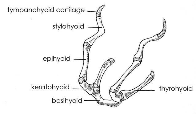
Draw and label the bones and cartilages of the hyoid apparatus.
What is the function of this structure?
Which structure is unique to the horse?
The hyoid apparatus forms a suspensory mechanism for the tongue and larynx. Lingual process of the basehyoid bone
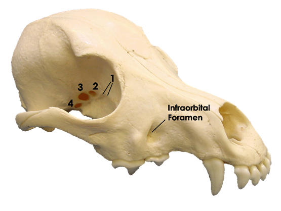
Name the foramina of the skull (x9)
- Ethmoidal foramina 2. Optic canal 3. Orbital fissure 4. Rostral alar foramen 5. Caudal alar foramen and oval foramen 6. Internal carotid foramen 7. Jugular foramen 8. Stylomastoid foramen 9. Hypoglossal foramen
What problems arise from the abnormal shape of the brachycephalic dog skull?
- Stenotic nares - occluded nostrils - > cartilage and soft tissue for the underlying tiny nasal bones to support. Difficulty breathing through nose.
- Long soft palate. Soft palate occludes larynx = difficulty breathing
- Often, have crowded teeth at unusual orientations. Difficulty eating and increased dental disease. Malocclusion due to mandible being less reduced than maxilla so mandible protrudes (prognathism)
- Exopthalmic eyes – bulgy (reduced facial bone). Can also be more prone to proptosis (acute forward displacement of the eyeball.
- Wrinkly skin - Infections
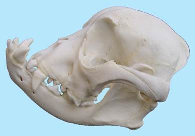
What are the three main dog and cat head shapes? Name a breed with each.
- Dolichocephalic - greyhound, oriental.
- Mesaticephalic - GSD, DSH.
- Brachycephalic - Bulldog, persian.
What is the function of the nasal cavity and how are these achieved? (x3)
- Warm and humidify air going to lungs. - Air brought into contact with large surface area for water and heat exchange.
- Filtering particles from air going to lungs. - Contact surface covered in mucous to trap small particles - hairs at entrance to nasal cavity trap large particles
- Detecting odour molecules in air - Many sensory receptor cells to detect odour molecules
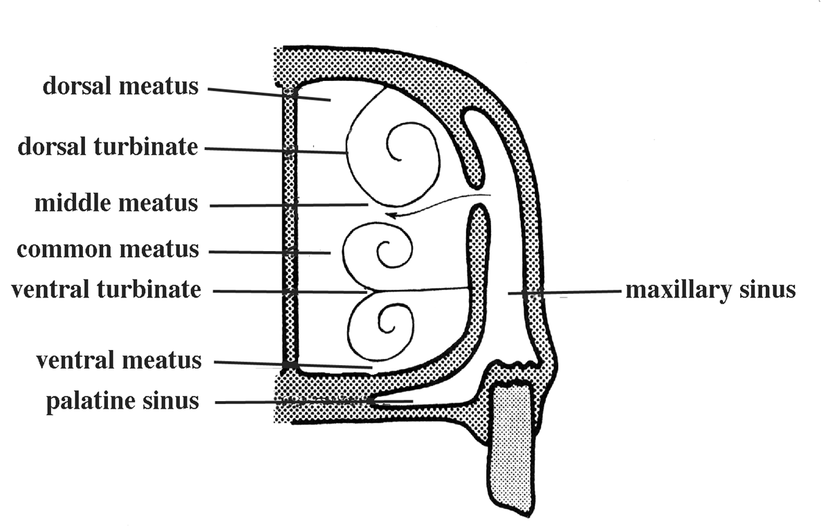
Describe the turbinate bones (x3) (basic description)
- Ethmoturbinates - small and lie towards the back of the nasal cavity. They are attached to the nasal septum, lateral nasal wall and cribriform plate.
- Dorsal Turbinates - single scroll attached to the nasal wall.
- Ventral Turbinate - double scroll attached to the maxilla.
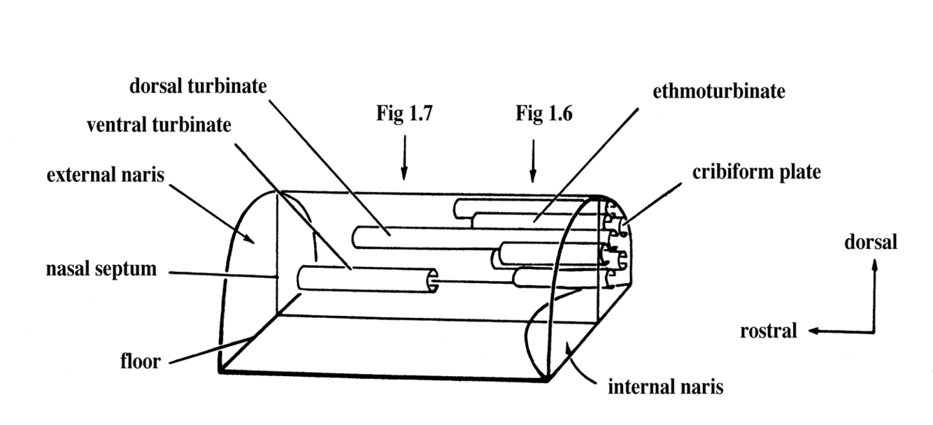
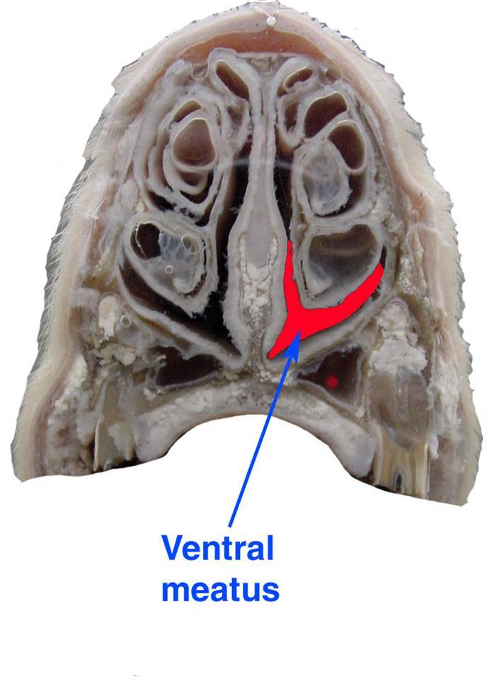
Name the four meati found within the nasal cavity. What are their functions?
- Dorsal meatus – to olfactory mucosa.
- Middle meatus – to the sinus system.
- Ventral meatus – principal airway.
- Common meatus – the medial communicating part
What is a paranasal sinus? Name the main ones of clinical importance. What functions do they serve?
Air-filled diverticula (out pocketing) of the nasal cavity. Frontal and maxillary. Mechanical protection, Thermal protection, Enlarge skull without adding weight
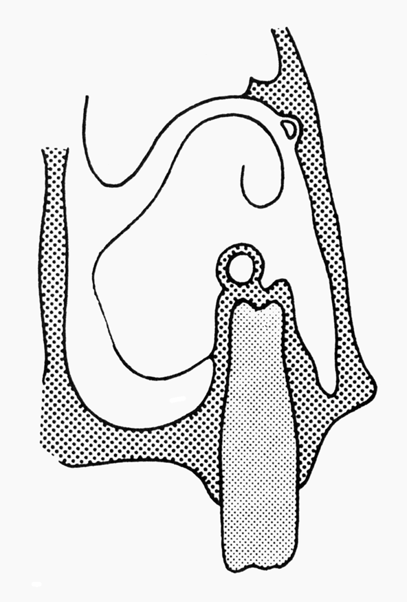
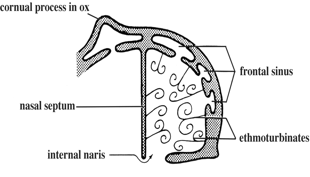
Draw and describe the frontal sinus. (of the dog)
- Lies within the frontal bone
- Has up to five separate compartments
- In the ox and sheep one of the 5 compartment is the CORNUAL PROCESS. This extends into the horn.
- In all domestic species except the horse the frontal sinus communicates directly with the nasal cavity by way of small openings between the ethmoturbinates.
What rules two rules are vital to remember when euthanasing animals via shooting?
- Aim for MEDULLA OBLONGATA – the respiratory and cardiovascular centre of the brain
- Avoid midline on species with a strong bony midline septum - species dependent
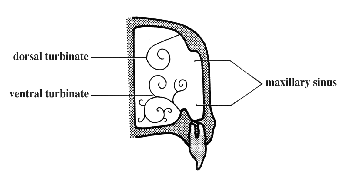
Draw and describe the maxillary sinus of the dog and cat.
Often known as the maxillary recess since it communicates so freely with the nasal cavity.
How do the palatine and maxillary sinus communicate in the horse?
(HINT: Draw)
Draw the arangement of the sinuses in the horse.
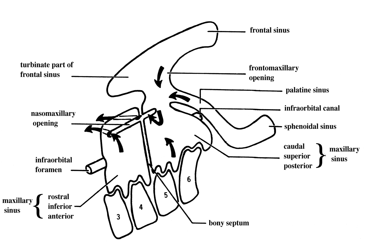
How does the maxillary sinus of the horse alter with age?
Young horse: Lateral parts of the maxillary sinus are much smaller due to the unerupted cheek teeth occupying the maxillary space.