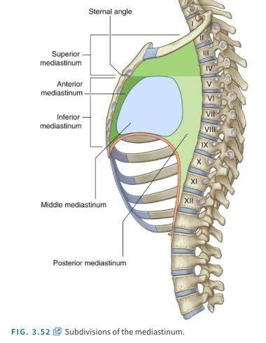
Describe the mediastinum
Mediastinum is the space between the pleura. It can be anatomically located:
- Between the vertebral bodies and sternum
- Between the superior thoracic aperture and diaphragm
It can be split into the superior and inferior mediastinum by this landmark:
- Sternal angle and TIV/TV
Inferior mediastinum can be further split into anterior, middle, and posterior mediastinum
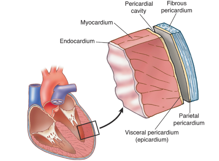
Describe the 3 layers of the heart
- Pericardium
- Myocardium-heart muscle
- Endocardium-inner most layer
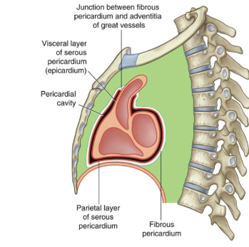
Describe the pericardial layers
Pericardium is divided into outer fibrous pericardium and inner serous pericardium
- Outer fibrous pericardium-attached to thoracic cage via central tendon of diaphragm and sternopericardial ligaments
- Inner serous pericardium- parietal and visceral layer containing serous fluid which allows for cardiac distention
What are the pericardial sinuses and what is their clinical importance?
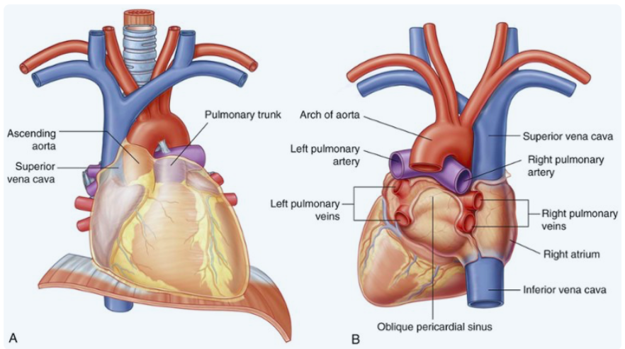
- Transverse pericardial sinus- behind aorta and pulmonary trunk and in front of SVC. Place finger through this during surgery to separate arteries from veins, during cardiac surgery-> where clamp goes
- Oblique pericardial sinus-around pulmonary veins. Place finger up from apex during open cardiac surgery to gain access

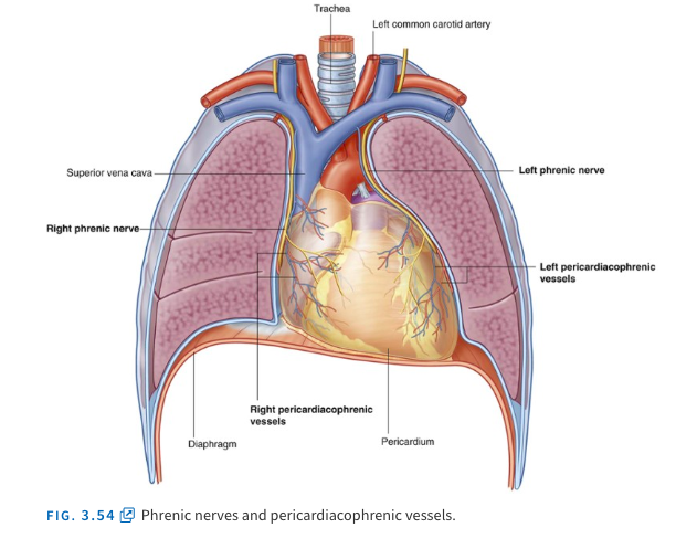
What is the neurovascular supply of pericardium?
The arterial blood supply is from the internal thoracic artery and its branches: pericardiophrenic, musculophrenic as well as thoracic aorta and its branch: inferior phrenic artery.
The venous drainage is via the internal thoracic vein and inferior phrenic vein.
The nervous supply is via phrenic nerve (C3, C4, C5) which carry somatic afferent fibres(pain) that also supply the dermatomes in lateral neck and supraclavicular region of shoulder.
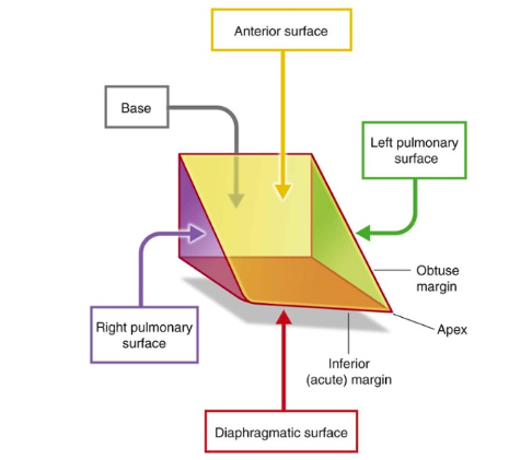
Describe the heart (apex, base, right and left pulmonary surfaces and anterior surface; diaphragmatic surface)?
Describe the margins of the heart?
Imagine heart as fallen down pyramid (pg185)
- Base-right and left atrium with pulmonary veins and vena cavae
- Apex (opposite base)- tip of heart in 5th ICS lt, 8-9cm from sternum
- Right pulmonary surface (facing right lung)-right atrium
- Left pulmonary surface (facing left lung)-left atrium and left ventricle
- Anterior surface-Right atrium, right ventricle,and a bit of left ventricle
- Diaphragmatic surface-right and left ventricle
Margins of heart
- Right and left margins of heart (same as PS)
- Inferior margin- sharp line between anterior and diaphragmatic surface
- Obtuse margin-curve between left auricle and apex between anterior and left pulmonary surface
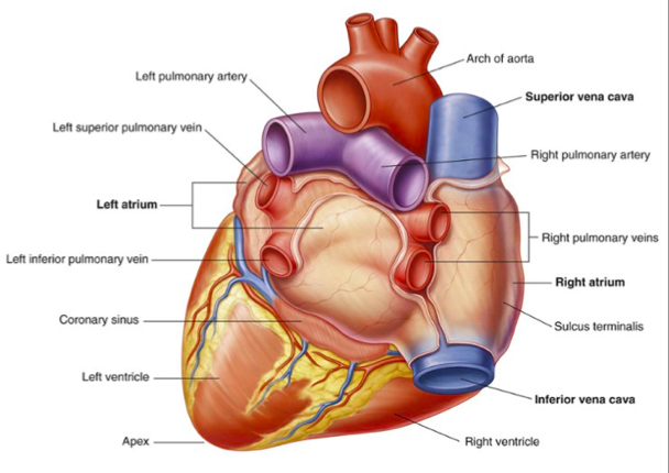
Describe the base of the heart?
Base of the heart consists of the:
- right atrium-w SVC and IVC
- left atrium-pulmonary veins
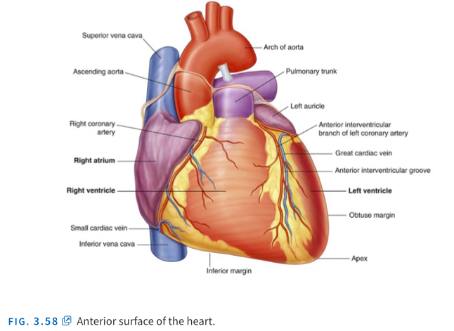
Describe the anterior surface of the heart?
Where is the apex located?
Anterior surface of the heart consists of:
- right atrium and right ventricle
- left ventricle
Apex is located 8-9cm from the midsternal line
Describe the right and left pulmonary surface?
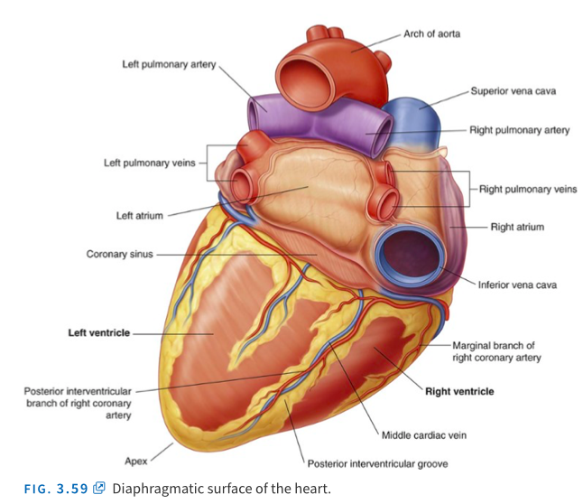
Describe the diaphragmatic surface?
Right and left pulmonary surface face the right and left lungs:
- right surface-right atrium
- left surface-left atrium
Diaphragmatic surface:
- right and left ventricle separated by interventricular groove
What does the coronary sulcus contain and what does it separate?
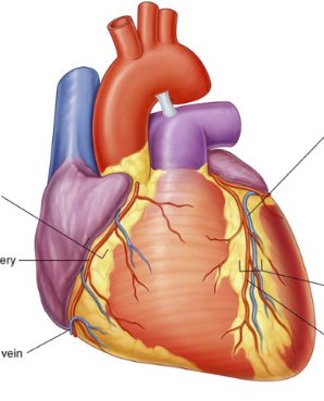
The heart has 3 sulci that help partition the chambers of the heart.
Coronary sulcus separrates the atria from the ventricles:
- contains the small cardiac vein, right coronary artery, coronary sinus, circumflex branch of the left coronary artery
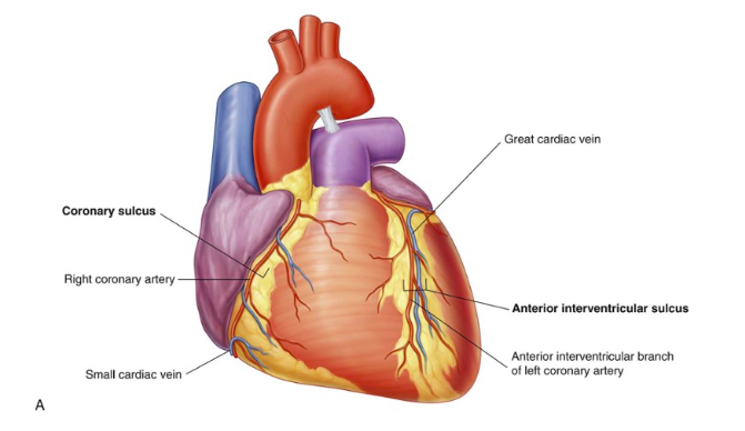
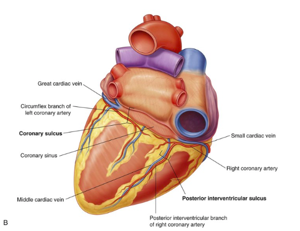
What does the anterior and posterior interventricular sulci contain?
Anterior interventricular sulci contains:
- anterior interventricular branch of the left coronary artery
- great cardiac vein
Posterior interventricular sulci contains:
- posterior interventricular branch of the right coronary artery
- middle cardiac vein
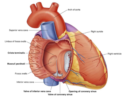
Describe the right atrium?
The right atrium forms the right border of heart, and is covered by the right auricle “ear”. It receives deoxygenated blood from the superior and inferior vena cavae as well as the coronary sinus.
The right atrium pumps blood to the right ventricle via the right atrioventricular orifice, guarded by the tricuspid valve.
The interior of the right atrium has both muscular and smooth walls which can be divided by crista terminalis:
Space anterior to the crista-muscular pectinati (rough)
Space posterior to crista-smooth
Separating the right and left atrium is a wall called the interatrial septum. A depression called the fossa ovalis. It is derived embryologically from the foramen ovale, which hole in the fetus heart that allowed oxygenated blood to flow from right to left atrium, bypassing the lungs (since lungs are non-functional before birth).
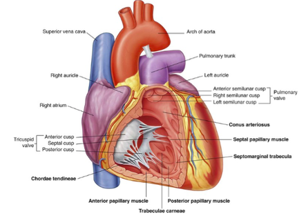
Describe the right ventricle?
The right ventricle forms majority of anterior sternocostal surface and portion of diaphragmatic surface. It receives deoxygenated blood from the right atrium via the tricuspid valve:
- Tricuspid valve
- it consists of fibrous ring that surrounds the right atrioventricular orifice. Three cusps are attached to the ring: anterior, posterior and septal cusp which are attached to the anterior, posterior and septal papillary muscles via the chordae tendinae, “heart strings”
The right ventricle pumps blood to the lungs via the pulmonary valve.
- Pulmonary valve
- consists of 3 semi lunar cusps: anterior, right and left cusp. The edge of the cusp is called the nodule (thickened edge in middle) and has 2 lateral portions called lunula of the semilunar cusp. Each valve has a pulmonary sinus.
The ventricular inflow portion has trabeculae carnae (rough) where papillary muscles originate from compared to the smooth outflow tract, infundibulum.
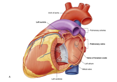
Describe the left atrium?
The left atrium forms most of the base and diaphragmatic surface of the heart, covered by the left auricle. It receives oxygenated blood from the 4 pulmonary veins.
Like the right atrium, the interior has smooth and muscular walls however there is no crista terminalis division:
- Space anterior-is rough with musculi pectinati derived embryonically from primitive atrium.
- Space posterior-is smooth and is derived embryonically from the tissue that forms the pulmonary veins. The 4 pulmonary veins open into this space.
The left atrium drains into the left ventricle via the left atrioventricular orifice, guarded by the bicuspid/mitral valve.
Separating the left atrium and right atrium is the interatrial septum. On this side, the valve of the foramen ovale can be seen and is opposite the floor of the fossa ovalis. In fetal development, it closes to prevent the backflow of blood from left to right atrium.
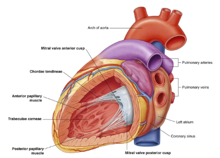
Describe the left ventricle?
The left ventricle contributes to the anterior, diaphragmatic and left pulmonary surface as well as apex. It receives oxygenated blood from the left atrium via the mitral/bicuspid valve:
- Mitral/bicuspid valve
- consists of a fibrous ring that surrounds the left atrioventricular orifice. Anterior and posterior cusps attach to the ring like a mitre hat: and are attached to the anterior and posterior papillary muscles respectively via the chordae tendinae.
The left ventricle pumps blood to the body via the aortic valve:
- Aortic valve
- consists of 3 semi lunar cusps: posterior, right and left cusp. Similar to the pulmonary valve, the cusp has a nodule in the middle and lunula at the sides. These for aortic sinuses which blood fill. In particular, right and left aortic sinus contain the openings for the right and left coronary arteries.
The walls of the inflow portion of the left ventricle are similar to that of the right ventricle as they are rough and filled with trabeculae carnae.
What is the coronary supply of the heart?
Vascular:
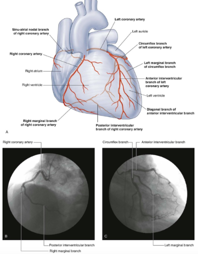
The heart is supplied by the right and left coronary arteries which give off several branches.
Right coronary artery arises from the right aortic sinus of the ascending aorta and gives off several branches:
- Sinoatrial node branch-arises at the groove between the right auricle and ascending aorta. Passes to supply the sinoatrial node.
- Right marginal artery-courses along the inferior margin of the heart to the apex
- Atrioventricular node branch
- Posterior interventricular branch of right coronary artery-found posteriorly, in the posterior interventricular sulcus along with the middle cardiac vein
Overall, it supplies the right atrium (+interatrial septum), right ventricle, as well as the sinoatrial and atrioventricular nodes and posterior 1/3 of the interventricular septum.
Left coronary artery arises from the left aortic sinus of the ascending aorta. It passes between the pulmonary trunk and left auricle to give off several branches:
- Anterior interventricular branch of left coronary artery-found in the anterior interventricular sulcus along with the great cardiac vein. During this course, it gives off diagonal branches which goes diagonally across the left ventricle.
- Left circumflex artery-passes posteriorly by circling around the heart. It gives off the left marginal artery which courses down the obtuse margin.
Overall, it supplies the left atrium and left ventricle, anterior 2/3 of the interventricular septum.
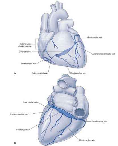
What is the venous drainage of the heart?
The heart is drained by the four main veins: great, posterior, middle, small cardiac veins which drain into the coronary sinus in the right atrium.
- Great cardiac vein-ascends in the anterior interventricular sulcus along with anterior interventricular branch of left coronary artery as the anterior interventricular vein. Loops left to the posterior of heart, where it is associated with left circumflex artery, before it enlarges to form the coronary sinus.
- Posterior cardiac vein- Ascends posteriorly on left ventricle to either join the coronary sinus directly or great cardiac vein.
- Middle cardiac vein-Ascends posteriorly in the posterior interventricular sulcus along with the posterior interventricular branch of right coronary artery.
- Small cardiac vein-Starts anteriorly in coronary sulcus and is associated with right coronary artery and loops posteriorly to join the coronary sinus. It receives right marginal vein tributary.
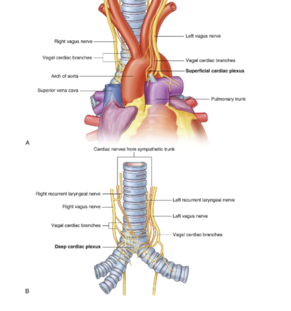
Describe the innervation of the heart?
Heart receives sympathetic (from T1 to T4) and parasympathetics (from vagus nerve) which join to form the :
- Superficial cardiac plexus-in front of aortic arch
- Deep cardiac plexus-in front of the tracheal bifurcation
Function:
- Sympathetics- causes increase in HR; and increase in force of contraction
- Parasympathetics- cause decrease in HR; decrease in force of contraction; constricts coronary arteries
Also receive visceral afferents (sensory neurons) branching from vagus/sympathetics:
- Visceral afferents from vagus-sense changes in blood pressure/blood chemistry and send it to CNS
- Visceral afferents from sympathetics- sense pain (such as MI) and send it to CNS. Join with somatic sensory afferents from T1, T2, T3, T4. which explains referred pain to left arm.
Describe the pulmonary trunk?
Pulmonary trunk is contained by the visceral layer of the serous pericardium. It splits at the level ot T4:
- right pulmonary artery (supplies right lung)-goes behind the SVC and ascending aorta
- left pulmonary artery (supplies the left lung)-goes inferior to arch of aorta and in front of descending aorta
Describe the ascending aorta and at which point does it become the arch of aorta?
The Ascending aorta starts from the left ventricular outflow tract, at this point it is known as the aortic vestibule, at the level of the 3rd costal cartilage.
Aortic vestibule has 3 cusps:
- posterior cusp
- right and left cusp-where the right and left coronary arteries originate
This ascends to the level of the 2nd costal cartilage. At this point it is known as the arch of aorta
