Identify the Image
Top, right side, left side

- Ocular
- Stage
- Focus
- Objective
- Condenser diaphragm
- Field diaphragm
Identify the Image
Left, Right

- Manufacturer
- Magnification
- Tube length
- Magnification Color Code
- Lens Correction
- Numerical Aperature
- Indicates Oil Objective
- Coverglass Thickness
Identify the Type of Light Microscopy

- Bright field microscopy
- Darkfield microscopy
- Phase-contrast microscopy
- Transmission electron microscopy (TEM)
Identify the Type of Light Microscopy

- Nomarski (differential interference contrast)
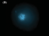
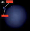
- Fluorescence microscopy
- Confocal laser scanning microscopy
- Scanning Electron microscopy (SEM)
What is the purpose of the objective? What is the range in magnification?
Provides the main source of magnification
Range in magnifications of 2.5-100x
What is the purpose of an eyepiece?
Used to view the specimen image magnified by the objective
What is the purpose of a stage?
Holds the specimen and allows for movement within the optical path of the micrscope
What is the purpose of focus knobs (fine and coarse)?
Raise and lower the height of the objective, thereby changing the plane of focus
What is the purpose of the condenser?
Focuses the light passing form the lamp to the specimen
Its height relative to the specimen is essential in establishing Köhler Illumination
What is the purpose of the condenser knob?
Raises and lowers the condenser
What is the purpose of a condenser diaphragm?
Regulates the amount of light passing through the condenser
Adjusting its diameter regulates how much light illuminates the specimens - closing condenser diaphragm can reduce scattered light traveling through the microscope, thus improving resolution
What is the purpose of the field diaphragm?
Located above the lamp housing/light source
It regulates the amount of light traveling form the lamp to the condenser
What is the light source?
Consist of a bulb which provides illumination
What is Köhler illumination?
A lighting technique so the specimen can be uniformly lit with minimal interference from the internal components of the microscope
What are the two focal planes established in Köhler illumination?
- The Conjugate Field Planes; back of the retina, field stop of injective, the specimen, and the field diaphragm are all in focus at once
- The Conjugate Aperature Plane; iris of the eye, rear focal plane of the objective, the front focal plane of the condenser, and the lamp filaments are all in focus at once
Light Microscopy
Light microscopy utilized light to illuminate the speciment
Bright-field microscopy
The specimen is illuminated with the full wavelengths of visible light and contrast in the image is provided by the specimen itself
Both live and preserved specimens can be viewed
How do you improve contrast with bright field microscopy?
If the specimens are preserved you can use a Hematoxylin and Eosin (H&E) stain
Dark Field Microscopy
The exclusion of unscattered light, thus regions that don’t scatter light (no specimen) appear black
If you need increased contrast of a live specimen, what microscopy technique should you use?
Dark Field Microscopy
Phase Contrast Microscopy
Utilizes the differences in refractive indices within a specimen to improve contrast
What microscopy is especially useful for viewing unstained specimens and viewing edges of structures where refractive index becomes more evident?
Phase Contrast
Differential Interference Contrast (DIC)
Aka Nomarski
Similar to Phase Contrast microscopy
It utilizes polarized light to create two separate coherent image components which are then recombined
The interference generated during the recombination allows for difference in refractive index to be detected, thereby creating increased contrast
What microscopy technique is especially useful for viewing intracellular components such as vesicles
Differential Interference Contrast (DIC)
aka Nomarski