What make up the External, Middle and Internal ear
- External Ear – Auricle, external acoustic meatus, external surface of the tympanic membrane
- Middle – Internal surface of the tympanic membrane, tympanic cavity, ossicle, pharyngotympanic tube
- Internal – auditory apparatus, vestibular apparatus, internal auditory meatus, vestibulocochlear nerve (CN VIII), oval window, round window
Describe the features of the External Acoustic Meatus
- External Acoustic Meatus
- Extends from deepest part of concha to lateral surface of tympanic membrane (~2.5cm)
- Does NOT follow straight course
- Need to pull the ear superior, posterior and laterally
- Lateral ~1/3 = Cartilagenous (lined with squamous epithelium)
- Medial ~2/3 = Bony Tunnel (but this is varied!!!!)
- Swimmer’s ear (otitis externa) is a painful condition resulting from an infection in external acoustic meatus.
What are the innervations of the external ear
- Auricle:
- Superifical = cervical plexus (lesser occipital, greater auricular), V3 (auriculotemporal branch of mandibular)
- Deeper parts = X**, **VII (more post. mainly near mastoid processes_)_
- External acoustic meatus: V3, V2, X
- Lower jaw is innervated by the same ear. Patient may come in with Ear pain + lower jaw pain. If they come in with lower jaw pain, you must also examine the ear
- Tympanic membrane:
- Outer surface = V3, V2, X** (+ small **IX).
- Inner surface = IX (glossopharyngeal)

The external acoustic meatus is not ___________ So you need to _______
IS NOT STRAIGHT
so you need to pull the ear superiorly, laterally and posteriorly to make it straight (other way around for bebies)
NOTE *he will examine the tympanic membrane
Learn the tympanic membrane inside out.
What is the middle ear lined with?
- Air-filled space with two parts (tympanic cavity and _epitympanic reces_s) within Temporal Bone
- Lined with Mucous Membrane
- Transmits, contains and communicates with important structures…

What makes up the boundaries of the middle ear?
Roof (“Tegmental Wall”)
Tegmen typmani bone separates middle ear from cranial fossa (Brain)
Floor (“Jugular Wall”)
Separates middle ear from Internal Jugular Vein below
On medial aspect, small branch of CN IX tympanic n. (parasymp.) enters middle ear to join tympanic plexus
Lateral Wall (“Membranous Wall”)
Almost entirely Tympanic membrane except upper part
Separates Middle ear from External Auditory Meatus
Posterior Wall (“Mastoid Wall”) (partially complete)
- separates the middle ear from mastoid air cells
- continues to mastoid antrum via aditus superiorly (possible infection route)
Transmits:
- Tendon of Stapedius (pyramidal eminence)
- Chorda Tympani (CN VII)
Anterior Wall (“Carotid Wall”) (partially complete)
Separates Middle Ear from Internal Carotid Artery inferiorly
Connected to Pharynx via the Pharyngotympanic Tube (Eustacian/Auditory Tube) superiorly
Transmits:
- Tensor Tympani Muscle (enter superiorly)
- Lesser Petrosal n. (exiting to Parotid)
- Caroticotympanic n. (symp. from Int. Carotid plexus)
- Chorda Tympani n. (exiting to oral cavity inferiorly)
Medial Wall (“Labyrinthine Wall”)
Separates Middle Ear from Inner Ear (Vestibulocochlear apparatus)
Key Features:
- Promotory (covered by mucous membrane containing Tympanic plexus (formed by tympanic branch of CN IX (glossopharyngeal), and caroticotympanic nerves of internal carotid plexus))
- Oval Window – connected to Stapes
- Round Window (vibration exit from inner ear)
- Lesser petrosal nerve (branch of tympanic plexus) leaves the middle ear, runs along middle cranial fossa through foramen ovale to reach otic ganglion carrying preganglionic parasympathetic fibres.
- (Ridge of Facial Nerve Canal)
- (Ridge of Lateral Semicircular canal)

What sits posteriorly to the middle ear?
Matsoid air cells (sits posterior to the middle ear)
(also connects to the nasal phaynx via the adutus to mastroid antrum and pharyngotympanic tube)

What does the roof of the middle ear separate it from?
(Also called the tegmental wall)
Separates middle ear with middle cranial fossa
Roof (“Tegmental Wall”)
Tegmen typmani bone separates middle ear from cranial fossa (Brain)

What does the floor of the middle ear separate it from?
What structures goes through the floor?
(Jugular wall)
Separates middle ear from Internal Jugular Vein below
On medial aspect, small branch of CN IX tympanic n. (parasymp.) enters middle ear to join tympanic plexus

Lateral Wall = Membranous wall
What is found in the lateral wall of the middle ear?
What does the lateral wall separate the middle ear from?
Lateral Wall (“Membranous Wall”)
Almost entirely Tympanic membrane except upper part (Epitympanic recess)
Separates Middle ear from External Auditory Meatus
(the lateral wall cannot be seen in the box pic)

Name the parts of the tympanic membrane
- Pars Faccida
- Lateral process of Malleus)
- Anterior malleolar fold
- Cone of light
- Umbo
- Handle of malleus
- Posterior malleolar fold

Label (A-F)
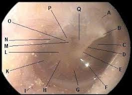
A = Pars Flaccida
B = Short process of Malleus
C= Pars Tensa
D= Manbrium of Malleus (Handle of Malleus)
E= Umbo
F = Cone of light
Label

- Pars Faccida
- Lateral process of Malleus)
- Anterior malleolar fold
- Cone of light
- Umbo
- Handle of malleus
- Posterior malleolar fold

Describe the Innervation of the Tympanic Membrane
- Outer surface = stratified squamous epithelium (like external auditory meatus)
- Innervated by _CN V3 (_auriculotemporal), ( and some from V2 and X )(auricular branch)
- Inner Surface = Mucous membrane (like tympanic cavity)
- Innervated by Tympanic branches of CN IX
What does the medial wall of the middle ear separate the middle ear from?
What are the features of the medial wall
Medial Wall (“Labyrinthine Wall”)
Separates Middle Ear from Inner Ear (Vestibulocochlear apparatus)
Key Features:
- Promotory (covered by mucous membrane containing Tympanic plexus (formed by tympanic branch of CN IX (glossopharyngeal), and caroticotympanic nerves of internal carotid plexus))
- Oval Window – connected to Stapes
- Round Window (vibration exit from inner ear)
- Lesser petrosal nerve (branch of tympanic plexus) leaves the middle ear, runs along middle cranial fossa through foramen ovale to reach otic ganglion carrying preganglionic parasympathetic fibres.
- (Ridge of Facial Nerve Canal)
- (Ridge of Lateral Semicircular canal)
What are the key features of the medial wall of the middle ear?
Medial Wall (“Labyrinthine Wall”)
Separates Middle Ear from Inner Ear (Vestibulocochlear apparatus)
Key Features:
- Promotory (covered by mucous membrane containing Tympanic plexus (formed by tympanic branch of CN IX (glossopharyngeal), and caroticotympanic nerves of internal carotid plexus))
- Oval Window – connected to Stapes
- Round Window (vibration exit from inner ear)
- Lesser petrosal nerve (branch of tympanic plexus) leaves the middle ear, runs along middle cranial fossa through foramen ovale to reach otic ganglion carrying preganglionic parasympathetic fibres.
- (Ridge of Facial Nerve Canal)
- (Ridge of Lateral Semicircular canal)

Describe the pathway of the facial nerve and the Chorda Tympani nerve
- Facial nerve goes into the internal acoustic meatus
- Runs into the Promenence of facial canal
- Then it goes out the styloid mastoid foramen
- On its way, it first gives off the Chorda tympani nerve
- Chorda Tympani Crosses the medial surface of tympanic membrane and handle of malleus
- Leaves the tympanic cavity via petrotympanic fissure
- Joins the lingual nerve
- Parasympathetic = sub-lingual and sub-mandibular salivary glands
- Special sense of taste for the anterior 2/3 of the tongue

What does the Chord Tympani nerve do?
Taste sensation from anterior 2/3 of tongue
Also parasympathetic activity from the submandibular to sublingual glands.
pic (look from inside out)

Describe the study done in Dunedin around the anatomy of the Chorda Tympani Nerve
Looked at lots of cadavers.
15-22% patients with middle ear surgeries, they get post-operative taste disburbances and mouth driness (damage to the chorda tympani)
15% of chorda tympani nerves branched AFTER the facial nerve left the skull (different to the textbooks)
Also, CT nerve enteres at 62% height of the Tympanic Membrane (textbook says enteres at upper malleus)
If you damage the Tympanic Membrane (going into middle ear) there is a chance of damaging…..
Chorda Tympani Nerve (Taste sensation to anterior 2/3 tongue and also parasympathetic activity from the submandibular to sublingual glands)
What does the anterior wall of the middle ear separate the middle ear from?
What is the anterior wall connected to?
What does it transmit?
Anterior Wall (“Carotid Wall”) (partially complete)
Separates Middle Ear from Internal Carotid Artery inferiorly
Connected to Pharynx via the Pharyngotympanic Tube (Eustacian/Auditory Tube) superiorly
Transmits:
- Tensor Tympani Muscle (enter superiorly)
- Lesser Petrosal n. (exiting to Parotid)
- Caroticotympanic n. (symp. from Int. Carotid plexus)
- Chorda Tympani n. (exiting to oral cavity inferiorly after it comes in via the posterior wall)

What is another name for the eustachian tube?
Pharyngotympanic Tube
Describe the Pharyngotympanic Tube
Runs from Nasopharynx to Middle Ear (Anterior Wall)
Lateral (1.2cm) = Bone canal
Medial (2.5cm) = Cartilagenous
Normally closed, but when tensor veli palatini contracts (i.e. during swallowing/yawning) the lumen opens
Equalises pressure on both sides of the tympanic membrane
Sensory innervation via CN IX (same as tympanic cavity)

-
Lecture 2: Meninges & Orbit65
-
Lecture 3 and 4: Opthomology50
-
Lecture 5 and (6?): Posterior Triangle of the Neck29
-
Lecture 1: Skull51
-
Lecture 7: Ears25
-
Lecture 8 and (9?): Anterior Triangle of The Neck50
-
Lecture 10 and (11?): Face and Parotid41
-
Lecture 12 and (13?) : Oral Cavity and Submandibular gland56
-
Lecture 14 and (15?): Nasal cavity56
-
Lecture 16 and (17?): Pharynx and Larynx67
-
Lecture 19: Cross Sections8
-
Lecture 18: Infratemporal and pterygopalatine fossae51
-
Cranial nerves33
-
Radiology39
-
Anatomy79
