II. The Visual System: The retina
A. The retina consists of several thin layers of cells distributed across the inside of the eye.
B. The fovea is the portion of the retina where light falls from an object that you are looking directly at. It is the portion of the retina with the highest acuity, the ability to resolve fine detail and patterns of light. Note: Acuity and sensitivity are different.
C. The optic disk is the retinal location where axons from a type of retinal cell collect and exit the eye and form the optic nerve. This is the blind spot because there are no photoreceptors in the optic disk.
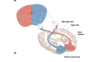
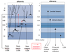
D. Below is a diagram depicting the eye and how a visual image is “mapped” onto the retina:
E. A neuron’s receptive field is the location in the environment (or the surface of the body) from which an appropriate stimulus will change that cell’s activity. For example light at ‘A’ (the tip of the flame in the diagram above) will affect the activity of retinal cell in location ‘a’ in the retina. Cells in different locations in the retina have receptive fields in different locations in the visual field.
F. There are five cell types in the retina.
- Photoreceptors – The first stage in the visual system.
a. Photoreceptors - the only cell type in the visual system that is directly sensitive to light.
b. There are two types of photoreceptors:
1) Rods
a) There are about 120 million rods in the human retina.
b) Rods are highly sensitive to light and are responsible for vision in very dim
light.
c) Rods are bleached in bright light and thus unresponsive in bright light.
d) Rods are not responsible for high acuity vision (not good for fine detail). e) Rods are achromatic (insensitive to colors).
f) Rods only exist outside of the fovea.
2) Cones
a) There are about 6 million cones in the human retina.
b) Cones are less sensitive to light intensity and are inoperative in dim light.
c) Cones are sensitive to color. There are three subtypes, selectively sensitive to
red, blue, and green wavelengths of light. d) Cones are most concentrated in the fovea.
c. Photoreceptors project to the bipolar cells. 2. Bipolar cells (BPs) - Retinal ganglion cells (RGCs)
a. RGCs are the only output cell type in the retina.
b. RGCs are the only means by which information from the eye gets to the rest of the
visual system and their axons form the optic tract. 4. Horizontal cells (HCs) - Amacrine cells (ACs)
G. Why is it that only the RGCs have axons?
1. Axons are needed for long-distance transfer of information. In the retina, the cells are very close together and so don’t need action potentials or axons. Also, communication by PSPs may be able to convey information that is more subtle than can be conveyed by the AP frequency code (recall that PSPs are graded).
2. Only the RGCs and ACs generate action potentials. The rest of the cell types use graded depolarization to release neurotransmitter to the next cell. A depolarization
increases neurotransmitter release. Small depolarizations cause small release of neurotransmitter; large depolarizations cause large release of neurotransmitter.
H. The relationship between different cell types in the retina.
- The retina is “inside-out” with the photoreceptors furthest away from the light (at the very back of the eye) and the RGCs the closest to the light. Thus light must pass through the other cell types to reach the photoreceptors.
- This works because all the cells in the eye, except the photoreceptors, are translucent.
- Also, at the foveal pit all cell types, except the photoreceptors, are pushed out of the way (see 9.15).

The retina consists of:
several thin layers of cells distributed across the inside of the eye.
What is the fovea?
The fovea is the portion of the retina where light falls from an object that you are looking directly at. It is the portion of the retina with the highest acuity, the ability to resolve fine detail and patterns of light. Note: Acuity and sensitivity are different.
What is the optic disc?
The optic disk is the retinal location where axons from a type of retinal cell collect and exit the eye and form the optic nerve. This is the blind spot because there are no photoreceptors in the optic disk.
diagram depicting the eye and how a visual image is “mapped” onto the retina:

What is a neuron’s receptive field?
A neuron’s receptive field is the location in the environment (or the surface of the body) from which an appropriate stimulus will change that cell’s activity. For example light at ‘A’ (the tip of the flame in the diagram above) will affect the activity of retinal cell in location ‘a’ in the retina. Cells in different locations in the retina have receptive fields in different locations in the visual field.
What are the five cell types in the retina?
Photoreceptors, bipolar cells, retinal ganglion cells, horizontal cells, and amacrine cells

Photoreceptors – The first stage in the visual system.
a. Photoreceptors - the only cell type in the visual system that is directly sensitive to light.
b. There are two types of photoreceptors:
1) Rods
a) There are about 120 million rods in the human retina.
b) Rods are highly sensitive to light and are responsible for vision in very dim
light.
c) Rods are bleached in bright light and thus unresponsive in bright light.
d) Rods are not responsible for high acuity vision (not good for fine detail). e) Rods are achromatic (insensitive to colors).
f) Rods only exist outside of the fovea.
2) Cones
a) There are about 6 million cones in the human retina.
b) Cones are less sensitive to light intensity and are inoperative in dim light.
c) Cones are sensitive to color. There are three subtypes, selectively sensitive to
red, blue, and green wavelengths of light. d) Cones are most concentrated in the fovea.
c. Photoreceptors project to the bipolar cells.

Retinal ganglion cells (RGCs)
a. RGCs are the only output cell type in the retina.
b. RGCs are the only means by which information from the eye gets to the rest of the
visual system and their axons form the optic tract.

Why is it that only the RGCs have axons?
- Axons are needed for long-distance transfer of information. In the retina, the cells are very close together and so don’t need action potentials or axons. Also, communication by PSPs may be able to convey information that is more subtle than can be conveyed by the AP frequency code (recall that PSPs are graded).
- Only the RGCs and ACs generate action potentials. The rest of the cell types use graded depolarization to release neurotransmitter to the next cell. A depolarization
increases neurotransmitter release. Small depolarizations cause small release of neurotransmitter; large depolarizations cause large release of neurotransmitter.
The relationship between different cell types in the retina.
- The retina is “inside-out” with the photoreceptors furthest away from the light (at the very back of the eye) and the RGCs the closest to the light. Thus light must pass through the other cell types to reach the photoreceptors.
- This works because all the cells in the eye, except the photoreceptors, are translucent.
- Also, at the foveal pit all cell types, except the photoreceptors, are pushed out of the way (see 9.15).
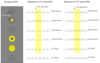
What is phototransduction?
Phototransduction is how light energy leads to a change in membrane potential.
Below is a diagram depicting how light changes photoreceptor membrane potential.
- The resting membrane potential of photoreceptors is -30 mV and this is in the dark.
- The maximum hyperpolarization is down to -65 mV and this is produced by bright light.
- Glutamate is the neurotransmitter used by photoreceptors. The greater the intensity of light, the less neurotransmitter released.

How does light produce the graded hyperpolarization?
A. The ligand-gated Na+ channels in the outer segment membrane are open in the dark, causing depolarization (to the “resting membrane potential” of -30 mV).
B. These ligand-gated channels are like receptors, but they are “inside-out”, meaning that they bind their ligand cGMP to a binding site on the intracellular face of the Na+ channel and this opens the channel.
How does light decrease the concentration of cGMP?
- The photopigment, which is highly concentrated in the membrane of the disks in the outer segment of the photoreceptors, is purple in the dark. When it absorbs light the photopigment is bleached to a pale yellow.
- The photopigment is called rhodopsin, and it consists of two parts…
a. Opsin is a protein.
b. Retinal, which is the only light sensitive molecule anywhere in the visual system.
The precursor of retinal is vitamin A. Retinal exists in 2 conformations:
1) In the dark it is 11-cis-retinal.
2) A photon of light will switch it to the all trans-retinal conformation.
- The steps in cGMP activation are as follows:
a. Opsin passes through the membrane seven times (i.e. it is a metabotropic or G- protein-coupled receptor).
b. The release of retinal from opsin allows opsin to change shape and this activates a G-protein (transducin).
c. The G-protein (G) dissociates and travels along the membrane and activates an enzyme (cGMP phosphodiesterase).
d. cGMP phosphodiesterase converts cGMP to GMP, and thus lowers the concentration of cGMP.
e. In the dark, cGMP is bound to the Na+ channel. Light decreases the concentration of cGMP, causing cGMP to disassociate from the channel. Consequently, the channels close and the photoreceptor cells hyperpolarize.

Why do we have this type of system (what is the advantage)?
a. The increased surface area and increased photopigment produced by having the photopigment molecules on the stacked disks, instead of on the Na+ channels, increases the chance of the light being detected by a rod. This system is so sensitive that a single photon can produce a detectable change in membrane potential of a rod type photoreceptor.
b. The use of G-proteins allows for amplification; each molecule of opsin can activate many G-proteins, each of which, in turn, can activate many enzymes of cGMP phosphodiesterase, each of which can, in turn, convert many molecules of cGMP into GMP.

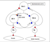
A comparison of events triggered by G-protein-neurotransmitter receptor and photopigment

Rods versus Cones
A. Rods contain rhodopsin and are more light sensitive than cones (a single photon of light
may be detectable by rods but not by cones).
1. One reason for this is that the process of signal amplification is greater in rods.
2. Cones have coneopsin instead of rhodopsin.
a. There are three different types of coneopsin: red, green, and blue. Meaning that
they are most sensitive to light in the red, or green or blue wavelengths.
b. The difference between the three forms of coneopsin is small. The difference is a small change in the amino acid sequence allowing for maximal sensitivity to different wavelengths of light. As an interesting tidbit, there are 2 versions of the red coneopsin gene in men and these produce red opsins that vary by a single amino acid. This difference results in the two red coneopsins being maximally sensitive to slightly different wavelengths of red light. Thus, men with different versions of this opsin do not perceive the same thing when they see a red object. This answers the philosophical question about whether different people see objects, colors etc. in the same way. They don’t.
3. Another reason that rods are more sensitive to light is that the outer segment of rods is larger; therefore they have a larger surface area to absorb light.
4. Rods also have more photopigment densely packed into the membrane of the optic disks, so they absorb more light.
B. In bright light the photopigment in rods (but not cones) is saturated (bleached). Therefore rods aren’t functional in bright light, while cones are.

1. This means that we have two parallel visual systems, one for bright light and one for very dim light.
2. At night colors appear to be muted. But the same spectral frequencies exist in bright and dim light. The reason we don’t perceive colors in dim light is that cones don’t work in dim light, so we don’t perceive color because we’re using a part of the visual system that is color blind.
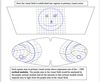
Below is a diagram exemplifying receptive fields of RGCs:

What is a receptive field?
A neuron’s receptive field is the location in the environment (or the surface of the body) from which an appropriate stimulus will change that cell’s activity. The term Receptive Field applies to cells in the visual system other that RGCs and also applies to neurons in other sensory modalities (e.g., touch, auditory).
Some features of receptive fields of RGCs:
- The receptive fields of RGCs are circular.
- The receptive fields of RGCs vary in size.
- Receptive fields of adjoining RGCs may overlap.
What are the mechanisms that account for the differences in size of receptive fields?
- RGCs on the edges of the retina (so their receptive fields are in the periphery of the visual field) collect information from a greater number of photoreceptors than do RGCs closer to the fovea. Therefore, their receptive fields are larger.
- Convergence is a situation where many neurons converge onto a few neurons. (There are about 120-130 million photoreceptors and only 1 million RGCs; therefore there is a lot of convergence of photoreceptors onto RGCs).
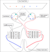
This is shown in the diagram below:

Convergence of synaptic input to RGC = large receptive field. Mostly in periphery of retina.
No convergence = small receptive field Mostly in fovea. - Divergence is few (or one unit) projecting onto many units. Divergence: One or a few units projecting to many units.
4. In the fovea there is less convergence of photoreceptors onto RGC (via bipolar cells) than in the periphery of the retina and this is a mechanism that explains the differences in size of the receptive fields of RGCs.

Divergence (image)

Light Sensitivity and Convergence
Convergence also helps to explain light sensitivity. There is less convergence in the cone system, therefore RGCs receiving input from cones are not as sensitive to light.