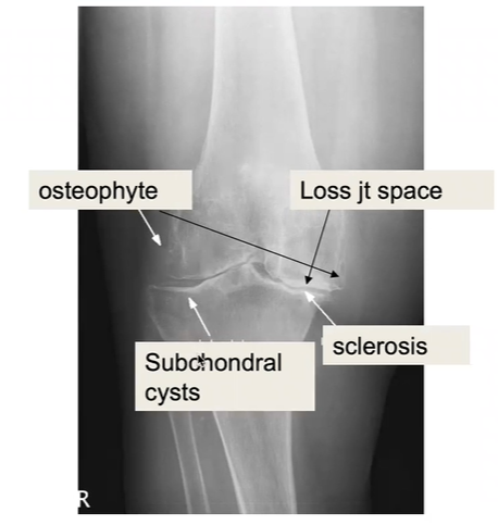
What do the labels show?
What is the underlying pathology
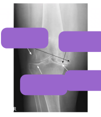
osteoarthritis
T1W MRI appearance
fat - bright
fluid - dark
1 thing is bright
most anatomical image
T2 weighted MRI appearance
Fat bright
fluid bright
2 things are bright - used more commonly
bone on MRI vs CT
MRI - dark
CT - bright
CT appearance of tissues
- air - dark
- coagulated blood - bright
- bone - very bright

What are the features and pathology depicted

rheumatoid arthritis
features on x-ray (LESS)
- loss of joint space,
- erosions,
- soft tissue swelling,
- subluxation & deformity
also may see deformity + deviations e.g. ulnar deviation

What are the features and pathology depicted

rheumatoid arthritis
features on x-ray (LESS)
- loss of joint space,
- erosions,
- soft tissue swelling,
- subluxation & deformity
also may see deformity + deviations e.g. ulnar deviation

What is adequate inspiration for CXR?
6 anterior ribs
What are differentials for cardiomegaly on CXR?
Heart failure Dilated cardiomyopathy Pericardial effusion Only assess on PA
how to determine roughly what lobe is affected on CXR?
- above horizontal fissure - R upper lobe
- heart border obstructed - R middle lobe
- R costophrenic - R lower lobe
infective causes of cavitating lesion on CXR
TB, S. Aureus, Klebsiella
type of imaging
what is shown

ERCP - Endoscopic Retrograde Cholangiopancreatography + cholangiogram
- side viewing scope is used
- ampulla of vater is cannulated + dye injected → cholangiogram
- shows multiple gallstones in the CBD
- bile duct very dilated, normally approximately to age of pt in decades/10 in mm once over 30s
- this can be a diagnostic and therapeutic investigation
normal bile duct diameter - rough guide
over the age of 30s
expect to be age in decades/10 as mm value
i.e. 40 → 4mm
what is the pathology here?
type of imaging modality

SBO obstruction
AXR
DSA vs CT angiogram
digital subtraction angiogram - x-rays of dye in vessels during interventional procedure e.g. stenting
CT angiogram - contrast given and CT done so can scroll through like slices
type of imaging
pathology

AXR
sigmoid volvulus
- coffee bean
modality of imaging
pathology

AXR
- PSEUDOOBSTRUCTION
- distension of the whole large bowel and rectum
- air in the rectum
- air all the way from R colon to the rectum
- presents like LBO
- not mechanical obstruction
what are the features of AS on x-ray? (spine)
- subchondral erosions
- diffuse syndesmophytes → bamboo spine
- sclerosis
- squaring of the lumbar vertebrae
- sacroiliac joint narrowing (wider in early disease)
- end stage may be narrow line or not visible
what is shown here?

hiatus hernia
sliding (fundus displaced above the diaphragm)
diagnosis

diverticulosis
-
Acute care40
-
Breast and vascular27
-
Cardiovascular152
-
Dermatology82
-
ENT + Ophthalmology49
-
Gastrointestinal111
-
Endocrinology188
-
Haematology61
-
Infection and Immunology43
-
Neurology147
-
Renal and urology70
-
Respiratory156
-
Rheumatology and musculoskeletal86
-
Miscellaneous84
-
PACES - Images20
-
PACES - Instruments96
-
DPD107
-
Scoring Systems21
-
Murmurs24
-
signs22
-
Research methods9
-
Core Content517
-
PPQ7
-
Viva20
