When evaluating 2D echo of LV, what are the things you want to specifically look at and evaluation
- wall motion
- wall thickness
- chamber size
- valve motion
- valve thickness
- over-all function
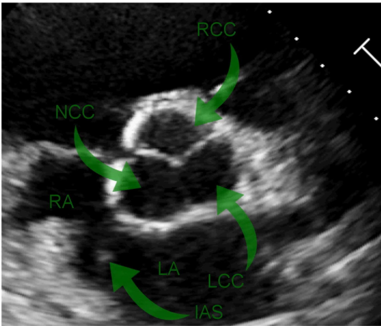
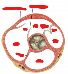
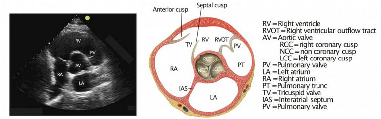
- Aortic valve - RIGHT coronary cusp
- Aortic valve NON-coronary cusp (but maybe LEFT)
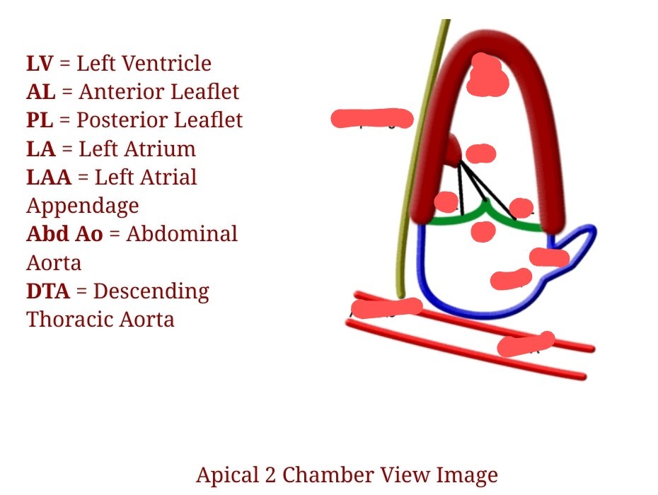
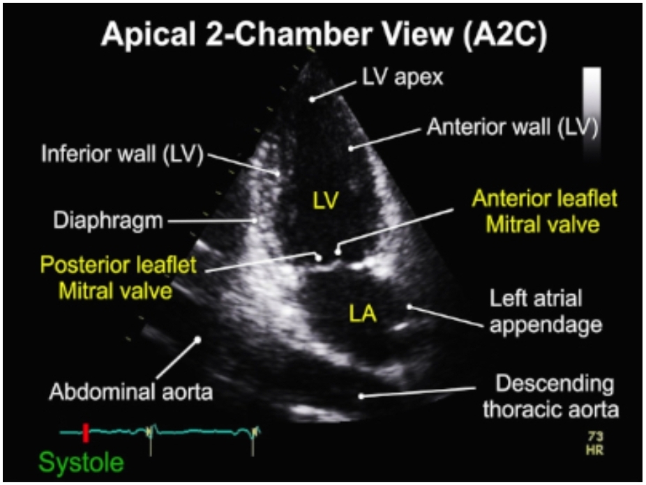
- Anterior MV leaflet
- Posterior MV leaflet
TAPSE
Abnormal Value
< 17 mm
MAPSE
Abnormal Value
< 8 mm
(associated with LV EF <50%; sensitivity 98%; specificity 82%)
12-15 mm is normal
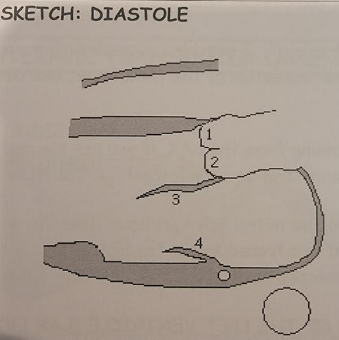
What are the name of the aortic valve cusps?
Apical akinesis with basal hyperkinesis.
Takotsubo’s CM
Echo appearance.
Takotsubo’s CM
Echo appearance.
Apical akinesis with basal hyperkinesis.
Pulmonary Doppler - Where to position the PW sector?
Pulsed wave doppler needs its sample volume placed 1cm proximal to the pulmonary valve to quantify pulmonary regurgitation.
Tricuspid regurgitation - incidence
90% of all individuals have mild TR
Normal VTILVOT
18-20
RV free wall thickeness
normal
≤ 5mm
When and where to measure RV wall?
Subxiphoid
During diastole
Why is there more movement with RV than LV during contraction?
Fibers are oriented more longitudinal with RV
TAPSE ≤ 16
in setting of PE
Increases mortality by
4.4x more likely to die from PE
(Lobo et al, 2014)
How to measure PA pressure?
- Identify TR jet.
- CW Doppler through the jet to create spectral waveform.
- Can see velocity (V)
VxVx4+CVP is an estimate.
Less than 25 is normal.
Where to measure ESN / PW for Pulmonic valve?
Just BEFORE the pulmonic valve
ESN Wave form
Can’t use in what chronic condition?
Chronic pulmonary hypertension
Why does the 2 point ultrasound work?
DVTs occur in areas of high turbulence.
Branching points are areas of high turbulence.
Nazerian et al (2014)
LR of POC Multiorgan U/S for Dx of PE in CHEST
If positive abnomrality in:
echo
lung
DVT
Echo 3.6x
Lung 15x
DVT 21.7x
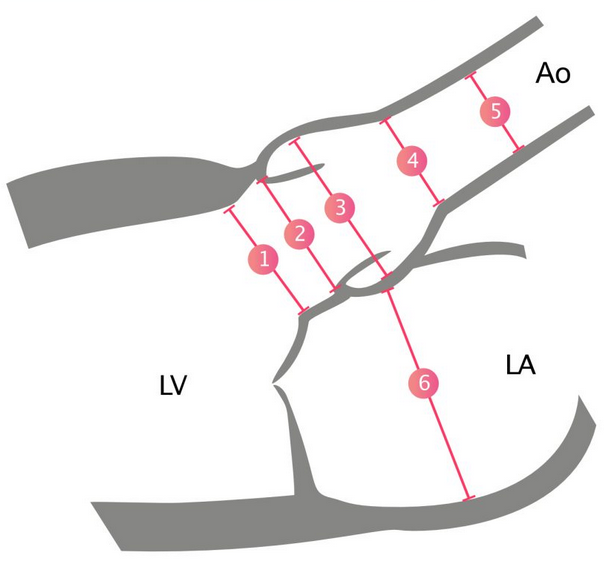
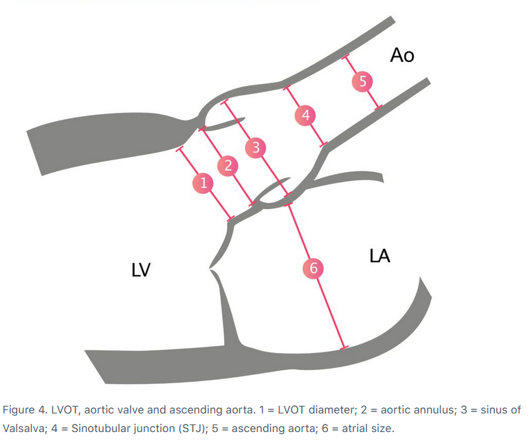
When to measure
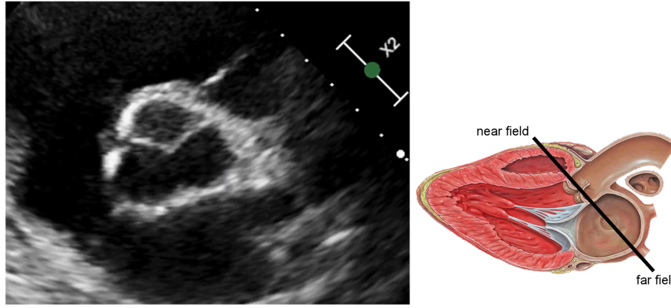
Aortic Valve / LVOT
mid systole
when valves are wide open

