Types of vasculitis are organized by. . .
. . . the size of the blood vessels affected by the pathology
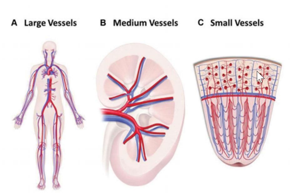
Vasculitis diagram: Where is the inflammation?
“Large” blood vessels
The aorta and its major branches
Medium blood vessels
Visible to the naked eye or by angiography, often named arteries that branch off of the “major” central arteries.
Small blood vessels
Arterioles, capillaries, and venules
Microscopic
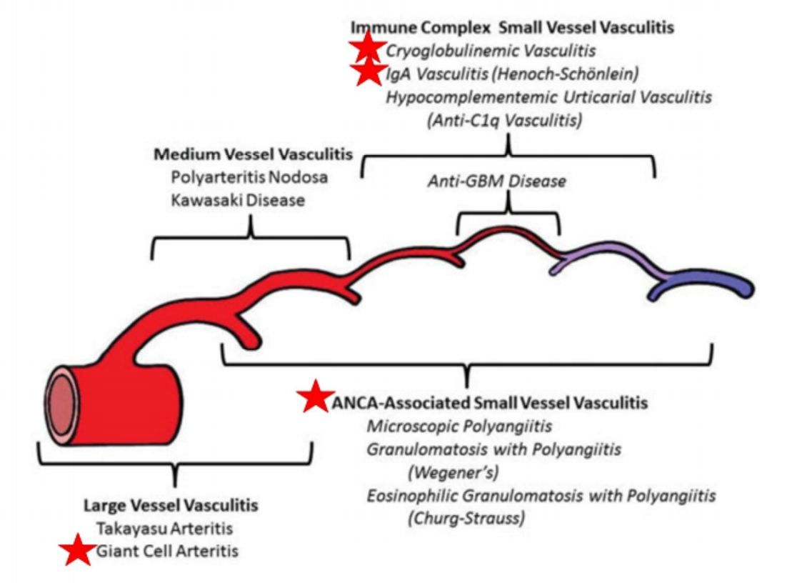
Etiologies of large vessel vasculitis
- Giant cell arteritis
- Takayasu arteritis
Etiologies of medium vessel vasculitis
- Polyarteritis Nodosa
Etiologies of small vessel vasculitis
- ANCA-associated vasculitis
- Cryoglobulinemic vasculitis
- IgA vasculitis
Organs selectively affected by small vessel vasculitis and corresponding symptoms
- skin - purpura
- nerves - neuropathy
- lungs - coughing, nodules, hemoptysis
- kidneys - hematuria
- joints - synovitis, arthritis, arthralgia
- eyes - inflammatory symptoms
Organs affected by medium vessel vasculitis and associated symptoms
- Mesenteric - abdominal pain
- Renal - kidney infarct, kidney pain
- Nerves - neuropathy
- Skin - ulcers, livedo, gangrene
- associated w/ insidious presentation
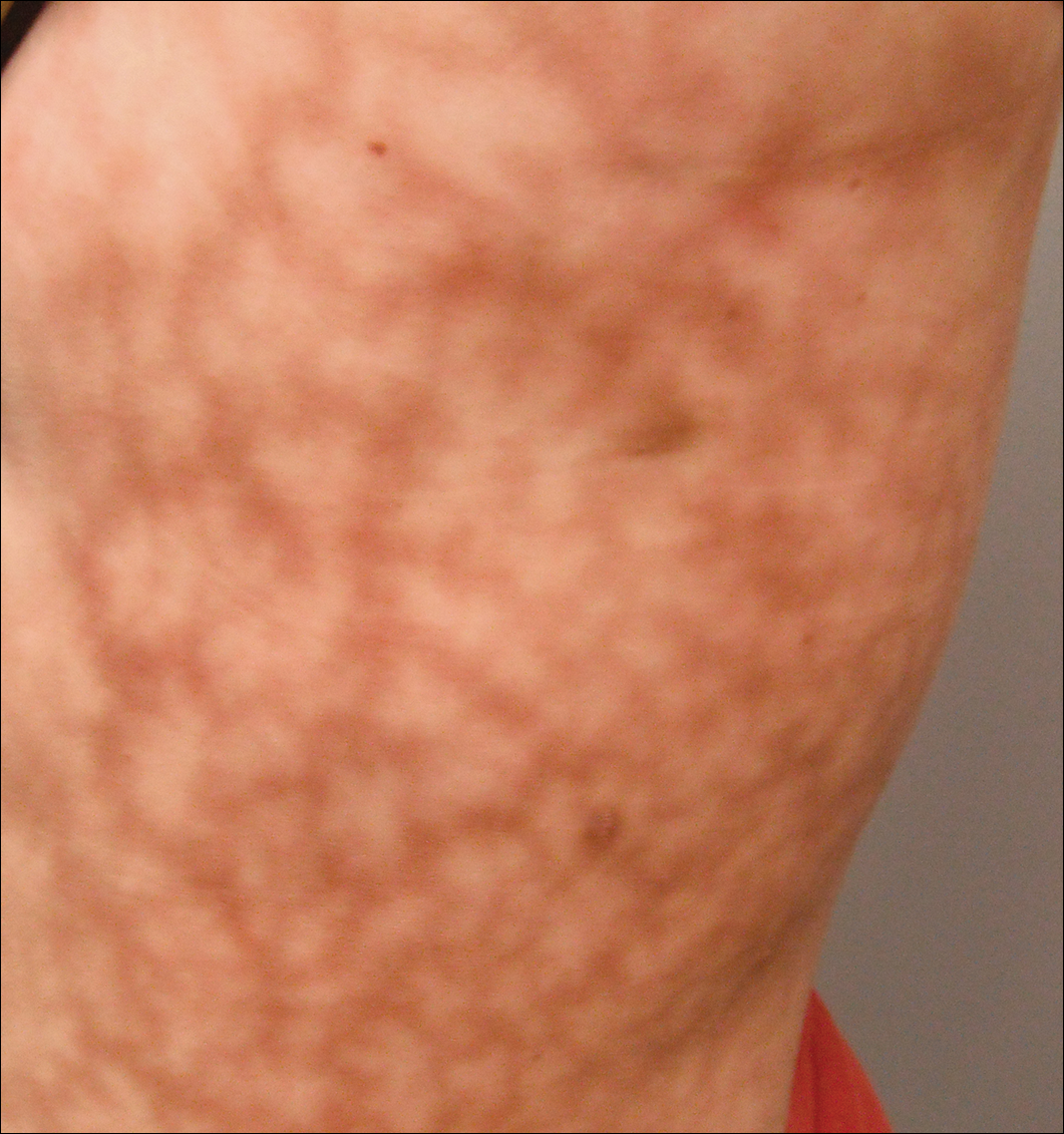
Livedo
Organs affected by large vessel vasculitis and associated symptoms
- Brain - headache
- Eyes - blindness
- Muscles of face - Jaw claudication
- Fever
- Extremity claudication
Specific lab tests are available to test for ___.
Specific lab tests are available to test for small vessel vasculitis, but not medium or large vessel vasculitis.
Small vessel vasculitis may be due to an underlying ___.
Small vessel vasculitis may be due to an underlying connective tissue disease.
Tests for small vessel vasculitis
- Usually involve testing for glomerulonephritis
- Test for proteinuria
- Stain H and E for RBC casts
- Kidney biopsy for “crescent” of immune cells surrounding a glomerulus
- Full-body observation for purpura
- CT scan for diffuse alveolar hemorrhage
Leukocytoclastic vasculitis
- When white blood cells attack blood vessels
- General term for any small-vessel vasculitis
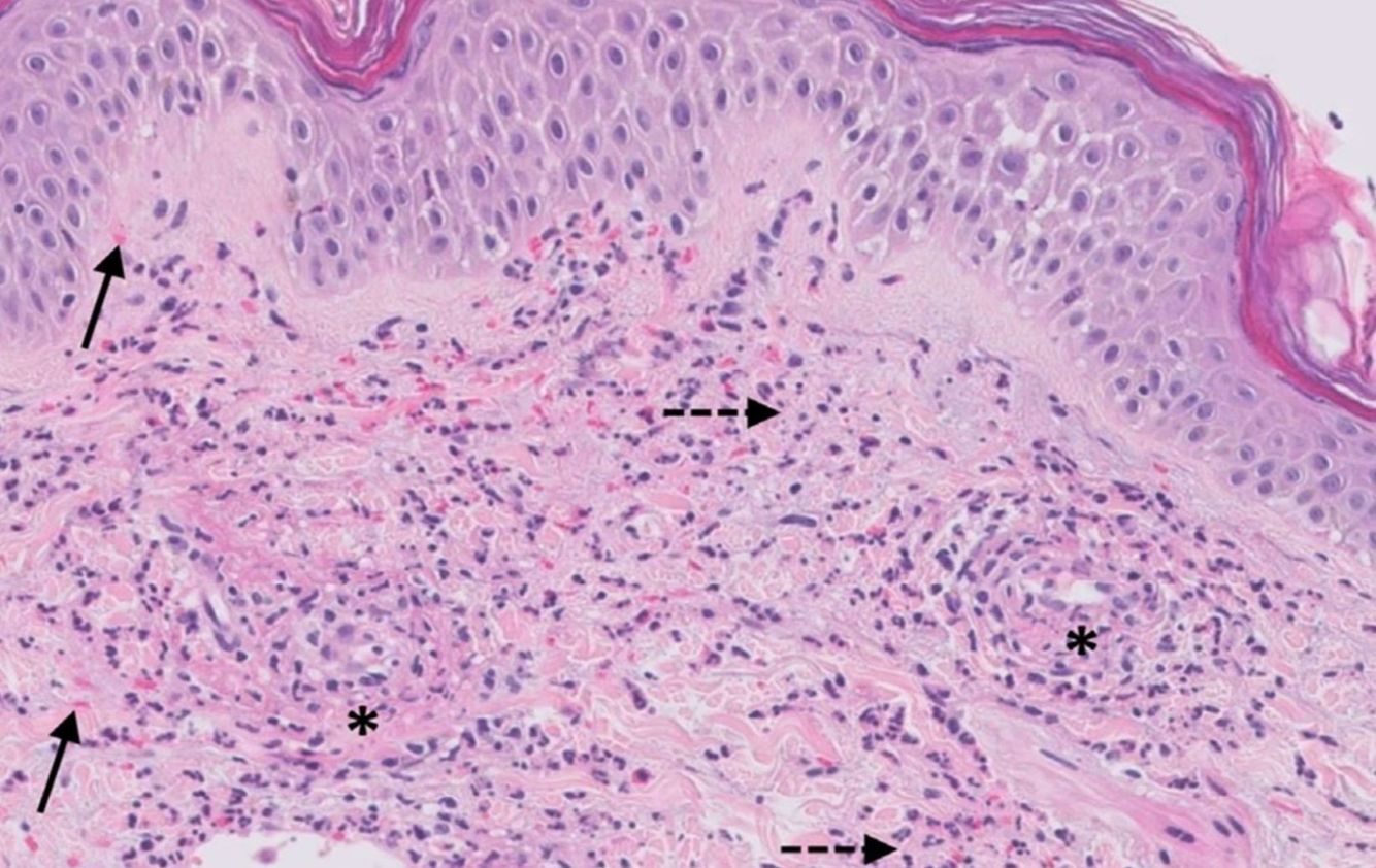
Small vesse vasculitis on H and E. Note the asterix in the vessel wall and extravasated RBCs (the histological equivalent to the finding of non-blanching erythema)
Asterix
Firboid necrosis in a vessel wall
Anti-neutrophil cytoplasmic antibodies (ANCA)
- Autoreactive antibodies against myeloperoxidase and proteinase-3
- Known to cause glomerulonephritis
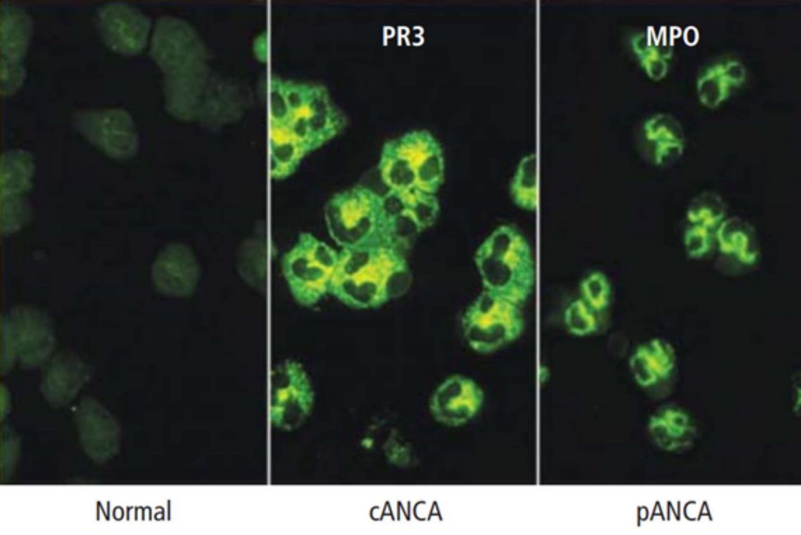
Indirect immunofluorescence test for ANCA
- Anti-MPO stains perinuclearly (pANCA)
- Anti-PR3 stains cytoplasmically (cANCA)
- Minor ANCAs such as elastase, Lamp-2, or lactoferrin may confound results (usually pANCA)
- ELISA used to confirm following indirect immunofluorescence screening test
Three phenotypes of ANCA
- Microscopy polyangitis (MPA)
- MPO > PR3, necrotizing vasculitis
- Granulomatosis with polyangitis (GPA)
- PR3 > MPO, necrotizing vasculitis, granulomas
- Eosinophilic granulomatosis with polyangitis (EGPA)
- MPO only, necrotizing vasculitis, granulomas, eosinophilia
____ is central to ANCA
NETosis is central to ANCA
NETosis model for ANCA
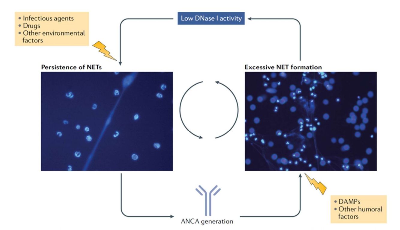
Role of DNAse I in ANCA
Impaired DNAse I activity leads to NET persistence following physiological NETosis. This leads to some damage which is then associated with anti-PR3 and anti-MPO antibody development. These ANCAs then activate further NETosis via FcγRI binding.
The cycle goes on.


-
Hypersensitivity34
-
Innate Immunopathologies of Skin57
-
Joint Pain and Approaches to Arthritis77
-
Sepsis19
-
Inflammasome Diseases49
-
Infections of Joints42
-
Common Cutaneous Infections94
-
Asthma and Food Allergy51
-
Urticaria, Angioedema, and Anaphylaxis41
-
Drug Hypersensitivity78
-
Type II Hypersensitivity Reactions in the Skin24
-
Spondyloarthritis46
-
Rheumatoid Arthritis33
-
Contact Dermatitis and Psoriasis45
-
Atopic Dermatitis32
-
Vasculitis39
-
Dermatomyositis19
-
T cell diseases of the skin59
-
Skin Cancers42
-
Lupus I42
-
Lupus II and Sjogren's Disease56
-
Fibrosing Disorders47
-
Granulomatous Diseases55
-
Tuberculosis52
-
Immunosuppressants41
-
HIV I40
-
Transplant Immunology68
-
Everything from POM (Work on over winter break)6
