Give 5 main functions of the vertebral column
- Carries and protects spinal cord and nerves
- Provides support to thoracic cage
- Transfers wright from upper body to lower limbs
- Shock absorbing
- Muscle attachement point
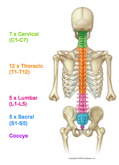
What regions are there in the vertebral column?
How many vertebrae in each?
- Cervical region (C1-C7)
- Thoracic region (T1-T12)
- Lumbar region (L1-L5)
- Sacral region (S1-S5)
- Coccyx
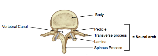
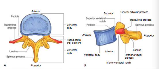
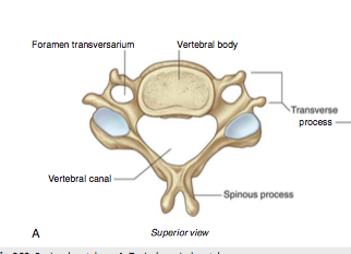
Label the image
Top to bottom (Right):
1) vertebral body
2) pedicle
3) transverse process
4) Lamina
5) Spinous process
Left:
Vertebral canal
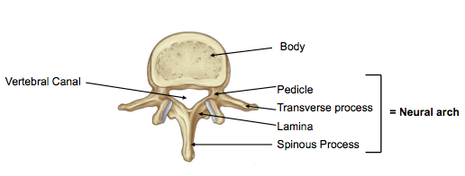
What forms the neural arch?
Pedicle, transverse process, lamina, spinous process
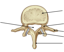
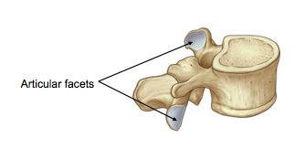
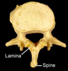
Label the image
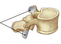
Arrows point to the articular facets
Describe the structure of a typical vertebra
- Largest most anterior portion is the vertebral body. Weight bearing portion, gets larger from C2-L5.
- Off the Body comes the neural arch
- Directly off the body are the paired pedicles either side
- These pedicles join the lamina which fuses in the midline
- Off the posterior of the arch is the spinous process
- Where the lamina and pedicle meet on either side is a transverse process (This articulates with ribs in thoracic region).
- In a similar region is a superior and inferior articular process that articulates with adjacent vertebrae.
- The neural arch encloses the vertebral canal which houses the spinal cord/ proximal spinal nerves/ meninges/ blood vessels/ connective tissue/ fat
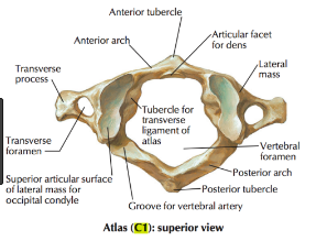
What is C1 called?
Why is its structure special?
What does it articulate with and what joint exists?
What movement does this allow?
C1 is also known as the atlas.
Its structure is special as C1 has no vertebral body or spinous process but is formed of only an arch and transverse processes,
On its arches it has two articulation points for the occipital condyles on the base of the skull.
The joint between the occipital condyles and atlas facets forms a compound synovial joint.
Permits movement- flexion and extension of the neck at the vertebral column.
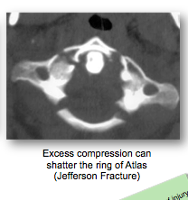
What is a fracture of the atlas called?
What can cause this?
A fracture of the atlas (C1) is called a Jefferson Fracture.
This can be caused by excess compression of C1 e.g. diving into shallow water or force transferred up the spine to C1 during a fall
(always check vertebral column in patients with lower limb/ calcaneal fracture).
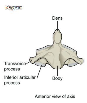
What is the name of C2?
Describe its structure
- C2 is also known as the Axis
- It is formed by a body with an attached dens/ odontoid process
- off the body come two lateral masses, transverse processes with foramina transversarium.
- It has a posterior neural arch formed by pedicles and a thick lamina
- And a large posterior spinous process
- Has superior and inferior articular processes.
What is a hangman’s fracture?
Fracture of the pedicles of C2 often caused by hyperextension of the neck during RTA/sports injury/ hanging.
Can cause compression of the spinal cord, respiratory failure and death.
What is whiplash?
Whiplash describes neck injury caused when there is sudden deceleration of the body that results in flexion and hyperextension of the neck
Describe the defining structural features of cervical vertebrae
- Smaller vertebral bodies that get larger from C1-C7
- Transverse processes with foramina called foramina transversarium
- Short, bifid spinous processes that get longer from C1-C7
- Triangle shaped verterbral foramen/canal
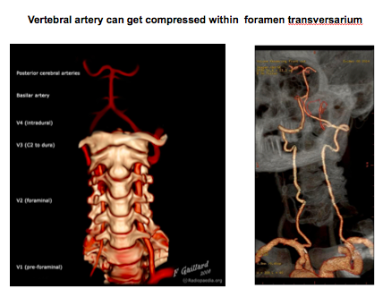
What runs through the foramen transversarium?
What can happen if the foramen transversarium is compressed?
The vertebral arteries running uptowards the brain to form the circle of willis which supplies the neural tissue.
Compression of the foramen transversarium can lead to compression of the vertebral arteries leading to defecit of blood supply to the brain producing stroke like symptoms.
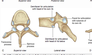
What are the defining features of thoracic vertebrae?
- Larger, heart shaped vertebral bodies that increase in size to accomodate more weight/ force applied
- Circular vertebral foramen/canal
- Articulation facets for the ribs: Demifacets on the vertebral bodies:
- Superior facet articulates with head of its own rib
- Inferior facet articulates with head of rib below
- Transverse process with articulation facet for tubercle of own rib.
What are the defining features of lumbar vertebrae?
- Large cylindrical vertebral bodies to cope with large forces applied
- Triangular vertebral foramen/canal
- Articulations aligned to prevent rotational movements which increases the stability of the lumbar region
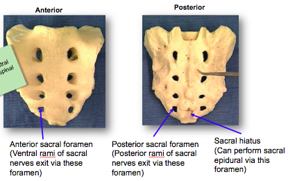
What are the defining features of sacral vertebrae?
What structures pass in this region?
What is the sacral hiatus and what can it be used for?
- Sacral vertebrae are 5 fused vertebrae
- Transmits weight to pelvic girdle
- Triangular in shape with apex pointing inferiorly
- Has a concave anterior surface with anterior sacral foramen for ventral rami of sacral nerves
- Has a convex posterior surface with posterior sacral foramen for dorsal rami of sacral nerves
- The sacral hiatus is a foramina at the distal tip of the sacrum and can be used for a sacral epidural.
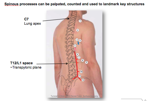
What is the clinical use of spinous processes?
Spinous processes can be palpated and used to count vertebrae to find key structures:
E.g. Transpyloric plane L1
E.g. Lung apex C7
How is the vertebral column inherently stable with little muscle activity?
- Vertebral column has a collection of ligaments and joints that interact with each other to keep vertebral column upright and stable.
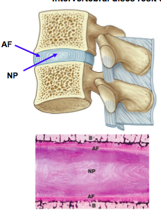
What structure exists inbetween vertebrae?
- Intervertebral discs exist inbetween the vertebrae
- Formed of outer annulus fibrosus = lamellar arrangement of fibrocartilage, limits rotation
- And an inner nucleus pulposus = rubbery central core made up of hydrated GAG’s that resists compressive forces.
What is the name of the joints between superior and inferior articular facets on the neural arches of adjacent vertebrae?
Zygapophyseal joints
What happens to the intervertebral discs as you descend the vertebral column?
They increase in thickness to accomodate the greater weight/ force applied as you descend the vertebral column.
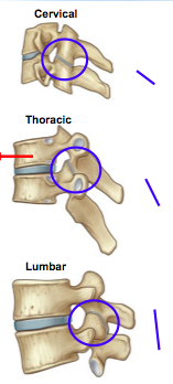
What happens to the zygaphyseal joints in different regions of the spine?
- The zygaphyseal joints change their orientation as you desend down the spine
- Cervical: close to horizontal, slope inferiorly from anterior to posterior. Allows wide range of movement, flexion, extension, rotation.
- Thoracic: Close to vertical, limits the amount of flexion and extension but rotation still possible.
- Lumbar: Pretty much vertical, joint surfaces are curved and interlocked which limits range of movement. Little rotation in lumbar region but some flexion and extension still.
what can happen to the zygapophyseal joints pathologically?
They can dislocate, discs displace away from each other.
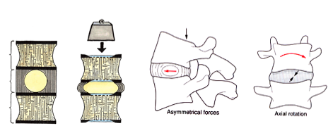
What happens when force is applied to the vertebral column?
This increases pressure in the nucleus pulposus.
The increase in pressure in the NP increases tension in the AF.
Increased tension in the AF pulls vertebral bodies together increasing stability and limiting further movement between vertebral bodies.


-
Block 2 Week 2 Thorax/ Tracheobronchial tree/ lungs39
-
Block 2 Week 2 Thorax Tracheobronchial tree/ lungs part 251
-
Larynx34
-
Heart and mediastinum69
-
Cardiac embryology33
-
Vertebral Column41
-
Introduction to CNS anatomy53
-
Block 3: Spinal cord and ascending tracts45
-
Block 3: spinal cord and descending tracts / reflexes33
-
Block 3: Basal ganglia19
-
Cranial nerves48
-
Brainstem46
-
Cerebral cortex and limbic system49
-
Intro to neuroimaging35
-
Eyes- orbit, movement and reflexes33
-
Gluteal region, hip and thigh46
-
Intro to musculoskeletal27
-
Proximal neurovasculature, Knee and Leg39
-
palpable masses32
-
distal neurovasculature, ankle and foot47
-
Pectoral Girdle/ Should/Arm/Elbow48
-
Injuries to bones and joints of lower limb38
-
Upper Limb Forearm and elbow39
-
Wrist and hand36
-
Abdominal viscera: Liver, Pancreas and Spleen36
-
Female Reproductive Anatomy64
