Describe the Ulcer 3
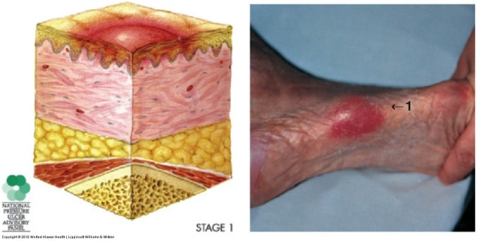
Stage 1
Skin is NOT broken (if broken at least stage 2)
Superficial tissue
Persistent erythem (unblanchable)
Stage
How deep?
What can be staged this without being an actual ulcer?
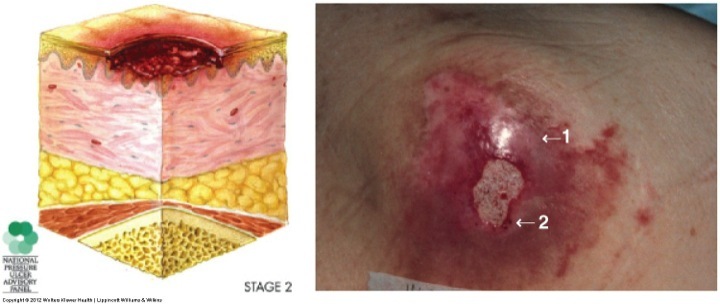
Stage 2
Pressure Ulcer
Partial Thickness
Epidermis and Dermis
includes skin tears and tape burns (silk tape).
(If pt has a tape reaction, use paper tape
Stage this
How deep?
What layers?
Stage 3 Pressure Ulcer
Full Thickness
epidermis, dermis, top of subcutaneous.
Stage this.
How deep?
What layers?
Painful?
Stage 4 Pressure Ulcer
Full Thickness
Epidermis, dermis, subcutaneous, bone/muscle
not painful
What wound healing phase is this in?
Stage this wound
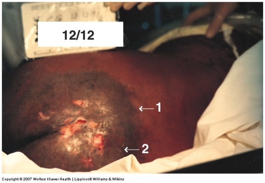
Describe 1, 2, 3.
What is absent?
Chronic Inflammation
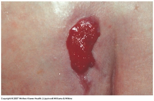
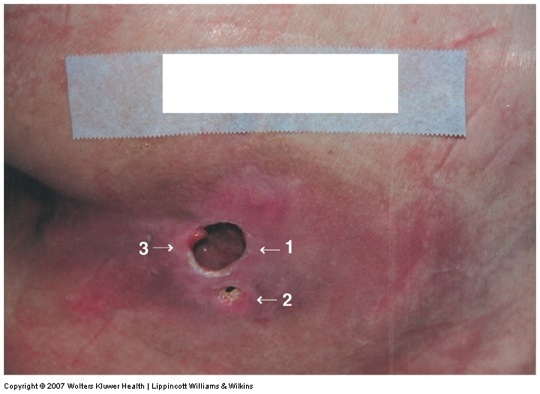
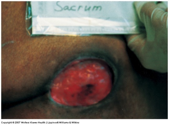
Stage 3 Ulcer
- This is over an old scar.
- Skin bridge Undermining/Tunneling
- Unblanchable Erythema. Edges are rolled under
Absence of proliferation due to no granulation tissue.
(wound edges are distinct and rolled)(wound bed is necrotic)
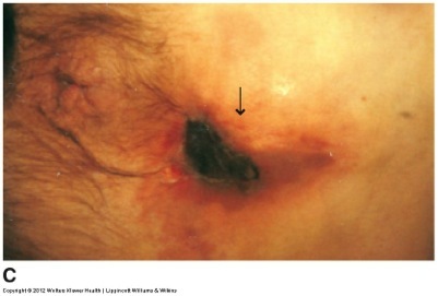
Stage this ulcer
Why is it staged that way?
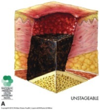
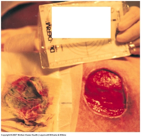
Unstageable Ulcer
The brown is a scab or eschar.
We don’t know what’s under the eschar.
Different that suspected deep tissue. Perhaps there isn’t anything, perhaps there’s a lot.
Where is the wound healing stage?
Identify the wound
Name characteristics
Unstageable.
could have chronic inflammation due to erythema.
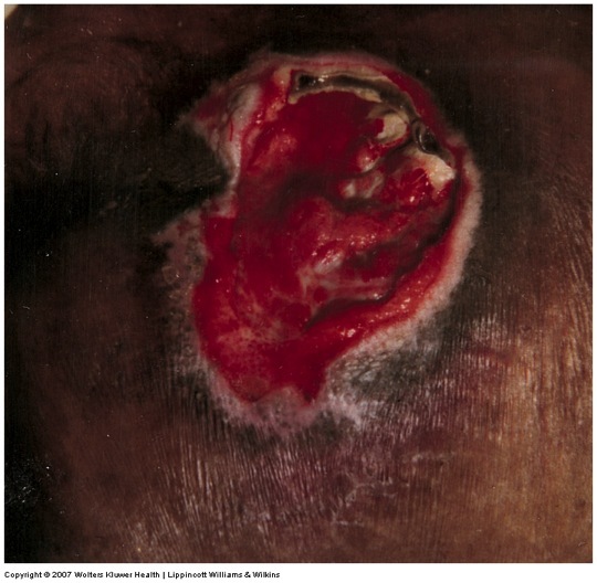
What wound healing phase is this in?
Stage it.
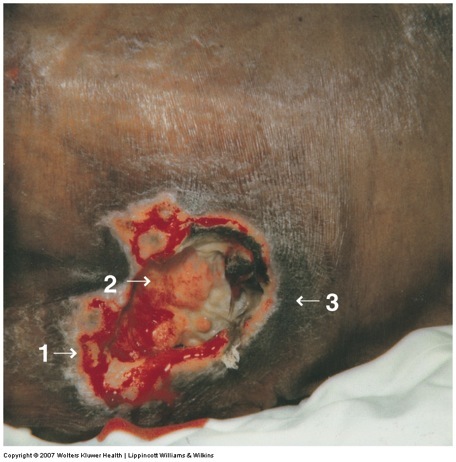
Analyze
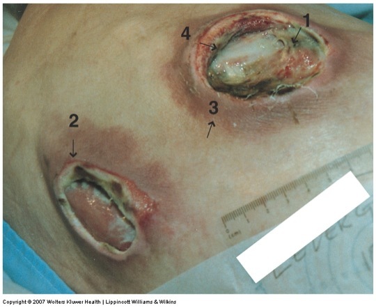
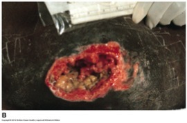
Chronic Inflammation
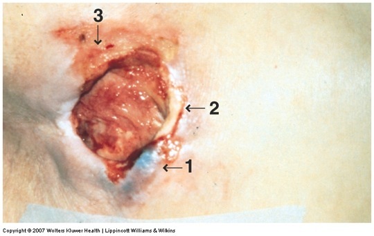
Stage 4 Pressure Ulcer
- Yellow slough
- Edema
- Discoloration of skin. Halo of erythema
- Rib bone
Rolled edges not connected to wound bed.
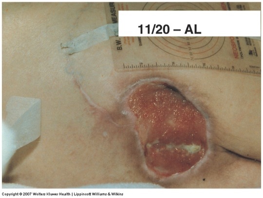
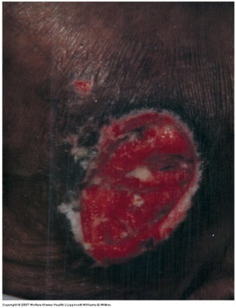
What phase of wound healing?
Analyze
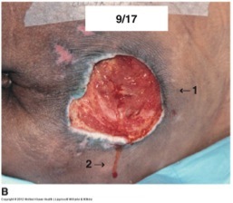
Acute Proliferative
- Edges are rolled but attached better to the bed.
A little slough
2.Granulation tissue–Indicates proliferation
Hemocidrin staining
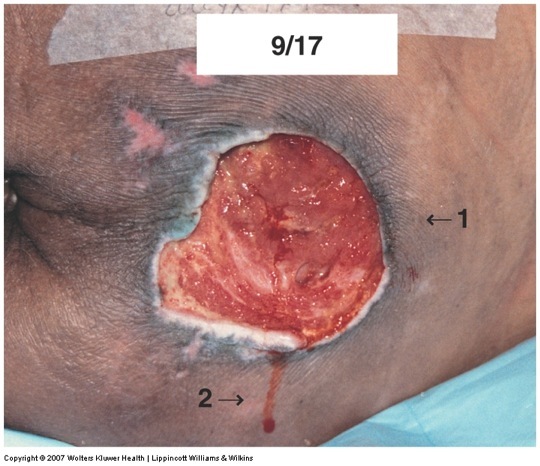
Stage this one
What healing phase?
Analyze
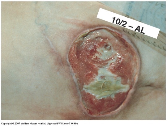
Stage IV Pressure Ulcer
acute inflammatory
Granulation tissue (slightly pale)
Clean; some rolled edges
Slough
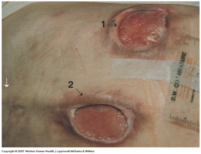
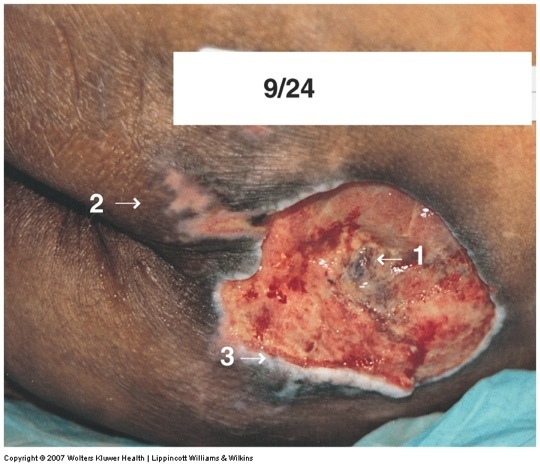
What phase is this wound?
Analyze
Acute Proliferative Phase
Contracting wound. Getting smaller.
Goood granulation.
Bed is attached to most edges.
Very little edge rolling.
What phase?
How can you tell?
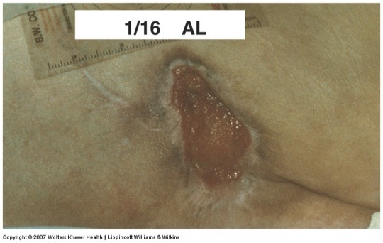
Acute Epithelialization
Good granulation
epithelia growing on the edges.
Hemociderin staining
Edges are very nice.
What phase of healing?
What can be staged?
Anylize
Acute Inflammatory
Stage II pressure ulcers.
- Clear line of demarcation between healthy and unhealthy tissues. Erythema
- Discoloration, edema, and induration (underlying tissue death)
Wound Healing phase?
What stage
Tissue depth?
Analyze
Chronic inflammation
Stage IV
Full Thickness
- Sanguineous drainage
- Muscles exposure
- Hemosiderin staining
(Necrotic tissue)
What Healing Phase?
Analyze
Acute Proliferation
New Granulation Tissue
Edges are mostly attached.
Color is good.
Epithelialization
What healing phase?
Analyze
Acute Proliferation
Wound edges attached.
Fibrin is attached yellow and adherant…do not take off.
Ready for epithelialization but still proliferative
Stage this one.
What Healing Phase?
Analyze
Stage III Pressure Ulcer
Acute Proliferative
Edges generally attached and not rolled.
Granulation tissue
Sanguiness drainage
What healing phase?
Analyze
Chronic Proliferation
- Slough and Hemorrhagic trauma
- Hypopigmentation.
- Granluation tissue color is not right. dull pink.
What healing phase?
Analyze
Chronic Proliferation
Hemosidering staining from prior bleeding surrounds ulcer.
Bruise or trauma on granulation tissue may necrose.
What healing phase?
Analyze
Chronic Proliferation
- New pressure-induced damage.
- Maceration from wound fluid.
- Friction injury (blood) with signs of inflammation.