mvmts @ TMJ: protrusion
protrude chin:
- lateral pterygoid (prime)
- masseter
- medial pterygoid
•Muscular triangle
–Bounded by anterior border of SCM, superior belly of omohyoid, midline of neck
–Contains infrahyoid muscles, thyroid and parathyroid glands
mucus membrane of larynx
respiratory epith
- except over true/aryepiglottic folds = stra-squa
pons
ant region of posterior cranial cav
4th ventricle
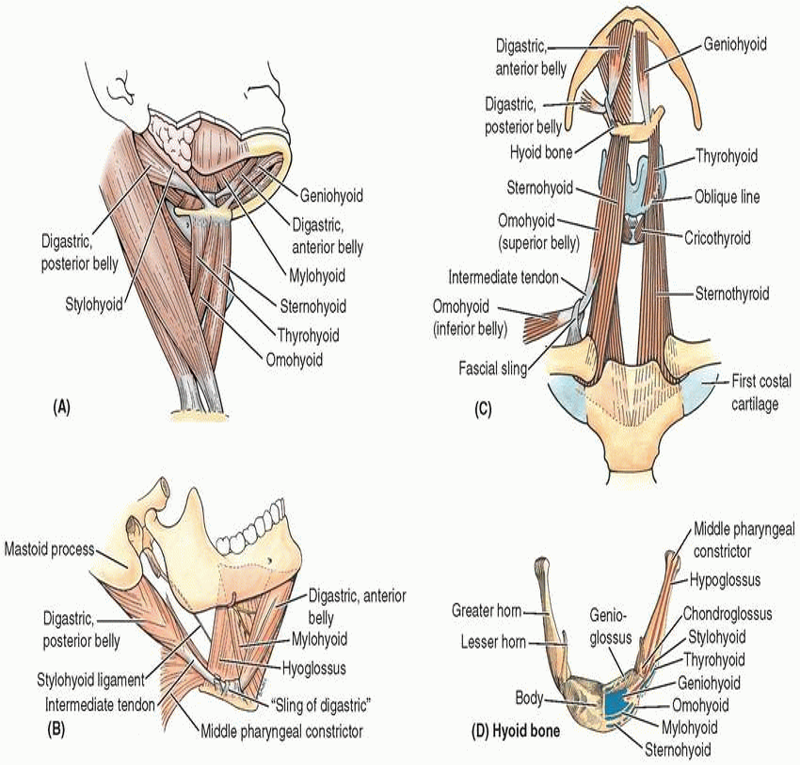
mylohyoid
N: N to mylohyoid - from inferior alveolar N - from CN V3 (madibular)
A: elevate hyoid, floor of mouth, tongue during swallow and speak
contents of infratemporal fossa
- Inferior part of the temporalis muscle.
- Lateral and medial pterygoid muscles.
- Maxillary artery.
- Pterygoid venous plexus.
- Mandibular, inferior alveolar, lingual, buccal, and chorda tympani nerves
- Otic ganglion.
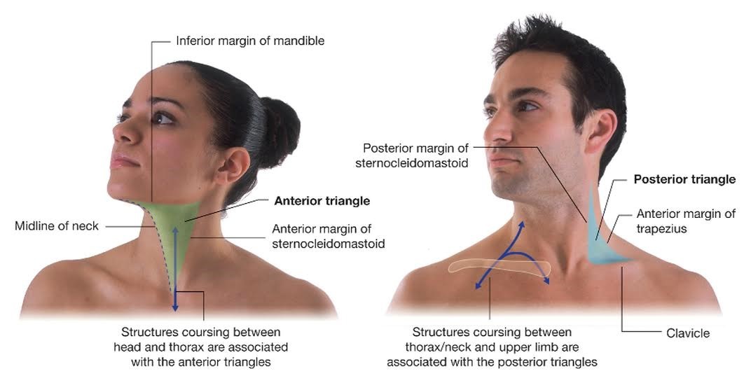
boundaries Anterior Triangle of neck
- Lateral—anterior border of SCM
- Anterior—anterior midline of neck
- Superior—inferior mandible
Pretracheal Layer of Deep Cervical fascia
Thin layer: muscular and visceral parts
laryngeal cartilages + hyoid bone –> (thorax) fib pericaridium
- Blends laterally with carotid sheaths
Encloses:
- thyroid
- parathyroid
- infrahyoid muscles
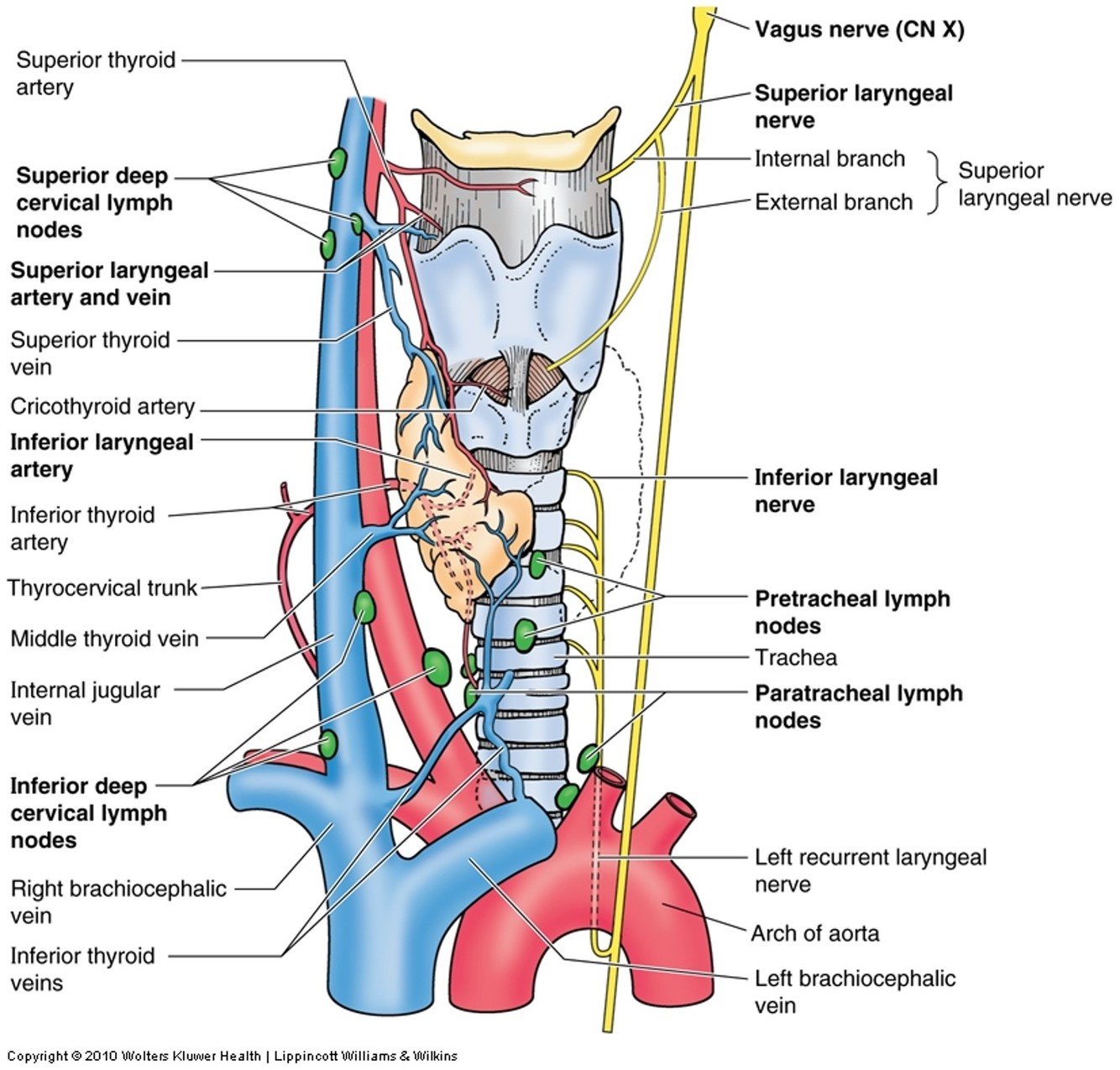
sensory and motor innervation of larynx
sensory
- above vocal folds: internal laryngeal (branch of superior)
- below vocal folds: inferior laryngeal (branch of recurr)
motor
- external –> circothyroid
- recurrent –> rest of intrinsic
dorsal scapular A
from either 2nd or 3rd part
passes backwards to supply levator scpulae, rhomboids
Thyroid cartilage
two flat laminae
- upper 1/3 = superior thyroid notch
- lower 2/3 = fused –> laryngeal prominence (Adam’s apple)
posterior horns
- Superior horns/laminae attach to hyoid bone by thyrohyoid membrane
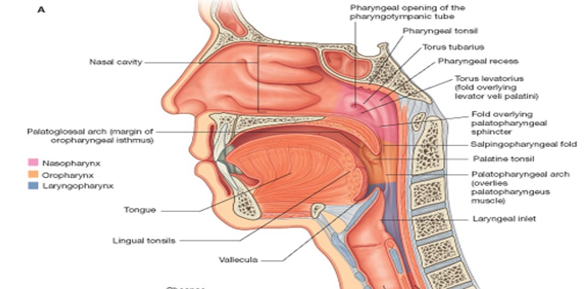
Pharynx
M tube post to nasal and oral cav: continuous with esop and larynx
ant to C1-C6, prevert M
retropharyngeal space: potential space b/w pharynx and prevert fascia
3 parts: naso, oro, laryngo
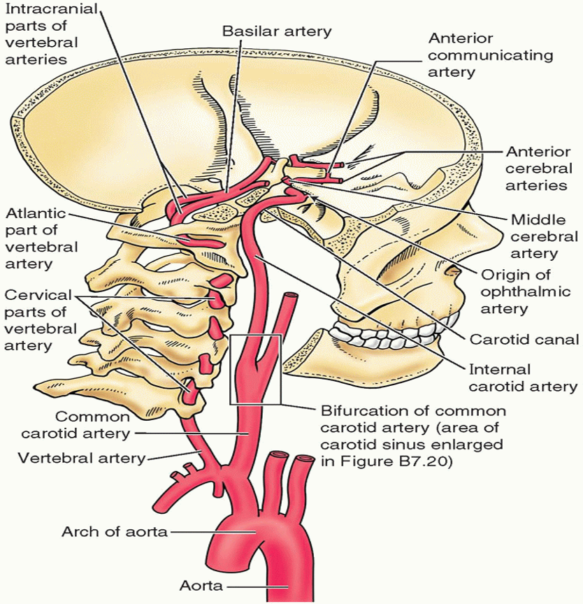
•Vertebral arteries
1st branche of subclavian –> txverse foramen (c1-c6) –> pierce dura –> foramen magnum –> basilar A –> clivus –> 2 posterior cerebral arteries –> circle of Willis
connect to anterior cerebral A via posterior commun A
vasc supply to cerv-SC and neck
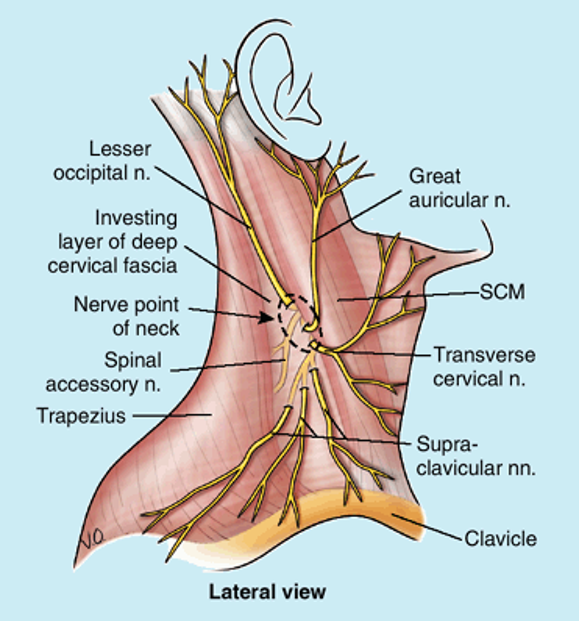
Cutaneous branches of the cervical plexus
posterior SCM
loop formed b/w anterior rami C2, C3
- –Lesser occipital (C2) to skin of neck and scalp posterior to auricle
–Great auricular (C2 and C3) to skin over parotid gland, mastoid process, auricle, and between angle of mandible and mastoid process
–Transverse cervical nerve (C2 and C3) to skin over anterior cervical region
•Supraclavicular nerves
- –Arise from C3–C4 loop
–Emerge from under SCM
–Supply skin over clavicle, superior thoracic wall, and shoulder
Cervical plexus block
posterior SCM @ around C2-C3
jugular foramen
b/w temporal & occipital
IX, X, XI
sigmoid sinus
inferior petrol sinus
posterior meningeal A
vertebral A
runs cranially in tx formaina of cerv-vert –> circle of wilis
Optic canal
lesser wing sphenoid
stuctures:
- optic N
- opthalmic A
- sympathetic plexus
Ventricular system
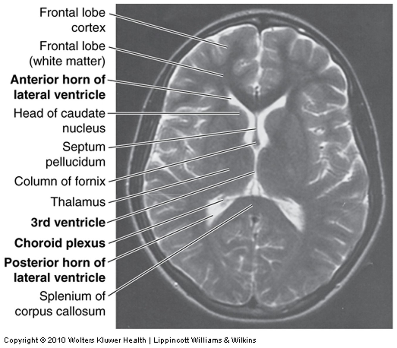
ECF in brain: like blood but less protein & diff [ion]
formed by choroid plexus in 4 ventricles of brain
- lateral ventricles –> intervent foramina –> 3rd ventricle –> cerebral aqueduct –> 4th ventricle –> apertures (2 lat, 1 midline) –>subarachnoid space
absorbed via arachnoid granulations (villi) into venous blood in dural venous sinuses
approx 400 mL/day CSF –> venous circ
general larynx
fx: phonation, sphincter guarding lower respir-tract
connects oropharynx with trachea
ant to prevert M, C3-C6 vert
otic ganglion
PNS
- pre: (glossopharyngeal N) –> otic ganglion
- post: –> auriculotemporal N –> parotid
location:
- infratemporal fossa
- inferior: foramen ovale
- medial: CN V3
- post: medial pterygoid
Cervical Ligaments
Stylohyoid:
- styloid process –> lesser cornu of hyoid
Stylomandibular:
- styloid process –> angle of mandible
Sphenomandibular:
- spine of sphenoid –> lingula of mandible
Pterygomandibular:
- hamular process of medial pterygoid plate –> posterior end of mylohyoid line of mandible
- attachment to superior constrictor & buccinator muscles

thyrohyoid membrane
txverse superior border and superior horns of thryoid cartilage
pierced by superior larngeal vessels, interal laryngeal N
geniohyoid
N: C1 via CN XII (hypoglossal)
A:
- pull hyoid ant-sup
- shortens mouth floor
- widens pharynx


