Exam 2 Flashcards

(505 cards)

1
Q

Stroma of breast

A
  • Fibrous tissue forms septae (ligaments of Cooper), which anchor skin to deep fascia
  • Involvement of these ligaments in the carcinoma lead to fixity of gland & wrinkling of skin
2
Q

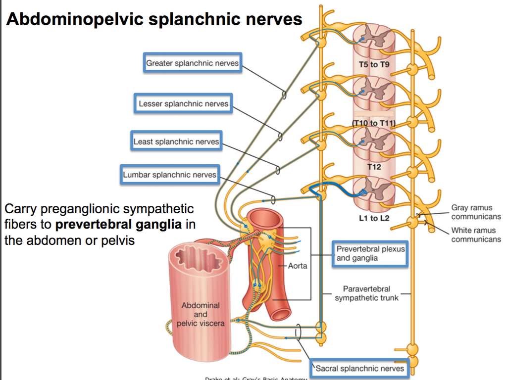
abdominopelvic splanchnic nerves

A

carry pregang sympathetic to prevertebral ganglia

3
Q

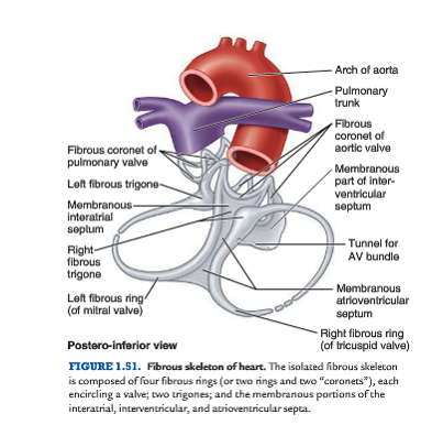
Skeleton of the heart structure

A
  • Four fibrous rings (L. anuli fibrosi) that surround the orifices of the valves.
  • A right and left fibrous trigone (formed by connections between rings), and the membranous parts of the interatrial and interven-tricular septa.
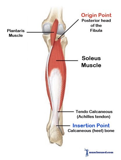
4
Q

Soleus

A

strolling M

deep to gastrocnemium

Origin:

  • Posterior head of fibula, proximal ¼ of posterior surface of fibula, soleal line of tibia

Insertion:

Posterior aspect of calcaneus via calcaneal tendon

Action:

Plantarflexes the ankle joint, assits in flexion of the knee joint

  • workhouse of plantarflex

N:

Tibial

Blood:

Posterior Tibial Artery

5
Q

Fractures of femur usually happen by

what accompanies it usually?

A

high E trauma

dislocation of hip often accompanies
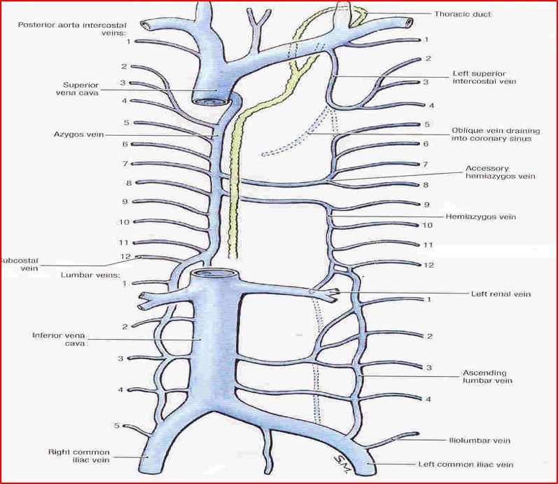
6
Q

Azygos vein

A
  • union of right ascending lumbar vein & right subcotal vein in the abdomen
  • Ascends up through aortic opening
  • Opens in to SVC
  • Connects IVC to SVC
7
Q

what kind of cartilaginous joint is the IV JOINT

A

Secondary

8
Q

Trendelenberg test

A

Injury to superior glutial n, trochanteric fracture, fracture of femoral neck, dislocated hip joint

when pt stands on affected limb, pelvis on opposite side will sag

  • rises in normal pt
9
Q

cardiopulmonary splanchnic nerves carry….

A

postgang to heart, lungs, esophagus

10
Q

Pleura

A

neg P = essential for breathing

2 layers continuous with hilum of lung
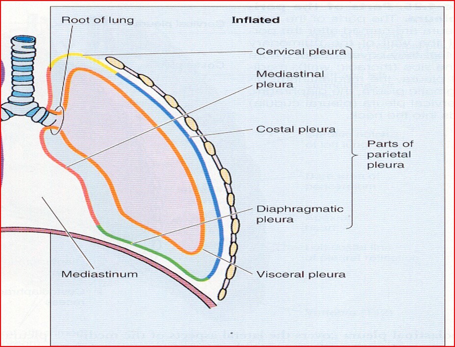
11
Q

borders of lung

A

anterior: cardiac notch
posterior: C7 - T10
inferior: rounded, sepates base from costal and diaphrag surf

12
Q

T12

A

•Body has a single large costal facet on each side, extending on to lower part of pedicle

13
Q

Sciatica

A

Pain caused by a compression/irritation of sciatic N by a problem in lower back

Common causes of sciatica:

  • Disc prolapse, spinal stenosis, spondylolisthesis
14
Q

spread of breast ca

A

communication of superficial lymph

  • midline = to opp side
  • lower = ab = liver = pelvis

spread to v-column by veins

  • viens drain breast go to venous plexus
15
Q

Plantar fascia

A

thick, central part of the plantar fascia forms the strong plantar aponeurosis

  • •flexor digitorum brevis, tendons of FHL, FDL, qadratus plantae, lumbricals, adductor hallucis and the lateral plantar nerve and vessels

has weaker medial and lateral parts

  • medial
    • abductor hallucis, flexor hallucis brevis, the tendon of the flexor hallucis longus, and the medial plantar nerve and vessels
  • lateral
    • abductor and flexor digiti minimi brevis
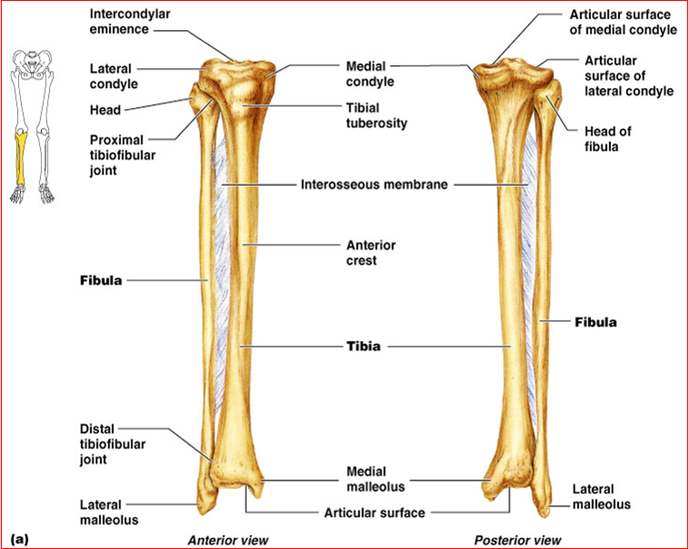
16
Q

Tibia and Fibula

A
17
Q

Laceration of Thoracic Duct

A

usually thin-walled/ dull white = easy to injury during procedures

leak of lymph into pleural cavity = chylothorax

18
Q

Echocardiography

A

graph position and motion of the heart

  • echo from beams of ultrasonic waves through thoracic wall

good for:

  • valve stenosis/regurg, esp L
19
Q

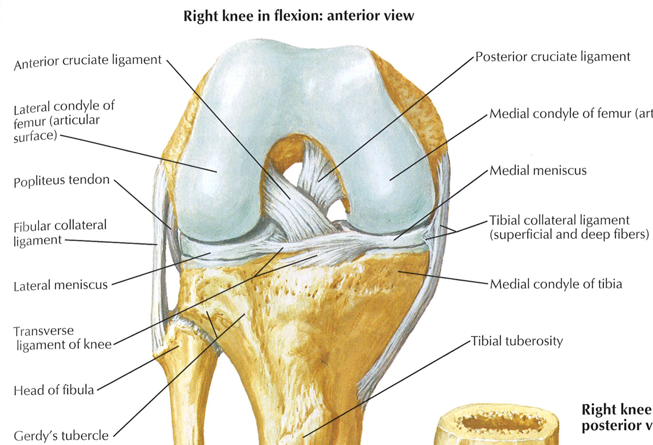
Anterior cruciate ligament

A

weaker of 2

poor blood supply

anterior intercondylar tibia –> posterior lateral condyle femur

func:

  • limit posterior roll of femoral condyles during flexion - convert to spin
  • prevents posterior displacement of femur
  • prevents hyperextension of knee
20
Q

tensor fascia lata

A

Origin:

ASIS

Insertion:

IT tract –> lateral condyle of tibia

Action:

Abduct and medial rotate, flex, stab truck on thigh

N:

superior gluteal

Blood:

lateral Femoral

21
Q

rotation of limbs during development

A

lower limb - rotates 90 degrees medially

upper limb - rotates 90 degrees laterally

22
Q

Extracapsular ligaments

A
  • Patellar ligament
  • Fibular collateral ligament
    • lateral epicondyle of femur –> lateral surf of fibular head
    • tendon of popliteus passes deep
      • separate from lateral meniscus
    • tendon of biceps femoris split into two
  • Medial (tibial) collateral ligament
    • medial epicondyle of femur –> medial condyle & surface of tibia
    • attaches to medial meniscus @ midpoint
    • weaker than FCL = commonly torn in contact sports
  • Oblique popliteal ligament
    • expansion of semimebranosus tendon
  • Arcuate collateral ligament
    • strengthens post-lateral
    • edge of capsule arches over politeus M
23
Q

Transposition of great arteries caused by

A

abnormal neural crest cell migration

non-spiral development of spiral septum

24
Q

R vagus N gives off branches that supply…

A

pulm, esophagus, cardiac plexuses

25
Sternal fissure and foramen ## Footnote : Developmental defect
26
Superior Mediastinum order of structures
anterior to posterior, is: (1) thymus, (2) veins, (3) arteries, (4) airway, (5) alimentary tract, and (6) lymphatic trunks.
27
Coarctation of Aorta
stenosis found at arch or thoracic aorta * usually near ligamentum arteriosum * if inferior = compensation by intercostal and interthoracic A collateral bv may become so large causing: * notable pulsation in intercostal spaces * erosion of adjacent surf of ribs
28
Superior aperture of thorax
•Clinicians refers this as Thoracic outlet • * Anteriorly: * Upper border of manubrium • * Posteriorly: * Anterior surface of body of T1 vertebra • * On each side: * 1st rib with its costal cartilage • * Partially covered by suprapleural membrane (Sibson's fascia) * Triangular in shape, apex on the tip of the transverse process of 7th cervical vertebrae and base to inner border of first rib * May have some muscle fibers called scalene minimus •
29
anterior Traumatic Hip dislocations
commonly * caused by a hyperextension force against an abducted leg less commonly * an anterior force against the posterior femoral neck or head limb not lengthened noticably but will be externally rotated
30
Tricuspid where found?
4/5 intercostal base of cusps attaches to fibrous ring free edge attached to pap M by chordae tendinea
31
Sciatic Nerve Anesthetic Block
Midpoint of line joining - posterior superior iliac spine (PSIS) + superior border of greater trochanter Midpoint of line joining – Ischial tuberosity + superior border of greater trochanter.
32
Vagus nerve
(X CN) carries parasympathetic fibers to thorax and abdomen
33
Dorsal M of foot
2 muscles blend together * extensor digitorum brevis * origin: superior surface, anterolateral calcenus * insertion: split into 3 M (blends with long extensor) on 2-4 toes * extensor hallucis brevis * largest and most medial bellow of extensor digitorum brevis * insertion: prox big toe * N: deep fibular * action: assist extensor digitorum longus in extending toes
34
Intercostal nerves
* Anterior primary rami of T1 to T11. * Anterior ramin of T12 is called subcostal * In addition to thorax, upper 3 supply upper limb and lower 6 supply ant. abd. wall (T7-12) * T4-T6 are called typical intercostal nerve * Nerve lies in the costal groove below posterior intercostal vessels [vein –artery – nerve. VAN] * Lies between internal intercostal and innermost intercostal
35
what prevents backflow in veins
valves in veins prevent backflow due to gravity or internal P contraction of skeletal M shortens (increases in girth) = compresses veins
36
External Intercostal Muscles:
•Extent: from the tubercles of the ribs posteriorly to the costochondral junctions anteriorly. Anteriorly they are replaced by the anterior (external) intercostal membranes. Origin: Inferior border of the rib above Insertion: Superior border of the rib below. The fibers run downwards and forwards. Action- Elevate the ribs – muscles of inspiration.
37
Meniscal tears
usually involve medial * pain on medial rotation of tibia * most occur in conjunction with TCL or ACL tear * unhappy triad: ACL, TCL, medial meniscus * direct blow to lateral aspect of knee with foot on ground lateral * usually not tear because of mobility * pain on lateral rotation
38
March fx
* Stress fracture occur in metacarpal bones * Often occur in military recruits after long marches
39
what causes widening of Mediastinum
usually from head-on collision malignant lymphoma hypertrophy due to CHF - inferior mediastinum
40
tracheosophageal fistula which is most common
imcomplete separation of trachea from esophagus proximal blind-end of esophagus and communication of distal part of esophagus @ carina
41
Thymus
primary lymph organ after •puberty, the thymus undergoes gradual involution replaced by fat art supply = anterior intercostal, anterior mediastinal branches of internal thoracic A
42
Structure passing through lesser sciatic foramen
* Pudendal nerve * Internal pudendal vessels * Nerve to obturator internus * Tendon of obturator internus
43
Congenital diaphragmatic hernia
Posterolateral defect most common anomaly defect =foramen of Bochdalek hernia = Bochdalek hernia
44
Superior extensor retinaculum
dorsum of foot continuous with upper part of flexor retinaculum structures: (medial to lateral) * •TP, EHL, ANTERIOR TIBIAL VESSELS AND NERVE, EDL AND PERONEUS TERTIUS
45
Subcostal muscles:
* Present in the posterior parts of the lower spaces only. * Extend from the internal surf of angle of rib to the internal surface of the rib below crossing one or two intercostal spaces. * Run in the same direction as internal intercostal muscles. * Action: depress the ribs - forced exhalation
46
artery supply to thymus
anterior intercostal internal thoracic branches - anterior mediastinum
47
Left coronary artery
branches: 1. SA nodal - 40% people supplies SA node 2. anterior IV (desccending) - anterior 2/3 IV septum 3. diagonal 4. circumflex 4. left marginal - L vent
48
AORTIC VALVE STENOSIS
most freq valve abnorm, usually degen calcification (\>60 y/o) LV hypertrophy
49
psoas major
Origin: Sides of vertebra T12 to L5 and transverse process of L1-L5 Insertion: Lesser trochanter of femur Action: Flex, stabilizes hip N: Ventral rami of L1-3 Blood: Medial Femoral Circumflex Artery
50
Patellofemoral Syndrome
* Excessive running – downhill “runner's knee” * Osteoarthritis of the patellofemoral compartment * Strengthening of vastus medialis corrects * Prevents lateral dislocation of patella
51
Causes of Horner’s syndrome
•A lesion in the following: –Brain stem – Midbrain,Pons, Medulla –Cervical region of the spinal cord –Cervical sympathetic chain –Injury to carotid arteries around which sympathetic fibers reach the orbit – * When there is cancer of lung, it may compress the inferior cervical sympathetic ganglion resulting in Horner’s syndrome. (Pancoast’s syndrome) * A lesion in the following: –Brain stem – Midbrain,Pons, Medulla –Cervical region of the spinal cord –Cervical sympathetic chain –Injury to carotid arteries around which sympathetic fibers reach the orbit – •When there is cancer of lung, it may compress the inferior cervical sympathetic ganglion resulting in Horner’s syndrome. (Pancoast’s syndrome)
52
Iliacus
Origin: Iliac crest, ilac fossa, ala of sacrum, and anterior sacroiliac ligaments Insertion: Tendon of psoas major and body of femur, inferior to lesser trochanter Action: Flex, stabilize hip N: femoral Blood: Medial Femoral Circumflex Artery
53
Ankle Joint
Talocrural Joint * uniaxial, synovial, hinge articulation between tibia (medial malleolus), fibula (lateral malleolus), and talus –Distal ends of tibia and fibula form a mortise –Trochlea of talus fits into mortise –Malleoli grip talus
54
Right lymphatic duct
•Drains lymph from the body’s right upper quadrant (right side of head, neck, and thorax plus the right upper limb). At root of neck, enters R venous angle: R internal jugular and R subclavian
55
Right vagus nerve
•Enters the thorax anterior to the right subclavian artery * gives off right laryngeal N posterior to the right brachiocephalic vein, SVC, and root of the right lung branches that contribute to the pulmonary, esophageal and cardiac plexus. .
56
transposition of great arteries presentation and result
* Aorta arises from RV and pulmonary trunk arises from LV * Associated with cyanosis (R-L shunting ) * Incompatible with life unless accompanying shunt like VSD, PDA, or patent foramen ovale is present
57
Opening in the diaphragm
* 3 large and many small openings * Vena-caval opening: at T8 * •Inferior vena cava * •Branches of the right phrenic nerve •Oesophageal opening: at T10 * •Oesophagus * •Vagus nerve * •Esophageal br. of left gastric artery •Aortic opening: at T12 Vertebra * •Aorta * •Thoracic duct * •Azygos vein
58
3rd layer plantar foot
flexor hallucis brevis * O: Plantar surfaces of cuboid and lateral cuneiforms * I: Both sides of base of proximal phalanx of 1st digit * Action: Flexes proximal phalanx of 1st digit * N: Medial plantar nerve (S2, S3) Adductor hallucis * O: Oblique head: bases of metatarsals 2-4; Transverse head: plantar ligaments of metatarsophalangeal (MTP) joints * I: Tendons of both heads attach to lateral side of base of proximal phalanx of 1st digit * Action: Traditionally said to adduct 1st digit; assists in transverse arch of foot by metatarsals medially * N: Deep branch of lateral plantar nerve (S2, S3 Flexor digit minimi brevis * O: Base of 5th metatarsal * I: Base of proximal phalanx of 5th digit * Action: Flexes proximal phalanx of 5th digit, thereby assisting with its flexion * N: Superficial branch of lateral plantar nerve (S2, S3)
59
Ribs characteristics
* 12 ribs on each side * Gaps between ribs are called intercostal spaces * Ribs are placed obliquely * •Upper ribs are less oblique * •It reaches to maximum at 9th rib •Length increases from 1-7 then decreases
60
Right Atrium
* Sinus venarum: Posterior wall of the right atrium that is smooth on which the venae cavae (SVC and IVC) and coronary sinus open. * Musculi pectinatae: Anterior wall is ridged with the pectinate muscles. * Right Atrioventricular (AV) orifice * Crista terminalis: which presents externally as shallow groove, the sulcus terminalis. * Fossa ovalis: represents the fetal foramen ovale and its valve. * Coronary sinus: a short venous trunk receiving most of the cardiac veins, is between the right AV orifice and the IVC orifice. * Oval fossa (L. fossa ovalis), which is a remnant of the oval foramen (L. foramen ovale) and its valve in the fetus.
61
Horner’s Syndrome
•Due to injury to the sympathetic fibers to the face. Symptoms: 1. Anhidrosis – Loss of sweating 2. Miosis- Constriction of Pupil 3. Ptosis – mild drooping of the upper eye lid
62
popliteal aneurym sx and femoral A
FA ligated in adductor canal
63
Plantar reflex
sole of foot stimulated with blunt instrument * tests L4,5, S1, S2 nerve roots reflex: downward of hallux (flexion upwards = babinski response! * can ID disease of SC and brain * primitive reflex in infants * due to cortico-spinal tracts not fuly developed * can be present in children up to 4 years old * except in infants with brain injury or cerebral disease
64
celiac plexus
SNS: greater splachnic PNS: vagus structures derived from foregut: * respir tract and esophagus * stomach * liver * gallbladdar/bile ducts * pancreas * upper duodenum
65
Blood Vessels in Popliteal Fossa.
popliteal A * adductor hiatus --\> inferior border of popliteus --\> anterior, posterior tibial A * deepest structure in fossa * 5 genicular branches * supply capsule, ligaments popliteal V * posterior tibial --\> inferior border of popliteus M --\> adductor hiatus --\> femoral V
66
digital deformities
•PolydSyndactylyactyly – extra digits * one or more fused * Brachydactyly – digits are shortened * Oligodactyly – digit is absent * Ectrodactyly – extreme case of oligodactyly, missing one, two or three of the middle digits
67
Nerves supply to the posterior compartment
68
PULMONARY VALVE STENOSIS
restriction of RV outflow --\> RV hypertrophy
69
Femur Structure
70
esophageal atresia
abnormal devision of trachea and esophagus * blind end pouch of esophagus instead of connecting down develops from foregut = endoderm * div posterior by thracheosophageal ridges * anterior = tracheal * posterior = esophagus
71
tibilais anterior
O: lat condyle, prox half of lat tibia I: medial cuneiform, base of 1 MT A: dorsiflex, invert foot N: deep fib blood: anterior tib
72
bv to hip anastamosis
cruciate anastomosis = resembles a cross * inferior gluteal * lateral circumflex femoral * medical circumflex femoral * ascending branch of 1st perforating A from profunda femoris trochanteria anastomosis: circulation around head of femus * superior and inferior gluteal * medial and lateral circumflex femoral * medial = main supply to femur * retinacular
73
obturator internus
Origin: Pelvic surface of obturator membrane and margins of obturator foramen Insertion: Medical surface of greater trochanter of femur Action: Laterally rotates thigh, abducts flexed thigh N: Nerve to obturator internus Blood: inferior Femoral \*bounded by 2 gemelli
74
Pulmonary pleura Blood supply/lymphatics
* Bronchial arteries & veins * Bronchopulmonary lymph nodes
75
Pericardiocentesis
drain fluid from pericardial cavity due to cardiac tamponade procedure: * wide-bore needed @ 5-6 intercostal space near sternum * cardiac notch in L lung and shallower notch of L pleural space exposes some of pericardial sac * intrasternal angle (see pic)
76
Claw Toe
hyperext @ MP joint flexion at DIP joint
77
Adductor Brevis
Origin: Body and inferior pubic ramus Insertion: Pectineal line and proximal part of linea aspera of femur Action: Adducts and flexes N: Obsturator nerve Blood: Femoral
78
ribs are placed \_\_\_\_\_\_\_\_ when is it maximum?
obliquely - increases inferior 9th
79
1st rib
* Broadest and shortest of the true ribs * Has a single articular facet on its head, which articulates with T1 * Has a scalene tubercle for the insertion of anterior scalene muscle * front of scalene tubercle for subclavian vein * behind the scalene tubercle for subclavian artery & trunks of brachial plexus
80
Ductus venosus
avoids portal circulation bypasses liver critical role in preferentially shunting o2-ed blood to fetal brain
81
Anterior mediastinum
* The smallest subdivision of the mediastinum. * Anteriorly : the sternum and the transversus thoracis muscles. * Posteriorly pericardium. * Superiorly: sternal angle * Inferiorly: the diaphragm. * In infants and children, the anterior mediastinum contains the inferior part of the thymus.
82
Surfaces of the Heart
* Anterior (sternocostal) surface, formed mainly by the right ventricle. * Right pulmonary surface, formed mainly by the right atrium. * Left pulmonary surface, formed mainly by the left ventricle; it forms the cardiac impression in the left lung. * Diaphragmatic (inferior) surface, formed mainly by the left ventricle and partly by the right ventricle.
83
Differences between the Right lung and Left lungs
84
Aspirated foreign objects tend to enter
•right bronchus * erect: right posterior of lower lobe * supine: right apical/superior lower lobe, or posterior seg of upper lobe
85
Inferior aperture of thorax: boundaries
* Anterior: Xiphoid process * Posterior: 12th thoracic vert * On each side: costal margin * 7, 8, 9, 10th ribs * 11th and 12th ribs * Closed by diaphragm
86
transverse acetabular ligament
joins labrum of acetabulum
87
Accessory soleus
present in approximately 3% of people muscle usu-ally appears as a distal belly medial to the calcaneal tendon may be associated with pain and edema during prolonged exercise
88
Transverse pericardial sinus
•The passage between the aorta and pulmonary artery in front and the superior vena cava behind
89
poptiteal a aneurysm
swell of left popliteal fossa "thrills" bruit detected when listening with stethoscope
90
Bronchial tree
•Right principal bronchus shorter, wider and more in line with trachea * 3 secondary •Left principal bronchus is longer, narrower and more oblique * 2 secondary •Each respiratory bronchiole provides 2–11 alveolar ducts, each of which gives rise to 5–6 alveolar sac
91
fibularis brevis
O: distal 2/3 lateral fib I: base of 5th MT A: weakly plantarflex, evert foot N: superficial fib A: fibular
92
during what stage of lung development can a fetus be viable?
wk 24 type I and type II cells develop - respiration is possible with ICU
93
SVC union of... @ what location receives blood from.... enters R atrium @
R and L brachiocephalic V (union of internal jugular and subclavian) unite at level of 1st right costal cartilage returns blood from all structures above diaphragm enters R atrium at level of 3rd costal cartilage
94
posterior border of the lung spans what vertebrae?
C7-T10
95
VENTRICULAR SEPTAL DEFECTS
membranous part of the IVS develops separately from the muscular part rank first on all lists of cardiac defects htn VSD on M part frequest closes spontaneously during childhood
96
Posterior intercostal veins: right side
Right side 1: brachiocephalic 2-4: R superior intercostal vein --\> azygos 5-11, subcostal: azygos
97
Extensor Hallucis Longus
O: middle 1/2 anterior fib I: base of distal great toe A: dorsiflex, extend great toe N: deep fib blood: anterior tib
98
piriformis syndrome
piriformis muscle in the butt irritates the sciatic nerve
99
Features of a typical rib: Posterior end
* Head has 2 facets separated by a crest. * Lower larger facet articulate with body of numerically corresponding vertebra. * Upper facet articulates with upper vertebra
100
Thoracic duct
largest lymph channel ## Footnote orig at cisterna chyli (chyle cistern) in the abdomen and ascends through the aortic hiatus in the diaphragm. •At sternal angle, the thoracic duct passes to the left, continues its ascent to the neck where it arches laterally to enter the left venous angle. empties at left venous angle : union of L internal jugular and subclavian
101
popliteal A at the inferior border of the popliteus branches into
posterior and anterior tibial A ANTERIOR TIBIAL A IS THE FIRST BRANCH goes through IO membrane to supply anterior compartment of leg
102
Sympathetic fibers of the face
pregang: T1 - superior cervical ganglion (SCG) * T1 - input form hypoT postgang: SCG - target tissues, around internal carotid A target tissues: dilator pupilla, muller's M, sweat glands
103
separation of digits: how and time
apoptosis - 8 weeks
104
Referred pain from diaphragm
2 directions: central: irritation of the diaphragmatic pleura or peritoneum * shoulder, c3-c5 peripheral: * skin over costal margins of anterolateral abdominal wall (lower intercostal nerves).
105
Contents of Posterior Mediastinum
thoracic desc A azygos, hemiazygos, accessory hemiazygos vagus N, splanchnic nerves (not sympathetic chain)
106
Inferior mediastinum
* Between the transverse thoracic plane superiorly and the diaphragm inferiorly. * Inferior mediastinum further subdivided by the pericardium into anterior, middle, and posterior parts. * Pericardium and its contents constitute the middle mediastinum.
107
Esophagus
T1–T4 inclines left --\> pushed medial by arch of aorta. pushed posterior by root of left lung In the superior mediastinum, the thoracic duct usually lies on the left side of the esophagus.
108
what kind of joint is the Sternoclavicular joint
•Saddle type of synovial
109
Sternum
* Flat bone with 3 parts * Manubrium: * •Superiorly has jugular notch, has clavicular notch on each side, also articulates with cartilage of 1st rib, upper half of 2nd rib * •Posteriorly related to arch of aorta & its branches AND left brachicephalic vein •Body: * •Articulates with 2nd to 7th rib * •Posteriorly related to heart & anterior borders of lungs & pleura •Xiphoid process: * •Cartilaginous at birth ossify slowly and unite with body of sternum after middle age •
110
Nipple
* A conical projection from center of breast * Has openings of 15-20 lactiferous ducts
111
dextrocardia
abnorm folding of embryonic heart * position of heart = reversed: apex is R instead of L * mirroring of great vessels and arch most common positional abnorm of heart * general transposition of the thoracic and abdominal viscera (situs inversus) * cardiac defects = low * transposition only the heart (isolated dextrocardia). * severe cardiac anomalies, ex: transposition of great arteries
112
development of diaphragm structures
1. septum transversum * central tendon 2. pleuroperitoneal membrane * primitive diaphragm 3. dorsal mesentery of the esophagus * crura 4. lateral body wall mesoderm * peripheral diaphragm
113
Gluteus medius
Origin: Lateral suface of ilium between anterior and posterior gluteal lines Insertion: Lateral suface of greater trochanter of femur Action: Abducts thigh, rotates thigh medially, Steadies the pelvis on the leg when the opposite leg is raised off ground. N: Superior gluteal nerve Blood: superior deep Femoral
114
Lymphatic drainage of lung
•Bronchopulmonary lymph nodes
115
Hip Joint Movement
•Movements –Flexion 140 degrees –Extension 10 degrees –Abduction 45 degrees –Adduction 30 degrees –Internal rotation 40 degrees –External rotation 50 degrees
116
knee joint strength
weak! stability depends on 1. strength of the muscles and tendons 2. strength of ligaments •Movements –Flexion and extension –Some gliding and rolling –Some medial and lateral rotation about vertical axis
117
Innermost Intercostal Muscles:
Deep to the internal intercostal muscles. § Not continuous. Has 3 parts. Intercostal nerves and vessels lie between these two muscles. They are present in the middle part of the intercostal spaces. The fibers run in the same direction as that of internal intercostal muscles. •Intercostalis intimi: Origin & Insertion-They pass between the internal surfaces adjacent ribs. Action: helps in inspiratio
118
Injury to Common Fibular nerve
foot = plantarflexed, inverted cannot stand on heels
119
Pus from the vertebral column
•tends to track around the thorax along the course of the neurovascular bundle and points at the site of exit of cutaneous nerves
120
articularis genu
deep to vastus intermedius retracts bursa during knee extension
121
Accessory hemiazygos: landmarks: beginning, end, joins...
beings at 4/5 intercostal, desc lefft from T5 - T8 crosses over at 7/8 and joins azygos
122
Increase in thoracic diameters: •Anteroposterior diameter
* Pump handle movement * Vertebrosternal ribs 2-6 * Partly by elevation of rib 7-10
123
Surgical Hip Replacement
indications * severe traumatic injury * degen disease osteoarthritis * erosion of articular cartilage * symptoms: pain, edema, mvmt limitations procedure: * metal prosthesis replaces head and neck * plastic socket replaces acetabulum
124
Deep transverse septum
separates superficial from deep compartment of posterior compartment of leg reforcing fibers * tip of medial malleolus and calcaneus ----\> flexor retinaculum
125
Pelvic parasympathetic fibers:
"•Nervi erigentes" pregang: S2-S4 hind gut, bladdar, sex organs empties bladder, erection
126
injury to apex of femoral triangle
huge bleeding 4 vessels lie on one another * FA * FV * profunda FV * profunda FA
127
Popliteus
Origin: Lateral surface of lateral condyle of femur and lateral meniscus Insertion: Posterior surface of tibia, superior to soleal line Action: * Weakly flexes knee * unlocks knee by rotation femur 5 degrees on fixed tibia * Medially rotates tibia of unplanted limb N: Tibial Blood: Posterior Tibial Artery
128
Femoral Vein Cannulation
Purpose: * blood samples * take pressure recordings from the chambers of the right side of the heart and/or from the pulmonary artery * right cardiac angiography * admin of fluids Procedure: * fluroscopic control: external & common iliac veins --\> IVC --\> right atrium
129
Thoracic aorta branches
paired branches as aorta desc thorax: * In descending order, these include the * Bronchial arteries * Mediastinal arteries * Esophageal arteries * Pericardial arteries * Superior phrenic artery * Posterior intercostals originate through out the length of the thoracic aorta
130
11th and 12th rib
* single articular facet on head with no neck, no tubercle * poorly marked in 11th rib and are absent in 12th rib
131
Features of a typical rib: Neck
• lies in front of transverse process of the vertebra
132
Gemellus inferior
Origin: Upper margin of ischial tuberosity Insertion: Medial surface of greater trochanter of femur Action: Laterally rotates thigh N: Nerve to quedratus femoris \*larger than superior
133
retromammary space
Between breast and deep pectoral fascia loose connective tissue allows some mvmt
134
unhappy triad
ACL, MCL, medial meniscus
135
Ankle Injury
Usually inversion * twisting of weight nearing plantarflexed foot anterior talofibular ligament
136
Sinus venosus
Right horn - sinus venareum Left horn - Coronary sinus Oblique vein of left atrium
137
R laryngeal recurrent N
hooks around R subclavian A ascends b/w tracheal and esophagus
138
internal thoracic A terminates as
superior epigastic musculophrenic
139
Flexor retinaculum
how M get to foot pneu: Tom, Dick, an Harry (anterior to posterior) * Tom- Tibialis posterior. M. * Dick- Flexor digitorum longus . M. * A- Posterior tibial artery. * N- Tibial nerve. * Harry: Flexor hallucis longus.M
140
Tibialis posterior
Origin: Interosseous membrane; posteror surface of tibia inferor to soleal line; posteror surface of fibula Insertion: Tuberosity of navicular, cuneiform, cuboid, and sustentaculum talki of calcaneus; bases of 2nd, 3rd and 4th metatarsals Action: * Plantarflexes ankle * inverts foot N: Tibial Blood: Posterior Tibial Artery
141
Femoral Triangle
NAVL: (medial to lateral) * N * A * V * Lymph node •Triangular depression on ant. aspect of upper 1/3rd of thigh below inguinal ligament Boundaries: * Laterally * Sartorius * Medially * Adductor longus * Base * Inguinal ligament * Apex * Faces downward where medial and lateral boundary meet * Roof * Skin, Superficial fascia, Deep fascia * Floor * Adductor longus, pectineus, psoas major and iliacus
142
iliofemoral ligament
ASIS, acetabulum --\> intertrochanteric line y-shaped A: prevents hyperextention
143
primordial atrium is represented in the adult by
right auricle.
144
Surgical Signifi cance of Transverse Pericardial Sinus
clamp and insert tubes of coronary bypass = divert blood during sx
145
Branches from the arch
* Brachiocephalic artery – supplying the right side of the head & neck and the right upper limb. * Left Common carotid artery – to the left side of the head & neck. * Left Subclavian artery – to the left upper limb.
146
CORONARY ATHEROSCLEROSIS and effect
lipid deposits in intima --\> luminal stenosis collateral channels may initially expand (compensation) may not receive enough o2 with increased amts of work = MI
147
T11 and T12 structure
•no facet on transverse process
148
Hip Joint-Capsule
increases stability of joint restricts extension of joint to 10-20 degrees beyond vertical postiion * flexion increasily unwinding spiraling ligaments/fibers
149
Central Traumatic Hip dislocations happen when...
lateral force against an adducted femur side impact MVCs
150
Intercostal nerves Branches supplies?
Branches: * Cutaneous (anterior and lateral) * Muscular * sympathetic chain Supplies: * skin * muscles * parietal pleura
151
Lungs
* Right lung is heavier (625 gm) than left (575 gm) * Adults lungs are spongy due to air in the alveoli and float in water * In stillborn child, they are solid and sink in water
152
Areola
pigmented mod sebaceous gland * enlarged during preg/lactation = Montgomery tubercles oily secr lub nippe nf areola to prevent cracking during lactation
153
Subcutaneous infrapatellar bursitis
“clergyman’s knee” * frequent flexing of knee * most common in roofers/floor tilers when not wearing knee pads
154
Gastrocnemius strain
"tennis leg" acute injury from partial tearing of medial belly of gastrocnemius * overstretch via full extension @ knee and full dorsiflexion @ ankle * stabbing pain --\> edema --\> gastrocnemius spasm often seen in people \> 40 y/o
155
4th layer of plantar foot
4. Plantar interossei (three muscles) * O: Bases and medial sides of metatarsals 3-5 * I: Medial sides of bases of phalanges of 3rd-5th digits * Action: Adduct digits (2-4) and flex metatarsophalangeal joints * N: Lateral plantar nerve (S2, S3) 4. Dorsal interossei (four muscles) * O: Adjacent sides of metatarsals 1-5 * I: 1st: medial side of proximal phalanx of 2nd digit;2nd-4th: lateral sides of 2nd-4th digits * Action: Abduct digits (2-4) and flex metatarsophalangeal joints * N: Lateral plantar nerve (S2, S3)
156
lobes of lung
R: * oblique and horizontal: 3 L: * oblique: 2 * lingula: tongue shaped projection
157
what types of joints are chondrosternal joints
* 1st-primary cartilaginous joint * 2nd-7th-plane synovial joint
158
hemimelia
absence of forearm or lower leg bones, most common absence = fibula
159
Mitral valve and relation to ribcage
anatom: 4th costal cartilage "bicuspid" attached by chordae tendinae (tighten before and during systole so cusps do not go into L atrium)
160
hemidiaphragm
Complete paralysis and atrophy of the corresponding half of the diaphragm
161
heart tube starts formation during... fully functional by....
wk 3 wk 4
162
Necrosis of Femoral Head in Children
traumatic dislocations of hip joint * disrupt artery to head of femur fx to separate of superior femoral epiphysis * =inadequate blood supply to femur head * post-traumatic avascular necrosis of head
163
Ankle jerk reflex
broad edge to tap calcaneal tendon plantarflex of ankle joint * S1/S2 N roots
164
fibrous septa - plantar foot
highly developed skin ligaments divide into fat-filled area * shock absorber anchor skin to plantar aponeurosis * inprove grip of sole
165
Shy-Drager Syndrome
multi sys atrophy - degen of autonomic N and motor sys orthostatic hypotension, dry mouth/skin, cold hands/feet, urinary incontinence, impotence can cause slow mvmts, mild tremors, loss of balance
166
Femoral Artery Cannulation
inferior to the midpoint of the inguinal ligament cardiac angiography * FA --\> exteral iliac --\> common iliac --\> aorta --\> left ventricle of heart * can be used to visualize coronary arties in coronary arteriography blood gas analysis
167
dorsal skin of foot
thinner, less sensitive than most of skin of sole of foot subcut tissue = loose * edema! especially anterior to and around medial malleolus deep fascia = continuous with inferior extensor retinaculum
168
Forced breathing M
•Inspiration: Diaphragm pec minor intercostal scalenii sternomastoids serratus ant erector spinae nasal * Expiration * Muscles of the abdominal wall
169
M of foot
2o individual * 14 plantar * 2 dorsal * 4 intermediate
170
PCL tear
how: * player lands on tibial tuberosity with knee flexed * knocked to floor in basketball usually occurs in conjection with TCL or FCL
171
Femoral Artery Palpation
supine: * midway b/w ASIS and pubic symphysis * little finger on ASIS * tip of thumb on pubic tubercle * midpalm, interior to midpoint of inguial ligament pulse usually strong * weak = partially occluded common or external iliac A
172
Oculomotor nerve
(III CN) –supplies the constrictor pupillae and ciliaris muscle
173
WHAT KIND OF JOINTS ARE Costovertebral, costotransverse, costochondral joint:
•Plane synovial joint
174
Joggers foot
compression irritation of medial plant * pass deep to flexor retinaculum * curves deep to abductor hallucis symptoms: * ache, burn, numb, tingle * medial sole of foot, region of navicular tuberosity repetitive eversion of foot * running, gymnastics
175
Artery of lungs
pulm A: deoxy blood bronchial A: R = 1, L = 2 - desc thoracic aorta right side can also orig from right 3rd post intercostal A
176
Hip Joint Structure
femur * head forms 2/3 of sphere * entire femur covered with articular cartilage except for fovea for ligament of femoral head * attaches fovea to transverse acetabular ligament * contains branch from obturator A acetabulum * imcomplete right of fibrocartilaginous labrum - completed by transverse acetabular ligament
177
Increase in thoracic diameters: vertical diameter
•Descent of the diaphragm
178
Right ventricle
•Conus arteriosus (infundibulum), which leads into the pulmonary trunk Trabeculae carneae : irregular muscular elevations Supraventricular crest:M ridge, separates inflow and outlow •three papillary muscles * anterior: largest, attachs anterior posterior cusps of tricuspid * posterior: smaller, attaches posterior and septal cusps * septal: attaches anterior and septal Moderator band * purkinje fibers from right bundle branch
179
left recurrent laryngeal N
comes off L vagus @ inferior border of the arch of the aorta, immediately lateral to the ligamentum arteriosum ascends to the larynx between the trachea and the esophagus.
180
Rib fractures
* Usually just anterior to angle because it is weakest part * Can damage the pleura and lungs * Lead to pneumothorax or hemothorax
181
PARASYMPATHETICs - Spinal cord segments ## Footnote * CN3 * CN7 * CN9 * CN10 * S2,3,4
* CN3 : EYE BALL * CN7 :SUBMANDIBULAR, SUBLINGUAL, LACRIMAL, NASOPALATINE GLANDS * CN9 : PAROTID, BARORECEPTOR * CN10 : HEAD + NECK, THORAX, ABDOMEN UPTO LEFT COLIC FLEXURE * S2,3,4 : REST OF ABDOMEN, PERINEUM PELVIS
182
Pericardium
* Has two layers. * An outer fibrous pericardium and an inner serous pericardium. * The serous pericardium is divided into outer fibrous parietal and inner visceral. * Between the two is a cavity called the pericardial cavity. * Cavity contains pericardial fluid.
183
purpose of ductus arteriosus
avoid pulmonary circulation in fetus connects pulmonary trunk and aorta
184
Patellar Dislocation
* Nearly always dislocates laterally * Angle of pull of quadriceps relative to axis of the patella and tibia * Tendency toward lateral dislocation is normally counterbalanced by medial more horizontal pull of powerful vastus medialis
185
sinus venarum
portion of M that is smooth in the R atria where the SVC and IVC enter
186
Fibrous pericardium
* dense connective tissue. * Blends with the tunica adventitia of the vessels entering and leaving the heart. * Is attached to the central tendon of the diaphragm-pericardiophrenic ligament * Attaches to the sternum.- Pericardiosternal ligaments. * It protects the heart against overfilling because it is fibrous and unyielding.
187
normal angle of femur in acetabulum
135
188
Foot bone fx
canlcaneal * fall on heels metatarsal/phalangeal * heavy obj falls/runs over food metatarsal * common in dancers, esp female ballet - losing balance
189
what drains into the azygos vein distally?
R ascending lumbar vein R subcostal V
190
•Interchondral joint
•7th to 10th costal cartilage articulate with one another by synovial joint
191
Pectineus
Origin: * superior ramus of pubic Insertion: * pectineal line of femus, inferior to lesser trochanter Action: * Adducts and flexes thigh, assist in medial rotation N: * femoral Blood: * Femoral
192
What cells are involved in the blood-air barrier
type I pneumocytes (squamous) * gas exchange type II pneumocytes (cuiboidal) * surfactant secretion - decrease SA * \<5% make up barrier but 60% of cells in alveoli capillary endothelium basement membrane
193
acetabular cavity
fusion of : pubis, illium, ischium
194
Semimembranosus
Origin: Upper and lateral ischial tuberosity Insertion: Posterior part of medial condyle of tibia Action: Flexes leg, extends thigh N: Tibial division of sciatic nerve Blood: inferior Femoral
195
what is commonly harvested for coronary bypass surgery?
great saphenous vein: * diameter =/\> coronary A * easly dissected * lengthy straight portions radial A has become increasingly more common
196
Artificial Cardiac Pacemaker placement pathway
electrode --\> SVC --\> RA --\> tricuspid --\> RV --\> fixed to trabeculae carneae
197
Bulbus cordis
Smooth part of right ventricle (conus arteriosus) Smooth part of left ventricle (aortic vestibule)
198
which side of the ________ intercostal A are longer?
posterior right side longer than left
199
Sternal Angle
* T4/T5 disc * 2nd rib articulation * Ascending aorta ends * Descending aorta begins * Arch of aorta ends and begins * Trachea bifercates * Azygos vein joins SVC * Highest point of arch of aorta is NOT at this level
200
Sternocostalis/ transversus thoracis
4 or 5 slips: anterior intercostal space Origin: Posterior xiphoid & inferior sternum. The fibres run upwards and laterally Insertion: costal cartilages ribs 2-6 Action- expiration (depression of costal cartilages)
201
lateral view of foot
202
Azygos system of veins
* On each side of the vertebral column. * Drains the back and thoracoabdominal wall.
203
Vitelline vein becomes
portal vein
204
abdominal splachnic N
1. Greater Splanchnic: T5-T9 - origin of celiac 2. Lesser splanchnic: T10-T11 - superior mesenteric 3. Least splanchnic: T 12 - inferior mesenteric 4. Lumbar (sacral) splanchnic : L1-L2 - bifurc of external/internal iliac **sympathetic splanchnic nerve will END in front of the ab\ AORTA ( at the PRE VERTEVBRAL GANGLIA)** * will run along branches of these A to reach organs
205
Adductor Longus
Origin: Body of pubis, inferior to pubic crest Insertion: Middle third of linea aspera of femur Action: Adducts thigh N: Obturator Blood: Femoral
206
innervation to knee joint
branches of sciatic, femoral ,obturator N
207
Fracture neck of femur
common in people 60 y/o and older * women - osteoporosis requires internal skeletal fixation: intracapsular Ischemic necrosis of neck + head * interruption of blood supply from retinacular vessels - trochanteric anastomosis - medial femoral circumflex artery * artery to ligament of femoral head - only remaining source of blood to proximal fragment - inadequate - aseptic vascular necrosis lower limb shortened * distal fragment pulled up by: * quad femoris, adductors, hamstrings * Psoas - lateral rotation
208
THORAX LANDMARKS: Costal margin:
•7th to 10th costal cartilages
209
vein supply of lungs
pulm veins: 2 from each long brochnial viens (2 of each side): R = aqygos, L = L superior intercostal, accessory hemizygos
210
Diaphragm structure
* Musculotendinous structure present between thorax and abdomen * Has 3 origins * Sternal: xiphoid process * Costal: originate from lower 6 costal cartilages * Vertebral: Right and left crus * •Right crus from right side of body of L1-L3 * •Left crus from left side of body of L1-L2 * •Medial and lateral arcuate lig. •All the parts insert into central tendon
211
x sec of hip
212
Facial nerve
(VII CN) –supplies all the glands of head except parotid. It is very important to know that there is drying of eye when there is facial nerve lesion but the loss of secretion of saliva may not be diagnostic since parotid is still functional.
213
thoracic duct orginates at
cisternae chyli in abdomen
214
Patellar Tendon Reflex
quadriceps should contract integrity of femoral nerve + L 2 3 4 spinal cord segments
215
•Two heart sounds
* S1: contraction of the atria and dumping blood into the ventricles. Closure of A-V valves. * S2: contraction of the ventricles dumping blood into the blood vessels. Closure of semilunar valves.
216
Endocardial Cushion
formed by neural crest cells separates R and L sides of heart
217
Posterior intercostal veins: Left side
Left side 1: brachiocephalic 2-4: L superior intercostal--\>L brachiocephalic 5-8: accessory hemiazygos 9-11, subcostal: hemiazygos
218
Plantar Fasciitis
inflammation plantar aponeurosis * repetitive strain on longitudinal plantar arch pain on prox plantar surface of foot tx: rest, analgesia
219
Aorticopulmonary septum (spiral septum)
appears within the truncus arteriosus dividing it into the Ascending aorta and the pulmonary trunk.
220
Primitive ventricle
Trabeculated parts of of R and L ventricle
221
Angina Pectoris
usually from narrowed coronary arteries usually relieved by period of rest (1-2min) * SL nitro = dilates coronary A and other A WARNiNG SIGN!!
222
Arterial supply to breast
* Branches coming from * Internal thoracic artery * Lateral thoracic artery * Thoracoacromial artery * Posterior intercostal arteries
223
quadriceps femoris
Origin: * rectus: Anterior inferior iliac spine and groove superior to acetabulum * vastalis lateralis: Greater trochanter and lateral lip of linea aspera of femur * vastus medialis: Intertrochanteric line and medial lip of linea aspera of femur * vastus intermedius: Anterior and lateral surfaces of body of femur Insertion: Base of patella and to tibial tuberosity via patellar ligament Action: Extends the leg at the knee joint, rectus femoris; steadies hip, helps iliopsoas flex thigh N: femoral Blood: Medial Femoral Circumflex Artery
224
Borders of Heart
* Right border (slightly convex), formed by the right atrium and extending between the SVC and the IVC. * Inferior border (nearly horizontal), formed mainly by the right ventricle and slightly by the left ventricle. • * Left border (oblique, nearly vertical), formed mainly by the left ventricle and slightly by the left auricle. * Superior border: formed by the right and left atria and auricles in an anterior view. •
225
Prevertebral ganglia
•Surround the main branches of abdominal aorta –Celiac ganglia –Superior mesenteric ganglia –Inferior mesenteric ganglia
226
Structure passing through greater sciatic foramen
Structure above pyriformis: * Superior gluteal nerve and vessels Structure passing below pyriformis: * Inferior gluteal nerve * Inferior gluteal vessels * Sciatic nerve * Post. cut. n. of thigh * Nerve to quadratus femoris * Pudendal nerve & internal pudendal vessels * Nerve to obturator internus
227
thorax landmarks: between T4 and T5
Sternal angle (angle of louis): * Manubrio-sternal joint * 2nd costal cartilage anteriorly and disc between T4 and T5 posteriorly
228
Ischiofemoral ligament
acetabular rim --\> greater trochanter * circles superior/lateral A: prevents hyperextension, screws femoral head into acetabulum
229
A 27-year-old woman involved in a car accident is brought into the emergency department. Her magnetic resonance imaging (MRI) reveals that she has a laceration of the spinal cord at the L4 spinal cord level. Which of the following structures will not be seen at this level? (A) Dorsal horn (B) Lateral horn (C) Ventral horn (D) Gray matter (E) White matter
Lateral horn
230
Cardiac Referred Pain
Anginal = radiating: substernaland left pec--\> left shoulder,medial aspect of the left upper limb ## Footnote cardiac pain referred to upper limb between SC segments of cutaneous N (T1-T3) common to the visceral afferent termination of coronary artieries
231
Biceps femoris
Origin: * long head: ischial tuberosity * short head: lateral lip of linea aspera Insertion: * long head: lateral head of fibula * short head: lateral supracondylar line of femur Action: Flexes and (LH) laterally rotates leg, extends thigh N: * long head: tibital - sciatic N * short head: common fibular - sciatic N Blood: Femoral \* short head does not cross 2 joints \* only hamstring that does a little rotation
232
Flexor digitorum longus
Origin: Medial part of posterior surface of tibia inferior to soleal line; Insertion: Bases of distal phalanges of lateral four digits Action: * Flexes lateral four digits plantarflexes ankle; N: Tibial Blood: Posterior Tibial Artery
233
Gluteus Maximus
Origin: Ilium posterior to posterior glutealline dorsal sacrum, sacrotuberous ligament Insertion: Iliotibial tract of fascia lata, gluteal tuberosity of femur Action: Extends thigh, assists lateral rotation * powerful extensor of thigh * largest M in body * climbing stairs N: Inferior gluteal nerve Blood: Superior Superficial and Inferior Femoral
234
. Right coronary artery
r aortic sinus branches: 1. SA nodal 2. R marginal - right border of heart (not apex) 3. AV nodal 4. post IV - adjanct sides of both ventricles, * right dominant: given at posterior iv sulcus 5. iv septal = post 1/3 of IV septum SUPPLIES DIAPH SURF
235
Recesses of pleura
•Reserve space for expansion of lung between two parts of parietal pleura costomediastinal costodiaphragmatic - first part of plueral cav to be filled up by plural effusion
236
Popliteal pulse
prone with knee flexed inferior of fossa where A is related to tibia weak/loss = FA obstruction
237
Gracilis
Origin: Body of pubis and inferior pubic ramus Insertion: Superior part of medial surface of tibia Action: Adducts thigh, flexes and medially rotates leg N: obturator Blood: femoral
238
Hamstring muscles characteristics
origin: * ischial tuberosity insertion * one of leg bones action: * extend hip, flex knee N: * tibial part of sciatic N \* common injury in athletes b/c it goes over 2 joints
239
Sural nerve graft
nerve grafts in procedures such as repairing nerve defects resulting from wounds locate near small saph V
240
Innervation and position of the diaphragm
wk 4: * septum transversum lies opposite upper cervical somites wk 5: * C3-C5 grow into septum and form phrenic N * nerves pass to septum via pleuropericardial membrane = N lie in fibrous pericardium wk 6: * rapid growth of ventral v dorsal = descent of diaphragm and nerves to thoracic somite lvl wk 8: * dorsal lies @ L1 - phrenic N follows
241
placement v best heart of pulm valve
Third left costal cartilage Second left intercostal space, just lateral to sternum
242
gluteal gait
pelvis droop to unaffected side with opposite leg is raise to walk paralysis of gluteus medius - stability pelvis when opposite leg raised
243
True/false /floating ribs
* Ribs that are connected to sternum through their cartilages are called true ribs * 1-7 ribs * Vertebrosternal / true ribs * 8-10 are false ribs: * 8, 9, 10 are connected to the costal cartilages immediately above them to form costal margin * Vertebrochondral * 11-12 are floating ribs * Vertebral * 3-9 are typical ribs * 1-2 & 10-12 are atypical
244
initialiting flexion of knee
unlocks knee joint popliteus (tendon) first 5 degrees via latereal rotation
245
position of leg after a traumatic posterior hip displacement
shortened internally rotated
246
Levels of Viscera Relative to Mediastinal Divisions
laying supine spreads ab viscera horizontally, pushing mediastinal structures superiorly usually will sag when standing due to gravity supine: * arch of aorta: superior to txverse thoracic plane * trach bifuc = txsected by txverse thoracic plane * central tendon of diaphragm = xiphisteral jxn and T9 standing: * arch of aorta: super txsected by txverse thoracic plane * trach bifuc = inferior to txverse thoracic plane * central tendon of diaphragm = middle of xiphoid process, T9-T10
247
which M abducts flexed thigh
piriformis, obturator internus, germelli
248
Hammar Toe
proximal phalax * dorsiflex (hyperextend) @ MP joint * plantarflex @ PIP joint weak lumbricals and interossei
249
Arch of the aorta
begins and ends @ posterior 2nd SC joint: right to left ascends anterior to the R pulmonary A , trachea bifurcation apex at the L side of the trachea descends posterior left root of the lung, T4
250
Middle mediastinum containts
heart ascending aorta lower 1/2 of SVC with azygos vein opening bifurcation of tachea, 2 bronchi pulm: A and right and left veins phrenic N
251
2nd layer of plantar foot
quadratus plantae * O: Medial surface and lateral margin of plantar surface of calcaneus * I: Posterolateral margin of tendon of flexor digitorum longus * Action: Assists flexor digitorum longus in flexing lateral four digits * N: Lateral plantar nerve (S2, S3) lumbricals * O: Tendons of flexor digitorum longus * I: Medial aspect of expansion over lateral four digits * Action: Flex proximal phalanges, extend middle and distal phalanges of lateral four digits * N: Medial one: medial plantar nerve (S2, S3); Lateral three: lateral plantar nerve (S2, S3)
252
•Secondary cartilaginous joint
•Manubriosternal joint:
253
medial compartment of thigh pic
254
Base of lung
rests of diaphragm separates: R lung from R lobe of liver, L lung from L lobe of liver, stomach and spleen
255
THORAX LANDMARKS : Apex beat/ apex of the heart
•Left 5th ICS ½ inch medial to midclavicular line
256
Adductor Magnus
Origin: * Adductor part: Inferior pubic ramus, ramus of ischium * Hamstring part: ischial tuberosity Insertion: * Adductor part: gluteal tuberosity, linea aspera, medial supracondylar line * Hamstring part: adductor tubercle of femur Action: * Adductor part: adduct, flex thigh * Hamstring part: extend thign N: * Adductor part: obturator * Hamstring part: tibial Blood: Femoral
257
Club Foot
subtalar joint * food = inverted * ankled = plantar flexed * forefood = adducted
258
gastrocnemius
long-jump M Origin: * Lateral Head- lateral condyle of femur * Medial head- popliteal surface above medial condyle of femur Insertion: Posterior aspect of calcaneus via calcaneal tendon Action: Plantarflexes the ankle joint, assits in flexion of the knee joint * cannot plantarflx when knee fully flexed N: Tibial Blood: Posterior Tibial Artery
259
Small Saphenous V
origin: * lateral dorsal venous arch of foot ascends with sural N * behind lateral malleolus * up lateral border of calcaneous tendon - up middle passes b/w 2 heads of gastrocnemius M in popliteal fossa * pierces deep fascia
260
Bronchoscopy
•Visualization of interior of bronchial tree • •Helpful to diagnose brochogenic carcinoma • •Biopsy can also be taken at the same time • •Inhaled foreign bodies are removed by this procedure
261
Saphenous Varix
localized dilation of terminal part of great saphenous vein * femoral triangle edema associated with varicose veins in other parts of lower limb may be confused with other groin swellings such as a psoas abscess
262
Coronary artery dominance
•artery that supplies the posterior descending artery (PDA) determines the coronary dominance. R: RCA - 70% L: CX (circumflex - branch of L artery) - 10% co: both - 20%
263
hip capsule - Intra capsular ligament
Ligament of the head of the femur synovial fold * conducts a bv * weak, little importance for strength of hip
264
Skin on plantar foot
thick over major weight-bearing areas * heel, lateral margin, ball of foot subcut tissue = more fibrous deep fascia = continuous with plantar fascia, deep fascia of sole
265
Popliteal lymph nodes drain into they drain...
drains into deep inguinal lymph nodes drains deep tissues of popliteal fossa, leg, knee joint
266
Tributaries of SVC
Left superior intercostal vein * 2-4 --\> left brachiocephalic vein. Supreme intercostal vein * first intercostal veins --\> brachiocephalic veins.
267
Calcaneal tendon rupture
most severe acute muscular problem of the leg * bruising in malleolar region * lump in calf = shortening of triceps surae lose func: * plantarflex against resistnace * raise heel * balance on affected side excessive passive dorsiflexion walking only when limb = latreal rotated, roll over transverse-placement of foot, no take off
268
gastrocnemius are largely composed of what type of fibers?
fast twitch II
269
where does IT band insert into
Gerdy's tubercle lateral side of tibia
270
Vertebral spines: THORAX LANDMARK
* C7 * Vertebra prominens * T3 * Root of scapular spine * T7 * Inferior angle of scapula
271
plantar foot M characteristics
function: * group: support stance, maintain arch of foot * most active in push off (time when forces tend to fallten transverse arch) 4 layers 1. abductors, short flexors of toes 2. long flexor tendons, 4 lumbricals, quadratus plantae 3. flexor little toe, flexor & adductor great toe 4. dorsal and plantar interosseous
272
endoderm origins of the lung
pulm epithelium larynx glands trachea bronchi
273
Features of a typical rib: Tubercle
outer surface of a rib at junction of neck and shaft. It articulates with its numerical vertebra
274
anterior compartment syndrome mostly due to?
fxs account for 75% soft tissue injury
275
Veins supply to breast
•Mainly to axillary vein, some goes to internal thoracic vein, posterior intercostal vein (communicate with vertebral venous plexus) •
276
what is associated with bruits and thrills?
aneurysm (usually popliteal a)
277
Pulmonary embolism
* Obstruction of pulmonary artery or its branches by an embolus * Usually arise in deep veins of lower limb * Symptoms are sudden onset of dyspnea, chest pain
278
Pubofemoral ligament
pubic ramus --\> joint capsule A: limits abduction, extension
279
thorax landmarks: t2
•Lower border of T2 Jugular notch / supra sternal notch
280
Congenital hip dysplasia
features: * •Hip dislocation, * •Asymmetry of leg positions * •Asymmetric fat folds * •Diminished movement on the affected side Ortolani test/maneuver * flex hips and knees in supine position to 90 deg. * index fingers placing anterior P on greater trochanters, abducting legs with thumbs * positive = "clunk" * head relocates anteriorly into acetabulum * posterior dislocation in infants Barlow's sign * bringing the thigh towards the midline of the body * positive = feeling femur head slip out of socket postolaterally
281
2nd rib
* Length is twice that of 1st rib * Angle and tubercle are very close * Shaft has no twist * Middle of the shaft has a large rough tubercle
282
Intra-articular ligaments, limitations on rotation
Cruciate ligaments * center of joint - cross obliquely * during medial rotation of tibia on femur, cruciate lig wind around each other = limited to 10 degrees * unwind during lateral rotation = 60 deg possible
283
insertion of IT band
lateral condyle of tibia
284
popliteal A
becomes popliteal A after FA passes through adductor hiatus end splits into anterior and posterior tibial A
285
placement v best heard of tricuspid valve
Inferior middle sternum Over inferior middle sternum
286
Apex of the heart
inferolateral part of the left ventricle, post to left 5th intercostal sounds of mitral valve closure are maximal (apex beat).
287
Obturator Externus
Origin: Margins of obturator foramen and obturator membrane Insertion: Trochanteric fossa of femur Action: Laterally rotates the thigh and stabilizes head of femur in acetabulum N: obturator Blood: femoral
288
membranous ossification what develops from this type of ossification?
mesenchyme cells replaced directly by the osteoblasts ## Footnote clavicle cranial vault some cranial base mandible
289
placement v best heard of aortic valve
4th L costal cartilage 2nd R intercostal, lateral to sternum
290
Left ventricle
Two valves one AV valve : mitral and one semilunar : aortic valve
291
Septation of Heart beings and completes at
wk 4, wk 5
292
skeletal system develops from:
paraxial lateral plate mesoderm - axial skeleton pareital lateral plate mesoderm - appendicular skeleton neural crest - part of skull
293
Features of a typical rib: anterior end
•Oval and articulates with costal cartilage
294
typical and atypical ribs
typ = 3-9 atypical = 1,2, 10-12
295
Internal thoracic vein drains into
bracheocephalic vein
296
alternate means of venous drainage when obstruction of the IVC occurs.....
azygos, hemi-azygos, and accessory hemi-azygos thorax, back, ab
297
thorax landmarks: T9
Xiphisternal joint
298
Pericarditis and Pericardial Rub
inflam of pericardium rustle of silk sound over L sternal border and upper ribs = pericardial friction rub
299
what makes up the lateral plate mesoderm in lung development
somatopleuric and splanchopleuric separate by the intraembryonic colemonic cavity
300
plantarflex
4x stronger than dorsiflexion major component of push-off parts when running, walking * thrust primarily at ball of foot * propel body forward and upward
301
Aortic valve placement
3rd intercostal, left side of sterum
302
Adductor canal
Hunter’s or subsartorial canal anoneuorsis * extend from apex of femoral trainge to adductor hiatus (oepning in adductor magnus) Bounded: * front and laterally * Vastus medialis * Behind * Adductor longus and magnus. * Roof * Sartorius Contents * Femoral vessels * Saphenous nerve * Nerve to vastus medialis
303
N of foot
304
•Paracentasis thoracis
7-9 intercostal space, midax lung may collapse, medialstinal shift
305
length relationship of ribs
increases from 1-7, then decreases
306
•T1:
•Superior costal facet on the body is complete (1st rib). Inferior costal facet is demifacet ( for 2nd rib)
307
Fat Wallet Syndrome/Wallet Neuropathy
lower back pain by sitting on bulky wallet common with men
308
saphenous N functions
innervation to knee, skin of anterior thigh and anteromedial leg
309
Injury to Recurrent Laryngeal Nerves
left recurrent laryngeal nerve winds around the arch of the aorta and ascends between the trachea and esophagus * may become stretched = loss of voice
310
•Crural fascia
deep fascia distal continuation of fascia lata thicker to prox leg continues as extensor retinaculum
311
causes of hemopericardium:
MI (weakened area of heart) bleeding into cavity after cardiac sx stab wounds
312
what develops from the Foregut splanchnic mesoderm in lung development
connective tissue cartilage smooth M
313
what type of gait results from damage to the tibial N?
loss of plantar flexion difficulty in getting heel off ground, shuffling of gait
314
most common sites of coronary artery occlusion
1. LAD 2. RCA 3. L circumflex
315
what kind of gait results from damage to the deep fibular N?
foot drop high-stepping gait
316
limb development is triggered by
interaction of apical ectodermal ridge (AER) with underlying mesenchyme
317
Thoracic vertebrae
* 12 thoracic vertebrae * Features: * Demifacets on side of body [may be one or two] * Facets on the transverse processes * Long backward and downward directed spine, heart shaped body and rounded small vertebral foramen * 2-8 are typical * 1, 9, 10, 11, 12 are atypical
318
Features of a typical rib: Costal groove
• has neurovascular bundle ( VAN)
319
posterior deep fibular N supplies
posterior leg
320
oval foramen
At birth, when the baby takes its first breath lungs expand with air = pressure in the R atrium falls oval foramen closes for its first and last time valve usually fuses with the interatrial septum
321
Thoracentesis needle procedure (laters pierced)
* The needle pierices the following layers in order: * Skin - subcutaneous tissue- ext. int. muscle- Int. Int muscle -innermost int muscle- endothoracic fascia- parietal pleura * Needle is placed just above the superior border of lower rib to avoid the damage to neurovascular structures
322
Flabella in gastrocnemius
sesamoid bone * close to prox attachment of lateral head * articulates with lateral femoral condyle visible in lateral radiographs in 3-5% of people
323
common fibular N location? divisions?
wraps around fib head deep: anterior compartment superficial: lateral compartment
324
Femoral Hernias
femoral ring = weak often a loop of small intestine bounded by FV (lateral) and lacunar ligament (medial) Initially, the hernia is small because it is contained within the canal, but it can enlarge by passing inferiorly through the saphenous opening into the subcutaneous tissue of the thigh common in females (wider hips) can be stragulated: * sharp, rigid borders of femoral ring (esp concave margin of lacunar ligament * interferes with blood supply to herniated intestine --\> necrosis
325
cardiac M develops from...
splachnic latereal plate mesoderm surround the dev heart tube
326
Paravertebral ganglia extend...
from the base of the skull (superior cervical) to tip of coccyx-ganglion
327
Foot Bones
Tarsus (7) * calcaneus, talus, cuboid, navicular, and three cuneiforms Metatarsus (5) * Numbered from medial side of foot * Has a base (proximally), a shaft, and a head (distally). * Bases of metatarsals articulate with cuneiform and cuboid bones, and heads articulate with proximal phalanges •Phalanges(14) * Each toe has 3 phalanges except big toe which has only two
328
Parenchyma of breast
* 15-20 pyramidal lobes of glandular tissue * Has clusters of alveoli and is drained by lactiferous duct * Each duct is having a dilation just beneath areola called lactiferous sinus * All ducts converge at the nipple so the incisions during surgery are given radially
329
hip anastomosis
Cruciate anastomosis * present on upper part of linea aspera, back of neck of femur trochanteric * formed at trochanteric fossa, gives off retinacular A to head of femur
330
absense of plantarflex
no push off ankle passively dorsiflexed walking = rotating foot as far lateral as possible to disable passive dorsiflexion
331
Posterior tibial pulse
posterior surface of the medial malleolus and the medial border of the calcaneal tendon tibial A passes deep to flexor retinaculum (medial ankle) * pt invert foot = relax retinaculum
332
Pericardial Effusion
fluid from capillaries into pericardial cavity heart = compressed = cannot expand and fill fully noninflammatory pericaridal effusions usually from CHF * venous return \> CO = right cardiac htn
333
Internal thoracic artery
* From 1st part of subclavian artery * Branches: * Pericardiacophrenic * Mediastinal * Anterior intercostal * •2 for each of the upper 6 spaces * breast [2-4] * Terminal * Superior epigastric * Musculophrenic
334
posterior compartment of leg
deep transverse intermuscular septum * superficial (larger and less confined) * deep subcompartment * all N and bv of posterior compartment and sole of foot * swelling = compartment syndrome actions: * plantarflex @ ankle * inversion at subtalar and transverse tarsal joints * flexion of toes
335
Posterior Mediastinum borders
* Anteriorly - pericardium * Inferiorly - diaphragm * Superiorly - transverse thoracic plane * Posteriorly - T5-T12 * Laterally - pleura
336
extensor digitorum longus
O: lat condyle tibia, IO membrane I: middle, distal phal of 2-5 A: dorsiflex, extend 2-5 N: deep fib blood: anterior tib
337
Increase in thoracic diameters: •Transverse diameter
* Bucket handle movement 7-10 ribs * Partly by vertebrosternal ribs
338
Movement of Knee Joint
extension: * 0 degress * muscles: quadrieps femoris, tensor fascaie latae flexion: * 120° (hip extended); 140° (hip flexed); 160° passively * muscles: Hamstrings Medial rotation: * 10° with knee flexed; 5° with knee * muscles: Semitendinosus and semimembra-nosus when knee is flexed; popliteus when non-bearing knee is extended Lateral rotation: * 30° * muscles: Biceps femoris when knee is flexed
339
Sciatic nerve
L4,L5,S1,2,3 thickest N in body enters @ greater sciatic foramen, blow piriformis ends at apex of poplitea fossa * divides into tibial and common fibular (peroneal) N branches * articular: hip joint * muscular: hamstring (except bicep short head = fibular component)
340
341
PULMONARY VALVE INCOMPETENCE
thick/inflexible semilunar valve = not close completely backrush of blood under high P into RV during diastole = murmur
342
Fetal Circulation
* Placenta – umbilical vein – ductus venosus – IVC (which recieves also from lower limbs and liver (portal vein) – Right atrium – Left atrium through F.Ovale – Left ventricle – Aorta – organs – umbilical arteries – placenta. * SVC – Rt. Atrium – Rt. Ventricle – Pulmonary Trunk – Ductus arteriosus – Aorta
343
Trachea
anterior to the esophagus and * Posterior surface = flat, pressed to esophagus right of the midline in mediastenum ends at sternal angle --\> right and left main bronchi. **not a component of the posterior mediastinum**
344
T9
•Absent inferior costal facet
345
Primitive heart tube dilatations
346
Psoas Abscess
Retroperitoneal TB Transversalis fascia continuous with psoas fascia * forms a fascial covering for psoas major in the anterior region of thigh May be mistaken for: * An indirect inguinal hernia or a femoral hernia * An enlargement of inguinal lymph nodes * Saphenous varix
347
Pes Planus
flat feet pre age 3 = normal Cause: * degen intrinsic ligaments (inaequate passive arch support) * secondary to dysfunc of tibialis posterior (dynamic arch support) * Plantar calcaneonavicular ligament fails to support the head of the talus- medial part of longitudinal arch
348
Tetralogy of Fallot
* Pulmonary stenosis * Interventricular septal defect * Overriding of the Aorta * Right ventricular hypertrophy babies born blue (R-\>L shunting)
349
Nerve supply of lung
•Autonomic: PNS from vagus: * •Motor to bronchial muscle = bronchospasm * •Secretomotor to the mucous glands * •Sensory for stretch and cough reflex * Sympathetic T2-5 segments * Stimulation causes bronchodilation * Form anterior and posterior pulmonary plexuses located anterior and posterior to roots respectively
350
Pubic
anterior acetabulum consists of: * body * articulates with body of other side * ramus
351
pott’s disease
TB of spine irritation of intercostal N
352
what is posterior to manubrium
arch of aorta and its branches L brachiocephalic vein
353
Coronary Angioplasty
1. balloon to crush blockade against wall 2. thrombokinase: dissolve clot \* intravascular stent may be introduced to maintain dilation
354
ACL TEAR
how: * hyperextension, severe anterior force when knee semiflexed * cross-body block in football * skiing
355
•Sternal biopsy importance?
•Bone marrow is commonly taken from this part due to exposed location
356
differentiate between all of the cranial malformations and which sutures are involved.
* Brachycephaly – early closure of coronal suture * Scaphocephaly – early closure of sagittal suture * Plagiocephaly – early closure of either the coronal or lambdoid suture unilaterally
357
MITRAL VALVE INSUFFICIENCY
prolapsed mitral valve - floppy valve blood regurg into LA when LV contracts = murmur
358
Thoracentesis placement
* Sampling of pleural fluid * Done below the 6th rib in the mid axillary line * 8/9 ICS in mid axillary line are ideal •
359
posterior Traumatic Hip dislocations
posterior * 80-90% in MVC * knees flexed = striking dashboard * leg extended and force transmitted from floorboard affected limb will be shortened and internally rotated
360
Sartorius
Origin: * ASIS (anterior superior iliac spine) and superior part of notch below it Insertion: * Superior part of medial surface of tibia Action: * flex, abducts, laterally rotate: thigh * flexes leg @ knee N: * femoral Blood: * femoral
361
Semitendinosus
Origin: Upper and medial ischial tuberosity Insertion: Superior part of medial surface of tibia Action: Flexes leg, extends thigh N: Tibial division of sciatic nerve Blood: inferior Femoral \* goose's foot (pes anserinus) * insertion tendon combines with tendons of gracilis and sartorius * medial aspect of knee joint
362
Hiccups
•involuntary spasmodic contractions of diaphragm due to irritation of nerve supply.
363
blood supply of foot
364
placement v best heart of bicuspid valve
Fourth costal cartilage and 4th intercostal space Over apex of heart (5th intercostal space at midclavicular line)
365
bones that form knee joint
lateral and medial femoral and tibial condyles intermediate femur-patella fibula not involved in knee joint
366
Right phrenic nerve
right side of the right brachiocephalic vein, SVC, and the pericardium over the right atrium. anterior to the root of the right lung descends right side of the IVC to the diaphragm
367
•Primary cartilaginous joint
•Xiphisternum joint
368
Pulmonary ligament
•dead space for expansion of pulmonary veins and other structures of root during increased venous return (exercise) and during descent of diaphragm
369
Anterior intercostal arteries
* 2 anterior intercostal arteries in each space * 1-6 arise from internal thoracic artery * 7-9 spaces from musculophrenic arteries (terminal branch of internal thoracic artery) * 10th-11th spaces do not have anterior intercostal arteries * They have corresponding veins
370
tibial N roots
s1, s2
371
Quiet breathing M
* Inspiration: diaphragm & intercostals * Expiration: passive recoil of alveoli and thoracic wall
372
fascia of leg
373
locking and unlocking knee
When the knee is in full extension, the femur slightly medially rotates on the tibia to lock the knee joint in place by biceps femoris * femur is medial, tibia is lateral (biceps femoris = only lateral rotator) Popliteus is often referred to as the "Key" to unlocking the knee since it begins knee flexion by laterally rotating the femur on the tibia.
374
Palpation of Dorsalis pedis artery
palpate: * lateral to extensor hallucis longus tendon * medial to the extensor digitorum longus tendon * distal to navicular bone used to assess peripheral vascular disease absent (uni/bi) in 2-3% of young healthy people
375
10th rib
•closely resembles a typical rib, but is shorter and has a single facet on head for T10
376
4 routes taken by presynaptic fibers
1. @ the level 2. ascend/descend in sympath chain at higher/lower level 3. pass through sympath chain through abdominopelvic splanchnic N to reach prevertebral ganglia 4. end in suprarenal medulla presynaptic sympath from head, neck, limbs, body wall, thoracic cavity = 1,2 presympath abdominopelvic = 3 presympth to suprarenal medulla = 4
377
Bakers cyst
posterior herniation of synovial membrane through joint capsule into popliteal fossa
378
ATRIAL SEPTAL DEFECTS
usually from imcomplete closure of oval foramen O2-ed blood from LA --\> RA, RV, dilation of pulmonary trunk * overloads pulm vascular sys = hypertrophy of RA, RV, pulm A
379
Roots of lung
* Bronchus, 1 Pulmonary artery, 2 Pulmonary vein[sup & inf] * One bronchial arteries on right side and 2 on left side * Bronchial veins * Pulmonary plexuses * Bronchopulmonary nodes
380
Lower Leg Bones
381
Flail chest due to...
•multiple rib fractures
382
Calcaneal tendon
achilles tendon most powerful (stronger) in body : 15 cm Inserts on the posterior surface of the calcaneal tuberosity two bursaes: * subcutaneous bursa * retrocacaneal bursa.
383
1st layer of plantar foot M are supplied by what N?
medial plantar N
384
endochondral ossification
mesenchymal cells are first replaced by the cartilage cells which form a cartilaginous model which later is replaced by the osteoblasts ## Footnote limb bones (except clavicle) part of cranial base
385
Quadratis femoris
Origin: ischial tuberorsity Insertion: Quadrate tubercle - intertochanteric crest femur Action: Laterally rotates thigh N: Nerve to qudratus femoris Blood: inferior Femoral \* Help stabalize head of femur in acetabulum
386
N supply to hip joint
* Femoral nerve supplies the anterior aspect of the hip joint * Obturator nerve supplies the posterior and inferior aspect of the joint. * Superior gluteal nerve supplies the superior aspect of the joint
387
Plantar aponeurosis
* Plantar aponeurosis arises posteriorly from the calcaneus * Distally, the plantar aponeurosis divide into five bands * continuous with the fibrous digital sheaths, that enclose the flexor tendons
388
blood supply to anterior compartment of leg
389
SYMPATHETICs - Spinal cord segments SYMPATHETICs - Spinal cord segments ## Footnote * HEAD AND NECK * UPPER LIMB * THORACIS VISCERA * ABDOMEN * PELVIS PERINEUM * LOWER LIMB
* HEAD AND NECK T1-T4 * UPPER LIMB T1-T4 * THORACIS VISCERA T1-T5 * ABDOMEN T5-T10 * PELVIS PERINEUM T11, T12, L1, L2 * LOWER LIMB T12, L1, L2
390
Flexor hallicus longus
Origin: Inferior 2/3 of posterior surface of fibula; inferior part of interosseous membrane Insertion: Base of distal phalanx of great toe Action: * Flexes great toe at all joints * weakly plantarflexes ankle N: Tibial Blood: Posterior Tibial Artery
391
embryonic origins of ANS: pre and post gang
pre = neural tube post = neural crest
392
Pott's fx
dislocation of ankle * foot forcibly everted pull on medial ligament * tears off of medial malleolus lateral mvmt of talus * tears off of lateral malleolus * more common: break of fibula superios to tibiofibular syndesmosis * tibia moved anterior
393
Popliteal fossa boundaries
boundaries * •Superomedial: Semitendinosus and semimembranosus muscles. * •Super lateral: Biceps femoris muscle. * •Inferior. Medial and lateral heads of gastrocnemius muscle. * •Floor. Popliteal surface of the femur, posterior aspect of the knee joint capsule, and popliteus muscle. * •Roof. Deep fascia of the thigh (fascia lata) and leg (crural fascia)
394
Blood supply to the layers of the pericardium
pericardiacophrenic vessels, from the internal thoracic arteries and veins
395
most common cause of unhappy triad injury presentation of leg after injury?
lateral blow to knee, especially in contact sports lateral rotation, valgus
396
Gluteus minimus
Origin: Lateral surface of ilium between anterior and inferior gluteal lines Insertion: Anterior border of greater trochanter of femur Action: Abducts thigh, rototates thigh medially, Steadies pelvis when opposite leg is raised and chief abductor of hip with medius N: superior gluteal Blood: superior deep Femoral
397
N supply to diaphragm
* Phrenic Nerve (C3, 4, 5- root value)- Motor to the entire muscle & sensory to the central part * Lower 5 intercostal nerves & Subcostal nerves- Sensory to the peripheral part
398
truncus arteriosus develops into
aorta pulmonary trunk
399
Left phrenic nerve
Descends between the left subclavian and left common carotid arteries. anterior to the root of the left lung. runs inferiorly --\> pierces diaphragm
400
Lymphadenopathy
infection of lateral side of foot: * enlargment of popliteal lymph nodes * then inguinal * can be without infection to popliteal lymph nodes due to infection of medial side of foot/leg/thigh * can also be due to infection/tumor in inguinal region
401
Chylothorax
•Lymph from the thoracic duct
402
Empyema
•pus in the pleural cavity
403
thigh compartment x-sec
•Anterior compartment * Muscles: * quadriceps femoris (knee extensors) * Iliopsoas, pectineus ( flexors of the hip) * sartorius * pectineus * Nerve supply is femoral nerve * Main arterial supply is femoral •Medial compartment * Muscles are adductors of hip * Nerve supply obturator nerve * Main artery supply is profunda (deep) femoris •Posterior compartment * Muscles are hamstrings * Nerve supply sciatic nerve * Blood supply mainly from profunda (deep) femoris
404
405
406
Gynaecomastia
* Enlargement of breast in man * Klinefelter’s male 40%
407
Left vagus nerve
descends neck posterior to left common carotid artery curves medially at inferior border of the arch of the aorta, immediately lateral to the ligamentum arteriosum * gives off the left recurrent laryngeal nerve.
408
Cardiac Tamponade and what causes it
heart compression * pericaridal effusion *
409
diaphragm actions
* on contraction, it pulls down the central tendon & increases the vertical diameter. * It is the Main muscle of inspiration. * Muscle of abdominal straining * Weight lifting * Thoraco-abdominal pump
410
Popliteal aneurysm
edema, pain in popliteal fossa distinguish from other masses * thrills * pulpable pulsations * bruits * abnormal A sounds - detect with stethoscope
411
Femoral sheath
funnel shaped, fasical tube * siurrounds proxial part of femoral A (lat), femoral V(intermed), femoral canal(medial) Base is called femoral ring * wider in females: wider pelvis, smaller vessels * closed by condensation of connective tissue (femoral septum)
412
Primitive atrium
pectinate muscles of R and L atrium
413
Rib articulation
Typical rib articulates with two vertebrae: •With the numerically corresponding vertebra and the vertebra above.
414
great saphenous vein joins the femoral vein....
3cm inferior to inguinal ligament
415
Posterior cruciate ligament
stronger of 2 posterior intercondylar tibia --\> anterior medial condyle femur functions: * limit anterior rolling * prevents anterior displacement * prevent hyperflexion main stabilizer when walking downhill (wt-bearing flexed knee)
416
ankle jerk
tests tibial N
417
Prepatellar bursitis
friction between skin and patella or fall on flexed knee housemaids knee * chronic imflammation * bursa = distended with fluid * swelling anterior to knee
418
Azygos vein
* Forms a collateral pathway between the SVC and IVC. * It ascends in the posterior mediastinum. * arches over the superior aspect of the root of the right lung to join the SVC. commun with venous plexus
419
Fracture of fibular neck
injure common peroneal N * winds lateral around neck of fibula Paralysis of all muscles in anterior + lateral compartments of leg (dorsiflexors + evertors of foot) * foot drop
420
micromelia
shortening of all segments of limb
421
1st layer of plantar toes
Abductor hallucis * O: Medial tubercle of tuberosity of calcaneus; flexor retinaculum; plantar aponeurosis * I: Medial side of base of proximal phalanx of 1st digit * Action: Abducts and flexes 1st digit (great toe, hallux) * N: Medial plantar nerve (S2, S3) Flexor digitorum brevis * O: Medial tubercle of tuberosity of calcaneus; plantar aponeurosis; intermuscular septa * I: Both sides of middle phalanges of lateral four digits * Action: Flexes lateral four digits * N: Medial plantar nerve (S2, S3) Abductor digiti minimi * O: Medial tubercle of tuberosity of calcaneus; plantar aponeurosis; intermuscular septa * I: Lateral side of base of proximal phalanx of 5th digit * Action: Abducts and flexes little toe (5th digit) * N: Medial plantar nerve (S2, S3)
422
when do limbs start developing
limb buds start formation at end of wk 4 lower limb buds lag 2-3 days behind
423
Left Atrium
* Larger smooth-walled part and a smaller muscular auricle containing pectinate muscles. * Four pulmonary veins. * Thicker wall than that of the right atrium. * An interatrial septum. * A left AV orifice. •
424
Accessory Obturator Artery
enlarged pubic branch of inferior epigastric A * replaces obturator * joins as acessory obturator A in 20% people could be involved in strangulated femoral hernia * close to/across femoral ring * warning for surgeons placing staples during endoscopic repair of inguinal/femoral hernias
425
Course of a sympathetic motor fibers
lateral hort -\> ventral root -\> commun with sympath as white rami -\> synapse as pre/paravert ganglia --\> grey rami comunicans -\> ventral/dorsal rami -\> postgang N run with spinal N or bv to reach targe organs
426
Dislocation – Hip Joint (4)
Congenital dislocation * femoral head is not properly located in acetabulum * Inability to abduct thigh, shorter limb Acquired dislocation * Automobile accident - norm position riding in car * hip is flexed, adducted, and medially rotated * Posterior dislocations - most common * A head-on collision * knee to strike the dashboard may dislocate the hip * femoral head is forced out of the acetabulum Sciatic nerve injury Anterior dislocation * forces hip into extension, abduction + lateral rotation * catching a ski tip when snow skiing
427
regions of foot
hindfoot: * talus, calcaneus midfoot * navicular, cuboid, cuneiforms forefoot * metatarsals, phalanges
428
comminuted calcaneal fx = increase pain via contraction which M?
gastrocnemius
429
abdominopelvic splanchnic N
430
sural N functions
sensory to calf, lateral foot, little toe
431
Bronchopulmonary segments of the lungs:
independent respiratory units pyramidal * base = periphery * apex = hilum
432
Hydrothorax
fluid in pleural cavity
433
ankle joint mvmts
–Dorsiflexion * Produced by muscles of anterior compartment of leg * Limited by triceps surae * More stable when dorsiflexed –Plantarflexion * produced by muscles of posterior compartment of leg * Some rotation, abduction, and adduction of joint possible in plantar flexion
434
ankle joint ligaments
medial (deltoid) ligament * originates on medial malleolus * different fibers * anterior and posterior tibiotalar * tibionavicular * tibiocalcaneal
435
knee bursa
articularis genu (deep to vastus intermedius) retracts bursa during knee extension
436
Pes Anserine bursitis pain happens when
pain on inside of knee: running, taking stairs spontaneous anteriomedial knee pain on climbing or desending stairs and tenderness @ PA
437
Skeleton of the heart func
* Keeps the orifices of the AV and semilunar valves patent and prevents them from being overly distended. * Provides attachments for the leaflets and cusps of the valves. * Provides attachment to the myocardium.
438
umbilical arteries become
proximal: * internal iliac arteries * superior vesical arteries distal: * medial umbilical ligaments
439
AORTIC VALVE INSUFFICIENCY
aortic regurg = murmur collapsing pulse (forcible impulse that rapidly diminishes)
440
•Notching or grooving of ribs:
* Seen in coarction of aorta * Due to collateral circulation
441
Mediastinum Is divided into a superior and inferior
imaginary line passing from the sternal angle anteriorly to the junction of the 4th and 5th thoracic posteriorly
442
Inferior extensor retinaculum
y shaped * stem attached to upper surface of calcaneous * limbs * up and medial to medial malleolus * interior to merge with plantar aponeurosis contains * peroneus tertius * extensor hallucis longus tendons
443
Premature closure of foramen ovale results in
Result in hypertrophy of right side of heart and underdeveloped left side of heart
444
Hallux Valgus
Foot deformity - lateral deviation of big toe * pressure from footwear * degen joint disease
445
•Diaphragm is highest in _____ position, higher in _____ and lowest in \_\_\_\_\_\_\_
supine standing sitting
446
what is most common valve defect? cause?
aortic valve stenosis dengenerative calcification usually in people \> 60 y/o
447
SNS v PNS locations of pregang N? length and pre and postgang N? size of ganglion? action? neuroT?
SNS lateral horn T1-L2 pre = short, post = long ganglia = large action = diffusion neuroT: pre = Ach, Post = NE (except Ach for sweat glands) PNS lateral horn S2-4 pre = long, post = short ganglia = small action = focal neuroT: all Ach
448
T11
•Body has a single large costal facet on each side, extending on to upper part of pedicle
449
Parietal pleura blood supply
A: internal thoracic, posterior intercostal V: internal thoracic vein, azygos
450
Nerves in Popliteal Fossa
tibial N: * medial * muscular brances: soleus, gastrocnemius, plantaris, popliteus * gives off medial sural N Common Fibular N: * lateral, smaller terminal branch of sciatic * gives off communicating sural to join medial sural to form sural N
451
knee capsule
452
referred pain to anterior chest or abdomen is a result of.... tx?
pott's disease, fx of rib, herpes zoster Intercostal nerve block
453
Menisci of Knee joint
function: * "shock absorbers" coronary ligaments are portions of joint capsule transversue ligament = joins anterior edges of menisci medial * c-shaped * adhered to libial collateral ligament * less mobile than lateral lateral * o-shaped * joined to posterior cruciate ligament and medial femoral condyle by posterior menisco-femoral ligament * tendon of popliteus passes between the lateral meniscus and the fibular collateral ligament * bursa
454
Coronary sinus develops from
left horn of sinus venosus
455
Hip Joint-extracapsular ligaments
Lower third of neck of femur is extracapsular ligaments –Iliofemoral (Y-shaped) - follow medial rotation –Pubofemoral - straight to pubis –Ischiofemoral ligaments - follow lateral rotation
456
Congenital hip dislocation
femoral head or acetabular dysplasia 80-85% affected = girls
457
T10 structure
•Body has single complete costal facet on each side, extending on root of pedicle
458
Posterior Compartment Arteries
459
Internal Intercostal Muscles
Lie deep to the external intercostal muscles. Extent: from the sides of the sternum to the angle of the ribs posteriorly. Medial to the angles they are replaced by the posterior (internal) intercostal membranes. Origin: Floor of the costal groove Insertion: superior border of the rib below. Fibers run downwards and backwards Action- Muscles of inspiration.
460
locking and unlocking
lock = knee @ full extension * medial rotation of femur during last stage of extension * quad femoris unlock * lat roation of femur during initial stage of flexion * popliteus
461
THORAX LANDMARKS: NIPPLE
•4th ICS midclavicular line, varies in females
462
vasa vasorum
compression on tibial N or its blood supply by a popliteal aneurysm usually referred pain * skin on medial side of calf, ankle, foot
463
formation of fossa ovalis
* Septum primum forms and anchors on the endocardial cushion forming foramen primum * Foramen primum seals off, foramen secundum starts forming, septum secundum originates * Foramen ovale forms in septum secundum * Upon first breath, septum primum and secundum fuse and fully separate atria * Foramen ovale becomes the fossa ovalis
464
Genu Valgum v. Varum
angle between ASIS and line of gravity
465
•Shiny layer of lung firmly adherent to lung and can not be separated
visc layer
466
ductus venosus becomes
Ligamentum venosum Intra-hepatic portion of the fetal left umbilical vein
467
Nerve supply to the pericardium
•phrenic nerves, primarily sensory fibers for pain, and the sympathetic trunks (vasomotor).
468
Illum
superior, largest of hip bone superior part of acetabular cavity •Consists of: * Body * Form acetabular cavity with pubis & ischium * •Ala * Upper margin form iliac crest * Externally has 3 gluteal lines * Anterior, posterior and inferior bone marrow harvest: iliac crest
469
which M steady femoral head in acetabulum
piriformis, obturator internus, gremilli, quad femoris
470
Breast characteristics and placement
Modified sweat gland extensions: vertically 2-6 rib, horiz from lateral sternal border to mid ax line * ax tail = normal exten of breast tissue in axilla 2/3 on fascia of pec major, 1/3 on fascia of serratus anterior
471
piriformis
Origin: Anterior surface of sacrum and sacrotuberous ligament Insertion: Superior border of greater trochanter of femur Action: Laterally roates thigh , abducts flexed thigh N: Ventral rami of S1 and S2 “Nerve to piriformis” Blood: Femoral
472
Oblique pericardial sinus
•(posterior aspect) of the heart, formed by the left atrium. bounded laterally by the pericardial reflections surrounding the pulmonary veins and IVC and posteriorly by the pericardium overlying the anterior aspect of the esophagus.
473
Coronary sinus
ovlique V of L atrium, great cardiac vein post to coronary sulcus receives: great cardia, middle cardiac, small cardiac, left post IV, L marginal
474
what do each of the parts and separations mean?
* White – aortic roots – aortic arches (6) * Black – truncus arteriosus * Green – conus cordis * Black + Green = Bulbus cordis * Blue – primitive ventricle * Red – primitive atrium Right and left horns – sinus venosus – receiving one of 3 veins * umbilical, cardinal, vitelline * Groove between the primitive atrium and ventricle – atrioventricular sulcus (atrioventricular canal inside) * Groove between ventricle and bulbus cordis – bulboventricular sulcus (interventricular foramen inside)
475
Superior mesenteric plexus
SNS: lesser splanchnic PNS: Vagus structures derived from midgut: * lower duodenum (reset of small intestine) * cecum * appendix * prox 1/2 colon
476
what is the long jump M?
gastrocnemius
477
fibularis longus
O: head, prox 2/3 lateral fib I: medial cuneiform, 1st MT base A:weakly plantarflex, evert foot N: superficial fib blood: anterior tib and fib A
478
apex of lung
1 inch above medial 1/3 of clavicle grooved by subclavian A
479
Hemiazygos
Rises on the left side crosses right to join azygos at T9
480
Phocomelia
hands and feet attached close to trunk absense of long bones
481
Direction of fibers - intercostal M
•Fibers of external intercostal muscle run downward, forward and medially • •Fibers of internal intercostal muscle run downward, backward and laterally • •Fibers of innermost intercostal run in same direction as those of internal intercostal •
482
Nerve supply of parietal and pulmonary pleura
Parietal * costal/peripheral: intercostal * mediastinal/central: phrenic pulmonary * autonomic: pain insensitive
483
Popliteal Fossa Contents
* Termination of the small saphenous vein. * Popliteal arteries and veins and their branches and tributaries. * Tibial and common fibular nerves. * Posterior cutaneous nerve of thigh. * Popliteal lymph nodes and lymphatic vessels.
484
oblique fissure: * SP t3 - 6th costal cartilage * medial border of scapula when upper limb elev above head horizontal fissue: * from oblique fissure lat to 4th rib
485
contraction of the gluteus medius and minimus on stance side...
prevents excessive pelvic tilt during swing phase on opposite side
486
sirenomelia
both lower limbs fused or not fully formed correlation with primitive streak
487
Veins of the heart
great cardiac: travels with anterior desc A middle cardiac: travels with posterior desc A oblique: remnant of SVC anterior vardia veins: 2-4 : drain directly into R Vent
488
Posterior intercostal arteries
Supply greater part of intercostal space 1,2: superior intercostal – branch of Subclavian 3-11: descending thoracic aorta •Right = longer than the left pass behind the esophagus, thoracic duct, azygos vein and symphatic chain
489
Three signs of Horner’s syndrome
anhidrosis, meiosis and Mild ptosis
490
Intercostal spaces
* Space between two adjacent ribs. * 11 pairs of intercostal spaces. * These spaces are occupied by intercostal muscles, vessels, and nerves. * The space has intercostal muscles arranged in 3 layers. * •External intercostal muscle * •Internal intercostal muscle * •Innermost intercostal muscles.
491
Semilunar valves
no chordae tedinae F = less than 1/2 of tricuspid and mitral lunule, nodule (point of lunule) and sinus (pockets of lunule) * closes like umbrella *
492
Gemellus superior
Origin: Outer surface of iscial spine Insertion: Medial surface of trochanter of femur Action: Laterally rotates thigh N: Nerve to obturator \* smaller than inferior
493
Plantaris
Origin: * Popliteal surface of femur Insertion: Calcaneal tendon Action: Weakly assists gastrocmenius N: Tibial Blood: Posterior Tibial Artery
494
Mediastinum boundaries
inferior = diaphragm superior = superir thoracic aperture, T1, 1st rib, 1st costal, sternum Lateral = parietal pluera anterior = sternum posterior = internal fascia over T vert and heads of ribs
495
support for hip joint capsule
M and ligaments pull femoral head medially into acetabulum medial flexors (anterior) = weak ---\> strong anterior ligaments medial rotatoes (posterior) = strong --\> weak posterior ligaments
496
Carcinoma breast
spread by lymphatics fixity = infilt of suspensory lig * contraction = retraction/puckering of skin peau d/orage = infilt superficial lyphm vess retraction of nipple = infilt of lac ducts = fibrosis mammography
497
in infants and children, the anterior mediastinum contains...
inferior part of thymus
498
Trochanteric Anastomosis of Femur
provides circulation around head of the femur Branches from anastomosis passes along femoral neck - retinacular arteries
499
runner's knee is also known as
patellofemoral syndrome lateral deviation of patella
500
•Lower trunk of brachial plexus, subclavian vessels are closely related to upper surface of 1st rib and clavicle as they enter upper limb • •These structures may be compressed by cervical rib or by variations in insertion of scalenus anterior • * Symptoms may be: * Vascular-compromise of upper limb * Diminished radial pulse * Neural-pressure on lower trunk of brachial plexus producing pain down medial side of forearm and hand and wasting of small muscles of hand
501
Glossopharyngeal nerve
(IX CN) carries parasympathetic fibers to parotid gland
502
Location Femoral Vein
not usually palpable * immediately lateral to FA In thin people * may be close to surface * may be mistaken for great saphenous vein imporant for varicose vein operations - find great saph versus FV
503
ankle sprain
tears in fibers of ligaments supporting the ankle * anterior talofibular ligar most likely to tear * calcaneofibular may also be torn usually from forced inversion
504
fibularis tertius
O: distal 1/3 anterior fib I: 5th MT base A: dorsiflex, eversion foot N: deep fib blood: anterior tib
505
Base of the heart
heart’s posterior aspect: mainly L atrium, small portion R atrium Vert T6-T9 •Receives the pulmonary veins on the right and left sides of its left atrial portion, and the superior and inferior venae cavae at the superior and inferior ends of its right atrial portion.