Describe the location of gas and/or barium in the stomach in the recumbent versus upright positions.
Upright: it has a flat fluid line at the top of the stomach or bowel. Gas will be at the top of the stomach (meganblase). Recumbent: the barium looks sloshy and hazy. Recumbent will see through all at once.
Describe the appearance of gaseous distention of the small versus large intestine
- SI: Middle of the abdomen, small haustra.
- LI: Periphery, larger haustra.
What is a sentinel loop?
A sign seen on a radiograph that indicates localized ileus from nearby inflammation. Dilation of a segment of large or small intestine. An isolated loop of bowel is seen near the site of injury viscus or inflamed organ.
Posterior displacement of the magenblase (gastric air bubble) is suggestive of enlargement of which organ?
Liver (liver is anterior to stomach and if gets big pushes things back more!)
What is the normal orientation and position of the kidneys?
- Retroperitoneal
- Visible dt perirenal fat layer surrounding kidneys
- Inferior pole is more lateral; superior pole is more medial
- Left kidney=higher=T11-L2
- Right kidney=lower=T12-L3
Four types of abdominal calcification patterns
Concretions, Conduit wall, Cystic, Solid mass
Describe Concretions
calcified mass formed in the lumen of a vessel or hollow viscus. Most commonly seen in pelvic veins, GB, and urinary tract
Describe Conduit wall
calcification forms in the walls of hollow tubes. Most common in abdominal aorta and it’s terminal.
Describe Cystic
any calcium deposition in the wall of an abnormal fluid-filled mass. Most common in epithelial-lined true cysts, pseudocyst, and spherical and ovoid aneurysms, porcelain GB
Describe Solid Mass Calcification
most common is the lymph node, followed by malignancies TB, adrenal gland abnormalities
What is a phlebolith?
Calcification within the venous system. Falls within concretion calcifications.
Is plain film the best modality for diagnosis of an abdominal aortic aneurysm? If not, what is/are the best choice(s)?
- Ultrasound is best; 98% accurate
- CT also OK, esp if leak suspected
- X-ray shows 50-80% calcifications (image)
Describe the appearance and location of pancreatic calcification. Give the most common cause.
- Numerous dense, discrete opacities that cross the midline at the level of L1-2 (conforms to the shape of the PN)
- Seen on plain film
- Dt chronic pancreatitis from alcoholism
What is a dermoid cyst?
- Cystic teratoma
- MC ovarian tumor -
20-40 y.o. females = peak
-Seen on plain film: tooth, bone or fat, mb rim of calcification seen in area of ovary
Describe the appearance and location of a calcified uterine fibroma.
- MC uterine tumor, 25% of women > 35yo have them!
- Solid-mass calcification (irregular border and complex inner architecture, scattered radiolucencies)
- Seen somewhat midline in pelvis
- When small may look like LN
- If it has a whorled pattern with incomplete bands and arcs of calcification around poorly defined lucent foci, which would be a uterine leiomyoma.
Describe the appearance and usual location of prostate calculi.
- Concretion calcification
- Dt chronic prostatitis, usu M>40, mb dt TB
- Sharply defined homogenous calcifications
- Seen at pubic symphasis (second film = oblique)
What is an injection granuloma?
- Scar tissue from injections (subcutaneous into fat, rather than IM) given routinely in the gluts
- Solid mass calcification (mixed appearance)
Describe the appearance of residual contrast material in diverticulum.
“Chocolate-chip sign”; dots across the whole pelvis
What is a staghorn calculus?
Triple phosphate renal stone that grows to accommodate to the dimensions of the lumen of the renal pelvis and calyces.
What are some causes of pneumoperitoneum?
Most common cause is recent abdominal surgery. Trauma, a perforated viscus from a gastric or duodenal ulcer, gastric carcinoma can also cause it.
What is the percentage of radiolucent vs. radiopaque gallstones?
- Radiolucent: 70 %, cholesterol stones, see on ultrasound, or on xray with oral cholecystogram (ingestion of radiopaque dye will make background white, stones are radiolucent, also shows function)
- Radio-opaque (calcified): 30 %, see on x-ray, calcium bilirubin stones
- Mercedes Benz sign: stones might look like a star
What is a porcelain gallbladder and its significance?
- Calcification of GB wall
- Carcinoma develops 10-20 % of cases
- Can visualize GB on plain film if it is porcelain (otherwise don’t see GB!) Images: X ray and CT show porcelain gallbladder, ultrasound and another X ray
What is a hiatal hernia and how may it appear on plain films?
- Protrusion of stomach thru portion of diaphragm
- See meganblase/gas above diaphragm
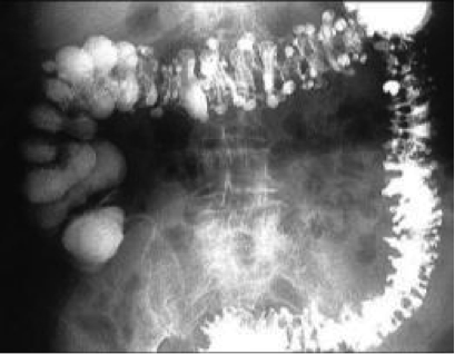
What is the difference in appearance in a contrast (barium) study of polyp, ulcer and diverticulum?
- Polyp: opacity inside of lumen (usu do colonoscopy though!). Less of them than diverticulosis and probably bigger
- Ulcer: often appears thickened or projecting outside wall (often in stomach or duodenum), often seen just below diaphragm as single excess pouch
- Diverticulum: opaque outpouchings (usually in sigmoid colon), multiple usually seen


