GI Physio Flashcards

(228 cards)

1
Q

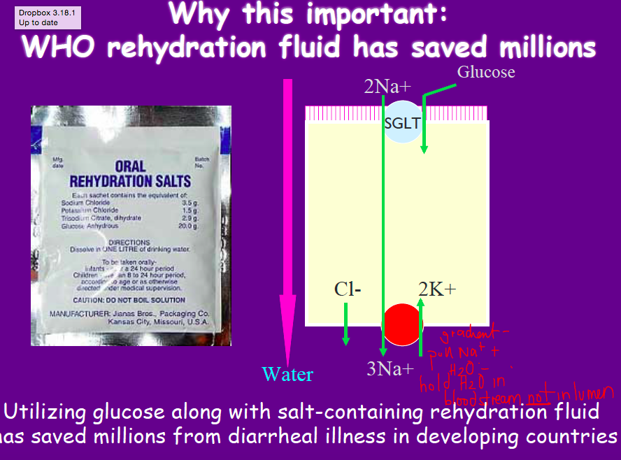
WHO rehydration fluid

A

SGLUT!

use glucose with salt - take up Na better

2
Q

cellulose digestion

A

So what ends up happening is that in your diet you have a whole bunch of indigestible carbohydrates. In particular cellulose. Cellulose your body actually has no native mechanism to break down into its constituent monosaccharides. There is a whole range. When you eat a green banana you can’t digest 75% of whatever you’re eating. Whereas in white rice you can digest a lot of it.

Now when cellulose is passed through your GI tract it’s not digested but when it gets to the colon the bacteria within your colon can actually digest it and break down the cellulose into not really monosaccharides but short chain fatty acids.

Short chain fatty acids are actually an energy source that your body can use. Cellulose it can’t use but it’s broken down into short chain fatty acids which it can use.

And in fact when you look at the cellulose products, butyrate, which is one of the short chain fatty acids produced is actually the primary energy source for the colonocytes.

In fact they almost get none of their energy from any blood supply.

3
Q

PAT1

A

classic amino acid transporter on brush border

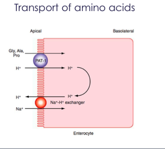
Again here this is just a picture that demonstrates that.

So PAT1 looks at things like glycine, alanine and proline.

So these are the smaller amino acids which symport with hydrogen.

4
Q

Ach receptor?

A

m3 - on parietal cells

5
Q

absorption of peptides

A

di and tri peptides appear to use the same transporter Pept 1

in the small bowel only!

only expressed on absorptive epithelium and driven by H+

6
Q

parasympathetic of GI

A

excitatory

cholinergic

vagus nerve controls motor function of upper gi tract

saccral nerves control distal colon and rectum

fewer neurons to small intestine

7
Q

Severe Combined immunodeficiency

A

stem cell defects in lymphoid maturation

effects seen in infancy and patients usually die young

malabsorption and diarrhea

8
Q

microtubules

A

composed of tubulin

imp for structural support

directs vesicles through cell

maintain polarity of polarized epithelia

9
Q

3 functions of CCK

A
  • 3 functions
    1. Increases secretion of pancreatic enzymes
    2. Relaxes sphincter of Oddi
    3. Conctracts gallbladder à bolus of concentrated bile à goes down the biliary tree à into the intestinal lumen!
10
Q

NSAIDs and mucosal damage

A

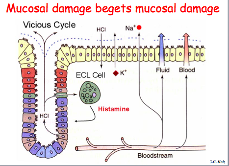
Now,clearly if you damage this mucous layer, it’s a problem. there are a number of things that can actually reverse this process. One of the most important is prostaglandin inhibitors PGI, and the PGI that you guys know best are NSAIDs. Asprin, Ibuprofen, those sort of things. And what those do is they inhibit prostaglandins. One of the effects is you produce less mucous. And now the HCl doesn’t necessarily just sit on top of a mucous layer, it actually can sit up against the surface of these epithelial cells and damage them. It can cross the mucosa into the lamina propria, where it starts hitting cells such as the ECL cells—and remember, I said that the ECL cells produce histamine, and we’ll discuss that a little bit more, but histamine is one of the primary secretagogues of acid secretion. So you set up a vicious cycle whereby HCl is allowed access to the epithelial cells; it can trigger a lot more acid production; you can damage this mucosa greatly, and actually develop peptic ulcers.
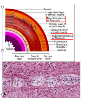
11
Q

Auerbach’s plexus

A

•Between the internal and external muscle layers of the muscularis propria is the myenteric (Auerbach’s) plexus
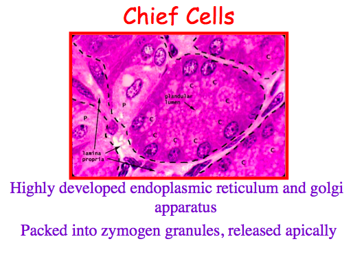
12
Q

chief cells

A

make pepsinogen

highly developed ER and golgi

packed into zymogen granules and released apically

Chief cellà Pepsinogenà Pepsinà breakdown proteins.

Let’s now turn a little bit to the enzymatic secretion, and the cell we’re concerned about here is the chief cell. So as you’ll recall, the chief cell is present throughout the oxyntic mucosa, as well as being present in the antral mucosa to a minor degree. And this is a gastric gland. And this little tiny white area here in the middle is actually the lumen. And unlike the G cells, all these granules containing pepsinogen are all present at the apical surface of the cell. So here you can tell that these cells are producing the substances that are going to be released out into the lumen, not into the bloodstream. These cells are have very highly-developed endoplasmic reticulum and golgi apparatus, because they’re spending a lot of time producing these enzymes, which are proteins.

So, what they release is pepsinogen. And whenever you hear something “-ogen,” like trypsinogen, chymotrypinogen, you know that you’re dealing with a pro-enzyme. So, like those pancreatic enzymes we discussed yesterday, pepsinogen is produced as a proenzyme—it’s not released in the active form, it has to be activated in the lumen of the digestive tractform, needs to be activated.

Ie why zymogen is released in packets. You don’t want it to be activated inside the cell and it will cause self digestion of the cell. Being in packet prevents activation inside the cell.

13
Q

H2 receptor

A

histimine receptor on parietal cells

(H1 is allergy)

14
Q

Vitamin K

A

utilized in liver to catalyze post-trnaslational modifications of blood clotting factors

15
Q

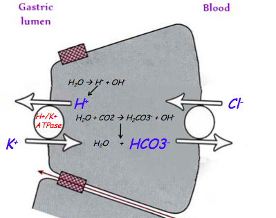
parietal cell - how secretes H+?

A
  1. Na/K creates gradient
  2. decreased Na in cell causes Na to enter the cell and huge decrease in Na in the lumen!
  3. Na electrochem gradient causes Cl to enter lumen
  4. Now, as Cl is pumping out into the lumen, it’s also coming in basolaterally through an anti-porter in exchange for bicarb. So you’ve got Cl coming in at this moment as well, and it’s getting replaced with HCO3. So this is an electroneutral process, right(Both Cl and HCO3 are negative). You haven’t changed the neutrality of the cell. But what you’ve done is you’ve pumped out HCO3, rendering this cell more acidemic, or more of an acid pH, because you lost HCO3, an alkaline agent, out basolaterally. This makes the cell more acidic, allowing you to then pump out the H+ ions that were liberated when you broke down carbonic acid into HCO3 and H+(Inside the cell using Carbonic anhydrase). The pump that does it is the H/K-ATPase. This is the 2nd important pump you have to remember, 1st was Na/K ATPase. you’re pumping all of this acid out, all of these protons out against a very steep gradient, so you still need a lot of ATP to do it. So you use the H/K-ATPase. Out of all the pumps and channels that you just heard about, this is the one to remember, because this is the most important pump in the parietal cell. This is the one that plays the major role in pumping out acid. So DO remember that. So Cl is exchange for the HCO3- and then Cl and H+ are pumped out into the lumen.
16
Q

L cells

A

GLP-1

inhibits acid secretion

17
Q

stimulant of pepsinogen secretion

A

acetylcholine!!

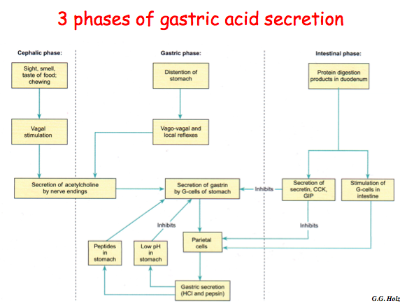
cephalic and gastric phase

acid triggers a local cholinergic effect

gastrin and CCK may also help

18
Q

functions of saliva

A

digestive (starch, taste)

protective (lubricate, buffer, clean)

immuno (IgA and other proteins)

19
Q

intestnial phase of gastric acid secretion

A
  1. protein digestion products in duodenum
  2. secretion of secretin/CCK/GIP
  3. inhibits parietal cells

turns off

20
Q

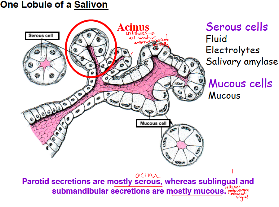
parotid vs sublingual/submandibular secretions

A

parotid = mostly serous (acini)

sublingual/mand = mostly mucous

21
Q

meissner’s plexus

A

•Below the muscularis mucosae is the submucosal (Meissner’s) plexus

–Relays information to and from the epithelia

  • Meissners plexus = immediately under submucosa
  • Role is to take information from lumen and send it to brain to affect function of epithelium
22
Q

gastric phase of gastric acid secretion

A
  1. distention of stomach
  2. vasovagal reflexes
  3. secretion of Ach by nerve endings
  4. secretion of gastrin by g cells
  5. parietal cells
  6. gastric secretion
23
Q

fat pancreatic enzymes

A

lipase, PLA2, choleteral esterase

breaks TGs and PPLs into 2 monoglycerides and fatty acids
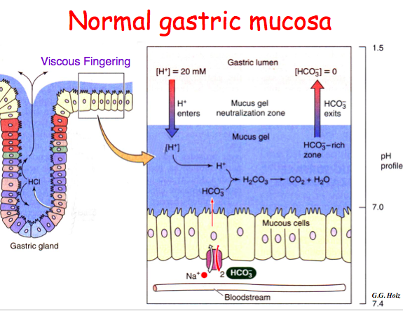
24
Q

viscous fingering

A

movt of acid through the mucos layer), where it just sort of chews its way through the mucous and reaches and sits on top (exact mechanism is unknown but very important that it happens as these surface cells are as prone to acid damage as any other cells in the body).

25
myoepithelial cells
contraction expels saliva from the acinus propels saliva in the main duct contraction by parasym and sym stimuli
26
osmotic diarrhea
occurs when poorly absorbed substance acts as an osmotically active solute draws water into the GI lumen and causes diarrhea improves with fasting!! fecal osmotic gap is presen
27
intralumenal fat digestion
dispersion of lipid into emulsion to maximize oil-water inerface lypolysis (pancreatic and other enzymes) uptake of products of lipolysis into micelles for transfer to epithelial surface
28
When do PPIs take effect?
3 days only affect activated parietal cells so take 30-40 min before meals inhibits more and more cells over 3 days
29
3 types of salivary glands
parotid sublingual submandibular all exocrine glands with a single duct!!
30
tight junctions
how much water gets through - tighter (more retentive) as you go through
31
deglutination
swallowing ## Footnote * Swallowing requires lots of coordination between oral cavity and esophagus/larynx etc to prevent aspiration * After voluntarily pushing food to back of mouth, oral pharynx, rest of swallowing process is automatic * Seal off nasal cavity * Raise the soft palate and hyoid * Close larynx * etc
32
How does IgA get from LP to lumen?
1. binding of IgA to receptor on basolateral face of epithelial cell 2. endocytosis 3. transport to apical face of epithelial cell 4. release of IgA dimer at apical face of epithelial cell
33
Where are T cels in MALT?
intraepithelium - almost all are CD8 cytotoxic! - destroy pathogens and infected epithelial cells lamina propria - most are CD4 (TH4) - mucosa has physio inflammation and don't want to destroy/over react
34
lactose
disaccharide of glucose and galactose
35
small bowel diarrhea
large volume of fluid! proble in small bowel absorbing water - deliver too much to colon (can only absorb 5-6 L) so obverwhelm the colon large in volume infrequent (3-4 movements per day)
36
Hartnup's Disease
defect in amino acid transporter amino acid depletion (esp tryptophan) can lead to deficiency in niacin photosensitivity, psychiatric, give supplements!
37
monosaccharide uptake
Alright so monosaccharide uptake. So from there they get transported into portal circulation. Galactose and fructose are almost completely metabolized by the liver within the first pass. They enter by facilitated transport and then they are converted to glucose derivatives like glycogen and other carbohydrates that your body will need. Glucose is also largely metabolized but some of it just manages to pass through. And people tend to think that that’s by design because it does allow for relatively quick access to glucose as an energy source for the rest of the body without it having to be reprocessed by the liver. It’s uptake by specific GLUT transporters and there’s a whole bunch of them (1-12) – in the liver that for uptake into hepatocytes there. That is beyond the scope of this lecture. You guys won’t be tested on that.
38
colipase
How does bile salt lipase actually work together when they should actually be working apart? - Lipase by itself will attach to fat droplets and start digesting - BUT Bile acids coat outside of fat droplets and displace lipase - Add on lipase, add on lipase - Minute start adding bile acid coats and protects fat droplet from lipase - Procolipase is an inactive precursor = can bind bile salts AND lipase - Brings lipase back to close proximity with fat droplet à allows it to continue to cleave of FA from TGs - Colipase is ssential for continued digestion of lipids
39
follicle associated epithelium (FAE)
inductive site present over organized lymphoid tissue contains M cells * M cells are particularly suited for just taking up antigen and passing them along to immunocytes * The main job of the FAE is antigen uptake - sampling of the environment of the GI tract – sampling of the lumen so the body knows what is going on there and knows what it has to react against
40
water uptake in the fasting state
electroneutral NaCl uptake - drives water absoprtion driven by output of H and HCO3
41
K secretion in colon
mostly in distal colon!
42
Mechanisms to avoid protein enzyme damage
1. proenzymes 2. keep in granules (not floating) 3. trypsin inhibitor is in the granules too 4. enterokinase - keep the activating factor far away in duodenum
43
where do bile acids come from?
liver! made from cholesterol secreted - in SB some are reabsorbed
44
Water secretion in intestine
centers around Cl- via CFTR
45
D cells
somatostatin inhibits acid secretion
46
unbranched starches (amylose) digestion
maltose + maltotriose maltotriose --\> glucose and maltose
47
IgA deficiency
most common congenital immune disease defcetive IgA B cell maturation most often asymptomatic no alteration in colon flora malabsorption due to small bowel bacterial overgrowth associated w celiac disease
48
digestion of branched starches
amylopectin form matose + limit dextrins limit dextrins NOT digested w pancreatic alpha amylase! digested with alpha dextrinase
49
2 stages of saliva generation
1. acinar generation of isotonic primary secretion (tight junctions! 2. ductal dilution of primary secretion so it's hypotonic (net reabsorption of Na/Cl and net secretion of K and HCO3 - alkaline)
50
acinar cells
secrete the salivary fluid and salivary proteins
51
antibody complement activation
lysis and ingestion
52
pepsin
once pepsin is formed it can cleave pepsinogen to form more pepsin pepsin is only cleaved at a low pH
53
types of cells in pyloric glands
endocrine cells make gastrin, somatostatin, pepsinogen
54
medium chain fatty acids
x ## Footnote We talked mostly about TGs and most of our diet is long chain fatty acids Medium chain fatty acids (6-12 C) are somewhat different - generally more soluble than any other lipids we see in our diet - have a measurable absorption - Paracellular à don’t have go to through forming of a micelle à brought to epithelial surface à transported across - Can just be absorbed paracellularly à portal circulation - Keep in mind that most lipids that go into intracellular space are actually exported through the lymphatic system - MCFAs taken up through portal system because they bypass the whole intracellular route - Why is this important for people who have steatorrhea secondary to Crohn’s? - Can supplement them with MCFAs to their diet - Don’t require bile salts so MCFAs will be sufficient dietary supplementation
55
digestion of carbs in the mouth
beginning of polysaccharide digestion alpha amylase! primary enzyme produces few monosaccharides So what does it do? It takes these polysaccharides, in this case amylose, and starts breaking them down (and this should be an alpha – points to salivary a-amylase – a should be alpha) into dextrins. Dextrins are really another name for oligosaccharides. But what it can do is break the linear branches down. But what it won’t do – like in the case of something like amylopectin where you have branched carbohydrates, it will break the linear structures but it won’t actually attack any of these branched structures. So these will remain within the mouth.
56
segmentation contractions
in colon mix contents facilitating absorption simultaneous contractions
57
3 muscle layers of the stomach
•3 muscle layers –Oblique –Circular –Longitudinal
58
sympathetic of GI
inhibitory adrenergic
59
blood supply to GI? Where does it go?
•Does not flow directly to the heart, but to the liver via the portal vein –Therefore the liver receives most of its blood from a venous, not arterial supply –Allows detoxification –Reduces bioavailability of oral drugs by 1st pass metabolism •
60
billiary tree
* Stuff exits liver through biliary tree * Bile exits through left and right hepatic duct à common hepatic duct àjoins the cystic duct to form the common bile duct * When not eating, ampulla of Vater (hepatopancreatic ampulla) is closed * Bile then goes backwards to be stored in gall bladder * Pancreatic duct from pancreas also does the same thing * When eating, the gall bladder squeezes which can have enough force to open the ampulla of vater * Bile comes out * Pancreatic juice comes out
61
3 things that stimulate acid secretion
1. histamine 2. ach 3. gastrin effect parietal cell which secretes HCl
62
chylomicron formation
What they do do is essentially traffic your products (FAs, monoglycerides) to smooth ER à products reformed to TGs - One of the things that’s different about fat metabolism is breaking down into constituent blocks and then reforms them into TG within the cell and that’s what your body will end up using - FAs and monoglycerides à reformed back to TGs - Synthesis of apoproteins on rough ER and those are combined with their TGs of golgi apparatus to form things called chylomicrons à excreted by exocytosis into the lymphatics
63
zymogen release
by acinar cells acetylcholine/cholecystokinin --\> PLC --\> increased Ca --\> K+ leaves the cell --\> increased negative charge --\> Cl leaves into the lumen, Na follows!
64
Interstitial Cells of Cajal
ICC mediate neurotransmission between enteric motor neurons and smooth muscle allow ENS to regulate smooth muscle!
65
absorption of fructose in SB?
GLUT5 (NOT ACTIVE) entry into cell is independent of glucose concentration much slower uptake and can be overwhelmed transported OUT of cell by GLUT2 only down concen gradient!
66
67
sodium substrate channel
in proximal SB
68
micelles
Now that you have some of your lipid products - You have your TGs, some broken down - Now should be broken down into monoglycerides or FAs à what do you do with those? - Still floating around in a hostile environment, not able to be solubized by themselves - - Bile acids secreted into biliary tre - Bile acids take all products of lipolysis (monoglycerides and FAs) - As add more bile salts, will hit critical micellar concentration of bile salts will develop something called a mixed micelle - Mixed micelle = allows products of lipolysis to be fully solubilized - Ex. Vegetable oil à blend it with water à looks murky à plus bile cells à at some point will be clear! All products of lipolysis into mixed micelles and actually go into true solution - -Probably most efficient way for body to take those products and transport them from the luminal aspect of your GI tract to the epithelial surface
69
ductular generation of secondary salivary secretion
1. Na/K ATPase makes driving force 2. Na gradient - Na/H counter transport - H leaves cell and the cell becoes alkaline 3. K is pumped out of the cell in K/H counter transport (H gradient driven) 4. stimulatied - increase intracellular Ca - K and Cl leave basolateral side 5. Now all the Cl is gone so Cl/HCO3 counter transorter used to bring Cl back into the cell and alkanilize the saliva \*\*FINAL: K and HCO3 in the saliva!!
70
gastric pacemaker
\*\* source is interstitial cells of cajal myenteric plexus generation of slow waves electrically coupled to smooth muscle cells via gap junctions receive input from enteric nerves
71
K secretion in colon
crypt cells! KCl secretion cAMP --\> open CFTR --\> secrete Cl Cl secretion paraleled by K secretion Na passes paracellularly NKCC1 on basolateral membrnae allows K and Cl into cell - controls rate of scretion!
72
phospholipase A2
stored in pancreatic acinar cells (inactive) breaks down dietary phospholipids by cleaving at 2 position of glycerol also degrades phosphatidylcholine in biliary secretions
73
MMC
during prolonged fasting (4h) - stomach exhibits stereotypical contractile patterns allows for undigested foods (like fiber) to leave stomach
74
Mechanism of H2RAs
trimeric g protein coupled receptor 1. binding of histamine 2. activates cAMP - activates H/K ATPase 3. increases parietal cell Ca - transloacts more pumps to membrane of parietal cell H2RAs block bloth of these effects - but do not prevent other pways of acid secretion (Ach or gatrin)
75
what is in pancreatic juice
sodium bicarbonate (ductular cells) digestive enzymes (acini) basic isoosmotic
76
enterokinase
made by duodenal epithelial cells converts trypsinogen to trypsin trypsin converts other pancreatic enzyme to active forms
77
sympathetic of GI - NT
adreneline
78
IPANs
long dendritic processes - synapse with other neurons local distention of intestinal wall and chemical contents cause activation of IPANs in submucosal plexus and myenteric plexus sequential activation of IPANs results in peristalsis\*\*
79
causes of osmotic diarrhea
carb malabsorption excessvie ingestion of poorly absorbed carbs magnesium induced diarrhea laxatives with poorly absorbable anions
80
3 phases of swallowing
–Voluntary stage •Push food to back of mouth –Pharyngeal stage •Raise –Soft palate- seal off nasal cavity –Larynx + hyoid –Tongue to soft palate –Esophageal stage * Contract pharyngeal muscles * Glottis closes off larynx * Open esophagus * Start peristalsis
81
intestinal digestion of lipids
meals moves to small intestine - pH rises fatty acids liberated y gastric lipase become ionized and orient on outside of oil droplets - stabilize fat emulsion (SOME LCFA dissociate and traverse lumen to epithelium) fatty acids are potent stiulaters of CCK release - Once in duodenum pH rises secondary to bicarb secretion - FAs liberated by gastric lipase become ionized à coat outside of oil droplets - Stabilize fat emulsion - Blockade for enzymes to work more efficiently - Even long chain fatty acids have some state of solubility - Some FAs will traverse themselves, make it to epithelial surface - When make it to epithelial surface à potent stimulators of CCK
82
parasympathetic stim to salivary glands
primary!! from vagal stim causes vasodilation and copious amounts of watery saliva facial and glossopharyngeal nerves
83
fat soluble vitamins
special class of dietary lipids present in trace amounts but absorption is critical A, D, E, K
84
microfilaments
composed of f actin atabilize apex of cell and villi in the turbulent environment of the gut
85
antibody opsonization
abs bind so it's digested by macrophages
86
M cells
These M cells sits in the FAE, take up these antigens, pass them off to the immune cells, the immune cells will then leave that area, go directly the organized lymphoid tissues to tell the other cells “hey, we’ve got a pathogen here specialized ells overlying lymphoid follicles function in ag sampling low lysosomal content (don't degrade!!) low microvilli pockets n basolateral surface for APCs for rapid ag transfer many vesicles
87
oxyntic segment
body plus fundus
88
secretion of SI - neuro
regulated primarily by sympathetic inhibitory pathways if hypovolemic --\> sympatehtic pathways activated --\> less secretion of electrolytes
89
water uptake in the fed state
nutreint dependent uptake of Na drives water uptake (sodium-substrate)
90
cephalic phase of gastric acid secretion
1. sight, smell, taste 2. vagal stimulation 3. secretion of Ach by nerve endings 4. secretion of gastrin by g cells of stomach 5. act on parietal cells 6. gastric secretion
91
intraepithelial lymphocytes (IELs)
located between epithelial cells attach to epithelial cells through an interaction between the integrin on the IEL and e cadherin in the epithelial cell first line of defense! mostly CD8 t cells can make cytokines/be cytolytic reside in paracellular space and don't allow antigents to pass
92
cholera toxin
A and B subunits B binds to R A activates adenylate cyclase alters intestinal permeability by acting on tight junctions and causing active Cl secretion no cell toxicity - small bowel looks normal on histology
93
carb digestion in the stomach
starches are partially hydrolyzed to detrinx and maltose \*\*residual activity from sailvary amylase which is terminated by low pH no other carb digestion!! Now starches are partially hydrolyzed within the stomach once it gets down there but really most dietary carbohydrates – there is no digestion that occurs within the stomach itself. Any salivary amylase that you have that you swallow – eventually you will swallow all of the amylase – it gets deactivated within the stomach in that acidic environment. And so there is some residual activity as the amylase is active but as it starts getting degraded within the acidic environment there is no other carbohydrate digestion that occurs. So really, a little bit starts in the mouth, it gets inactivated in the stomach, and then it gets passed through into the small bowel. So the stomach – at least for carbohydrate – and this changes, when we talk about things like fat there is a lot more activity within the stomach for things like fat digestion, but for carbohydrates there is very little.
94
overall innervation of esophagus
skeletal = cns smooth = cns + ens
95
pancreatic lipase
like gastric lipase BUT acts on both 1 and 3 positions of glycerol --\> FA and MG neutral pH \*\*inhibited by bile acids
96
Th2 cytokines
drive antibody production
97
components of the muscular propria?
outer longitudinal muscle inner cicular muscle * Majority of important processes occurs in mucous membrane (most important because this is where absorption takes place) * Layers of the mucus membrane: * Simple columnar epithelium (except in a few parts of GI tract) * Then lamina propria: loose connective tissue that also contains immune cells * Thin muscular layer à muscularis mucosae * Submucosa where nerves and blood vessels lie * Muscular propria for movement * Outer longitudinal muscle * Inner circular muscle * On outside, most organs have serosa (except for esophagus)
98
parietal cell
proton pump H/K - ATPase in the apical microvillus acid secretion begins in 5-10 min of stimulation This is an activated parietal cell because the tubulovesicles in the cytoplasm have coalesced to form villi and formed an expanded canaliculus Parietal cell is a Unique cell. Has this Crazy canalicular membrane. All these folds in the fig, folds always means a lot of surface area. In these canalicular fold are the pumps (H+/K+) to pump out acid. It has to pump against a great great gradient. To have a pH of the lumen of 1-2, the cell has to pump out tons of H+, that is why you see so many mitochandria in side the cell, as it needs all that energy to pump out acid. This what it looks like in an electron micrograph. It is a powerhouse! An absolute powerhouse—it produces 3x10^6 hydrogen ions pumped out into the lumen per second. But it does not secrete acid immediately on eating, there is a lag phase..
99
chronic pancreatitis
- Often you’ll see people with chronic pancreatitis complain of diarrhea - Reason is pancreatic insufficiency (can’t produce enough lipase, not able to produce enough bicarb to neutralize some of acidic contents coming from GI tract - Whatever pancreatic lipase is also inactivated by the fact that you’re not able to neutralize the pH - People will end up incompletely digesting fat in lumen - End up with steatorrhea - - This is just an image of someone with chronic pancreatitis - Characteristic = start developing calcifications within pancreas - Usually pancreas is a soft, fatty, mushy tissue - Even on a flat plate, can see calcifications (Ca deposits in pancreas) - Typically not see that - Light up on a CT scan - Most common cause of pancreatitis in this country is alcohol
100
ductual cells
a lot of mitochondria! powerhouse cells
101
secretory IgA
resistant to intestinal proteases doesn't bind complement (prevents inflammation) protects by inhibiting pathogen adherence - neutralizes viruses and toxins important in immne exc;usion - preventing pathogens out so immune response is not triggered
102
G cells
make GASTRIN numerous granules located at the BASE of the cell release into the blood - endocrine effect First, we’ll look at the G cells. G cells, again, produce gastrin, and they’re present in the antrum of the stomach, they have these dark granule in them. And this is the apical portion of the G cell(top in fig), and this is the basolateral portion with a lot of granules (Botton part of fig) and you see you’ve got all these vesicles and the vesicles contain the gastrin. But again, they’re at the basolateral portion, so they’re not really pumping them out into the lumen—they’re pumping them back into the blood stream. So this tells you again that gastrin functions as a hormone—it functions endocrinely-secretedintot he blood as opposed to directly on/near the cell. It’s not going to be pumped out into the lumen, it’s not going to be pumped next door to the neighboring cells, it’s pumped out into the bloodstream and comes back and acts on the parietal cells to trigger gastric release. So that’s the G cell.
103
Na transport in proximal colon
mostly electroneutral Na/H couter transport
104
3 regions of the stomach
–Cardia –Fundus/Body –Antrum (pylorus)
105
lactose intolerant
what really ends up happening is that you cannot digest your lactose into its constituent carbohydrates and so you end up having just osmoles of lactose floating around within your GI tract. When that gets to your colon you actually have bacteria that can digest the lactose and it breaks them down into its constituents glucose and galactose. Again these are extra osmoles so you start expanding, you feel the extra osmolarity within your colon because there is expansion. A lot of those are actually fermented by the gut bacteria into things like methane so that’s why people get very gassy and bloated and have diarrheal symptoms when they have lactose intolerance.
106
Vitamin E
vital antioxidant
107
Crohn's disease and steatorrhea
typically bile salts \>\>\> critical micellar concentration if ileum is resected, bile salts are not really reabsorbed via enterohepatic ciruclation micelle formation impaired becaue bile slalt deficiency and lipids are poorly absorbed Reason that’s important is bile salts are actually reabsorbed in terminal ileum - Something called enterohepatic circulation - Secrete bile salts à GI tract à mixed micelles à help with absorption - Primary absorbed in last 100 cm of ileum - Cut that out, bile salts not reabsorbed à secreted out in GI tract - Able to upregulate bile salt production to a certain degree, but not completely - Don’t have sufficient bile salts to digest fat à significant steatorrhea - -There are a few other causes of this such as biliary obstruction and PBC, commonly with Crohn’s disease
108
self regulation of HCL
low lumenal pH in the stomach inhibits gatrin release from G cells low lumenal pH in stomach and intestine stim somatostatin from D cells which acts on G cells to inhibit gastrin low lumenal pH in the intestine stimualtes release of secretin which inhibits gastrin g cells
109
H2 receptor agonists
-tidine inhibit histimine at H2 receptor resulting in reduced gastric acid secretion, reduced gastric volume OTC! If you turn down the secretagogues, you turn down the production of acid. There are H1 receptors, and those H1 receptors are what you block when you have sort of an allergic reaction and you’re all itchy and you take an anti-histamine. That’s the H1 receptor. This is the H2 receptor, and it’s on the parietal cells. As I mentioned, the primary secretagogue is histamine, because histamine is not only a direct stimulant of parietal cell secretion, but because both gastrin and ACh also act on ECL cells to increase histamine secretion, histamine is a sort of augmented cause of secretion. So histamine, as we know from before in the lecture, acts on the parietal cell through the H2 receptor. These were the first drugs, H2 receptor blockers. OTC So they developed H2 receptor antagonists—H2 receptor blockers. Which you guys have obviously heard abouve. Cimetidine which is Tagamet. Ranitidine which is Zantac. Famotidine which is Pepcid. Nizatidine which is Axid. And these all function by blocking the H2 receptor so that histamine cannot act on the parietal cell. And in doing so, they do an OK job of decreasing acid secretion. Not great as you’re only blocking one arm of the three of the secretagogues, right—you still have gastrin and ACh playing a role. Thus the effect is not complete. But it works ok and for mild cases it is good enough. Basically they bind to the H2 receptor, block the activation of cAMP, (which usually activates H+/K+ ATPase), also block the increase in pareital cell Ca+ in the cyto plasm that act on the secretory canaliculi. So, hitting histamine, u r blocking partly the acid production. You could try to block those other two; you could try using an anticholinergic to block acid and that will work, but anticholinergic drugs work systemically and you have very, very bad side effects. They cauase dry mouth. They cause constipation, dizziness. They make people feel really badly. So, people don’t really use anticholinergic drugs for something like this.
110
SGLT1
sodium-glucose transport (ACTIVE) Na/K dependent for glucose/galacgtose can not attach until 2 Na are attached Alright so absorption of glucose and galactose, like I said on the brush border you have lactase that will break down lactose into its constituent monosaccharides, in this case glucose and galactose. And again symporting with Na using SGLT-1 into the cytosolic side of the epithelial cells.
111
K cells
GIP inhibits acid secretion
112
parasympathetic of GI - NT
cholinergic excitatory
113
3 stimulants in intestinal phase of pancreatic secretion
1. secretin - acidic fluid froms stomach - stim duodenal S cells - release secretin - deuctal secretion of bicarb 2. peptides, AAs, fat in duodenum - triggers CK - stim release of enzymes from acinar cells 3. Ach from vagus acts on muscarinic receptors on acinar and ductal cells to release enzymes and bicarb
114
amylase
carbohydrate S: acts on starch, glycogen P: glucose, maltose, maltotriose, dextrins
115
limitations of PPIs
long time until peak effect no complete acid inhibition through the day substantial interpatient variability in inhibition (P450)
116
lamina propria in immunity
where B and T cells migrate following induction in the organized lympoid tissue diverse cell types * The lamina propria contains the vast majority of the lymphoid tissue in the body * It has a very diverse cell population * Most of the cells are going to be lymphocytes * Most of them will be B cells and CD4 cells * Contains a lot of macrophages, neutrophils, dendritic cells and mast cells
117
HIV and the gut
HIV optimally targets cells with CD4 receptor and chemokine Co receptor (ususally CCR5 which is on activated) and are pathed in pro inflamatory cytokines and surrounded by similar cells diarrhea with HIV! tons of diff pathogens
118
absorption of aminoacids
tons of diff transporters for amino acids H+ dependent! exit from epithelium by diffusion and active transport
119
carb digestion in the stomach
digestion of polysaccharides and dextrins continues in small bowel by pancreatic alpha amylase!! active because secretion of pancratic HCO3 raises pH \*\*end products = maltose and limit dextrins
120
gastrin receptor?
CCK-B ## Footnote 2 main forms of gastrin There are two forms of gastrin that are produced, because G cells are not only present in the stomach in the antrum, they’re also present in the duodenum. “little gastrin,” or G-17 gastrin, has 17 amino acids, which is produced in the antrum and the duodenum, and then you’ve got “big gastrin,” or G-34 gastrin, which is strictly produced by the duodenum. Regardless of which gastrin you produce, they both work perfectly well, and they both bind the same receptor, here the receptor is called the CCK-B receptor, and the reason why that’s confusing is that obviously yesterday we learned about the CCK-A receptor, and the CCK-A receptor, as you’ll recall, is there to bind, naturally, CCK. So, CCK-B receptor binds gastrin. And the CCK-B receptors are on the parietal cells and ECL cells like we would expect. * Gastrin can stimulate CCK A receptor and stimulate the pancreas a lil bit, but main action is on CCKB. * But you also find CCK-B receptors on ECL cells. And you recall that the ECL cells produce histamine. So this is a way of augmenting histamine production. The release of gastrin is directly stimulated by luminal peptides and amino acids, so the by-products of protein digestion. Once you start digesting proteins and liberating these protein by-products, you stimulate more gastrin and it actually stimulates more acid. Also, from the vagus you have a substance caused gastrin releasing peptide (GRP) and this will also basically trigger increased gastrin release from G cells.
121
secretory gland cells in oxyntic regions
\*parietal \*chief epithelial mucus neck stem cell
122
lactase deficiency
osmotic diarrhea brush border enzyme that breaks down lactose into glucose and galactose if not present - lactose reaches the colon and undergoes bacterial hydrolysis and fermentation osmotic diarrhea with bloating cramps flatulence
123
what inhibits gastric secretion from G cells?
somatostatin, PGE2, secretin, CCK, GIP low lumenal pH
124
MADCAM1
on endothelial lining of venule - binds integrin on lymphocyte for honing ## Footnote * Once a cell is activated in the mucosa and goes out into the periphery as it starts dividing and becoming more active, one of the things it starts doing is expressing these homing receptors and these homing receptors are going to be specific to mucosa * The most important one called a4B7 - present on immunocytes that are activated in the mucosa * It is triggered(?) in a vitamin D dependent fashion in the mucosa * Once the cells need to come back to the mucosa that a4B7 will bind to a receptor on specialized cells that are only present in the blood vessels of the mucosa called high endothelial venules and those venules will express something called MadCAM-1 that allows a4B7 to bind and then transcytose across the blood vessel into the lamina propria
125
how is pepsin activated?
in an acidic pH acid secfretion cleaves it Again, pepsin is produced by the chief cells down here in the crypts—mostly of the oxyntic mucosa, but also in the antral mucosa. ACh is the primary stimulant, but also secretin and CCK. They act on the chief cells, which will produce pepsinogen. Meanwhile, the parietal cells are producing the acid. Acid, again, will lower the pH down below 3 and will start partially activating the pepsinogen. This will go on for a little while until the pepsinogen actually becomes activated to form pepsin and then pepsin will start activating all the pepsinogens. So once you start getting pepsin formed, the process just takes off and you get a lot more pepsin produced from pepsinogen. This will then start digesting down the proteins into small fragments. These small fragments will then play a role in triggering gastrin, which will further increase HCl. So that’s the chain of events that leads to pepsinogen formation and activation. IN SHORT: I feel its easier to understand as a flow chart so I included this too As mentioned (points to lower left of fig) Ach - Secretin - CCK - Chief cells - Pepsinogen - (in acid env) - Pepsin (partially activatedà fully activated) -\> Now pepsin self stimulates activation of more pepsin and leads to breakdown of proteins into smaller and smaller particles(peptones) - which leads to more gastrin from G cells and repeats the process (points to Lower R of the fig)
126
How do the immunocytes target to the mucosa?
* Once a cell is activated in the mucosa and goes out into the periphery as it starts dividing and becoming more active, one of the things it starts doing is expressing these homing receptors and these homing receptors are going to be specific to mucosa * The most important one called a4B7 - present on immunocytes that are activated in the mucosa * It is triggered(?) in a vitamin D dependent fashion in the mucosa * Once the cells need to come back to the mucosa that a4B7 will bind to a receptor on specialized cells that are only present in the blood vessels of the mucosa called high endothelial venules and those venules will express something called MadCAM-1 that allows a4B7 to bind and then transcytose across the blood vessel into the lamina propria
127
CCK-B receptor location and effects
mediates actions of gastrin located on parietal and ECL cells receptor stimulation leads to: parietal cell acid secretion, ECL histamine release
128
129
acinar generation of primary secretion
\*\*saliva production 1. Na/K ATPase establishes gradient 2 NKCC1 - because of gradient, Na wants to enter - 2 Cl- enter and becomes very CONCENTRATED in cytoplasm 3. when the cell is stimulated - increase Ca - open Cl channels (apically) and K channels (basolaterially) 4. luminal Cl- --\> electronegativity draws Na through tight juntions and water follows passively (LOOSE epithelium(
130
I cells
CCK inhibits acid secretion
131
absorption in Sb of glucose and galactose?
SGLUT1
132
lamina propria lymphocytes (LPLs)
1. T cells - mostly CD4 differentiated effector cells raised threshold of activation prevents immune responses to harmless antigens activated due to continuous ag exposure produce high levels of cytokines 2. IgA producing moslty - responsible for local IgA and IgM production
133
secretin
secreted by S cells stimulates release of bicarbonate rich pancreatic juice by ductal cells \*\*stim by acid hitting duodenum
134
canniliculi of the parietal cell
And this is why there is the lag phase. Canaliculi initially sit as intracellular vesicels in the unstimulated state-Acid cannot leave the cell if secreted. Once stimulated, the canaliculi fuse with the intracelluar vescicles and form the mumbrane that communicates with the lumen. Very important as if this does not happen, the vesicles will release the acid inside the cell and it will start self digesting the cell.
135
GLUT4
facilitative transporter for frucose
136
pancreatic alpha amylase
leads to malltose and limit dextrins (branched) can work bc more basic pH So what does pancreatic alpha amylase do? It breaks down what have become dextrins (4 -5 sugars) into shorter, usually disaccharides. So pancreatic alpha amylase, really essentially what that does is it continues to work the same way the salivary amylase from the mouth works- it just kind of continues to do that work. And it breaks them down into in this case I just put them as maltose (2 glucose sugars attached to each other). And again same thing when you have amylopectins (shorter strands that are still branched) it will continue to break down alpha 1-4 (linear linkages) but will leave the 1-6, so you will continue to have branched carbohydrates, which are called limit dextrins.
137
vitamin d
regulates calcium absorption by intestine and Ca homeostasis
138
Hirschsprung's disease
absence of enteric neurons in terminal regions of gut problems w migration tonic contraction of affected segment (no parasym) - intestinal obsruction and megacolon
139
water transport in proximal small bowel
leaky epithelium! electrolytes accumulate in the paracellular space and water moves through leaky tight junction
140
Overal absorption/secretion in intestines
Na/Cl are absorbed K/HCO2 are secreted
141
Intermediate filaments
function as support cables associated w plasma membranes at tight junctional desmosomes - form a web linking desmosomes aroun the cell
142
actin and myosin
circumferential belts around the apices of the columnar cells - contractile ring contracts in the presence of ATP and Ca modulates barrier function
143
why does prune juice work as a laxative?
high levels of endogenous sorbitol So we talked about sorbitol before and a non-absorbable sugar. When you have non-absorbable osmoles within your GI tract it will pull in water into the lumen and if you have a lot if will give you diarrhea but if you have a little bit it will just give you some water into the GI tract and it will help you have a bowel movement. So if you look at certain things like prunes/prune juice you see they actually have a very high content of sorbitol. So that’s actually why when people have them they’ll have bowel movements – it helps them have bowel movements. On the other side if you have friends who are addicted to sugar-free chewing gum and they have diarrhea all the time that’s one of the things you can point out to them. So they’ll be like I have like loose stools or whatever, I think I have IBS or something like that but they are constantly chewing gum. That’s one of the things you can tell them to try and it might help.
144
colonic diarrhea
small in volume frequent (8-10) tenesmus
145
differences between triglycerides and phospholipids
- Some of the differences between TG and PL are glycerol (3 C backbone), which FAs are attached - TG = 3 FA attached by ester bond - hydrophobic - PL = 2 C groups attached to FAs and the third is attached to a phosphate group - One hypdrophilic bit
146
zonula adherens
adhesive of neighboring cells immediately below tight junction e-cadherin + Ca on the lateral surfaces of cells transduce signals in and out of the cell
147
pept 1
on brush border, absorbs peptides So how does this work? In general the images will be more helpful to you to understand what’s going on than just the text slides. So you have your endopeptidases that have broken down your proteins into shorter di/tri peptides or individual amino acids. What ends up happening for your di/tripeptides is that they’re transported at the brush border by this thing called PEPT1 – again symporting with hydrogen. And you have a Na/H exchanger here that helps you maintain the gradient. Once the peptides are brought in they’re either cleaved internally by additional peptidases into single amino acids or they stay as peptides and they’re both transported along the basolateral membrane into the blood. Na/K/ATPase is simply to maintain electrical neutrality with other exchangers here
148
fundus innervation
sustained tonic contraction! resting membrane potential above them mechanical threshold if inhibited - accomodation response
149
what happens to saliva with increased flow?
pH increases (more HCO3) levels of Na and Cl increase levels of K decrese
150
neuro of smooth muscle in the esophagus
more complex - relies on central and peripheral control (ENS)
151
OTC antacids
weak bases! neuralize gastric acid non-absorbed
152
S cells
secretin inhibits acid secretion
153
salivary alpha amylase
So what does it do? It takes these polysaccharides, in this case amylose, and starts breaking them down (and this should be an alpha – points to salivary a-amylase – a should be alpha) into dextrins. Dextrins are really another name for oligosaccharides. But what it can do is break the linear branches down. But what it won’t do – like in the case of something like amylopectin where you have branched carbohydrates, it will break the linear structures but it won’t actually attack any of these branched structures. So these will remain within the mouth.
154
pepsinogen II
produced in the oxyntic area as well as the antrum
155
vitamin A
retinoic reg gene transcription
156
common bile duct
–Tube-like structure formed by the union of the common hepatic duct and the cystic duct (from the gallbladder) –It is later joined by the pancreatic duct to form the ampulla of Vater –There, the two ducts are surrounded by the muscular sphincter of Oddi –When the sphincter of Oddi is closed, newly synthesized bile from the liver is forced into storage in the gallbladder –When open, the stored and concentrated bile exits into the duodenum
157
water flow in duodenum
hypertonic contents are brought into osmotic equilibrium with plasma by water flow into the lumen
158
composition of gastric juice
H2O, mucus HCl electrolytes intrinsic factor pepsinogen it’s mostly an isotonic solution of HCl, the pH you can get very low from 1-2. These cells also produces pepsinogen. Also the mucous that we’ve already learned sits up on the top, but you also have some minor electrolytes. In the mucous layer the pH is kept at about 7.0. Also present there is the intrinsic factor and the pepsinogen(primary proteolytic enzyme produced by the stomach), which will be converted to pepsin.
159
what stimulates gastrin secretion from G cells?
luminal peptides, AA GRP from the vagus
160
secretory gland cells in pyloric region
\*endocrine cell epithelial cell stem cell mucuous neck cell
161
absorption and activation of PPIs
weak base! easily passes through membranes when unprotonated so easily absorbed in duodenum circulates through bloodstream and into all cells as unprotonated compound protonated in the very acidic parietal cell canaliculus - becomes polarized and can't pass through membranes protonated form is activated and covalently binds H/K ATPase
162
gap junctions
scattered on basolateral membranes aggregates of particles called connexins create hexamer channels conduits for passage of small molecules between cells coordinates cells
163
exocrine pancreas
acinar cells (secrete ensymes) duct cells (secrete bicarbonate rich fluid)
164
neuro of striated muscle in the esophagus
central mechanisms are sole pthway! sequential activation of lower motor neurons in brainstem peristalsis sequentially down striated portion of esophagus
165
MHCI
presents proteins derived from cytoplasm (viruses)
166
isomaltase
limit dextrans --\> glucose
167
muscularis propria
* The inner circular muscle reduces the diameter of the intestinal lumen * The outer longitudinal muscle provides for colonic shortening * The muscle layers, working together can provide complex motility patterns * They work together due to signaling by adjacent nerve plexi * Most of the GI tract has 2 layers to the muscularis propria * Inner circular layer * Contracts and squeezes tube (decreases diameter of lumen) * Outer longitudinal layer * Runs length-wise à contraction shortens GI tract * Need a way to coordinate muscle layers * Therefore, between muscle layers = nerve plexi (Auerbachs plexus)
168
acute pancreatitis
activate enzymes in the pancreas digest pancrease and enzymes can flow out and damage other organs
169
MALT effector sites
reacts against the antigens ## Footnote * Intraepithelium * Lamina propria
170
cholecystokinin
secreted by I cells stimulates release of digestive enzymes by acini released when products of digestion are there - triggers I cells acts through PLC and mobiliation of intracellular calcium resulting in zymogen granule exocytosis
171
cholesterol esterase
nonspecific! degrades esters of dietary cholesterol, vit a, d, e broad specificity
172
small intestine in fed state (ENS)
digested food products primarily move by local paristalsis
173
How does the lumen become isotonic and neutral after a meal?
in the duodenum HCO3 - exits either through CFTR or HCO3/Cl exchange! water follows to make it isotonic (from hypertonic) (blood to lumen) lumen is isotonic and neutral pH from this point!
174
Components of the mucus membrane?
muscularis mucosae lamina propria epithelium * Majority of important processes occurs in mucous membrane (most important because this is where absorption takes place) * Layers of the mucus membrane: * Simple columnar epithelium (except in a few parts of GI tract) * Then lamina propria: loose connective tissue that also contains immune cells * Thin muscular layer à muscularis mucosae * Submucosa where nerves and blood vessels lie * Muscular propria for movement * Outer longitudinal muscle * Inner circular muscle * On outside, most organs have serosa (except for esophagus)
175
mass action contraction
in colon simultaneous contraction of smooth muscle over large confluent areas to move material from one portion of the colon to the next
176
GLP-1
secreted in ileum and colon inhibits gastric emptying
177
zonula occludens
= tight junctions maintain polarity of polarized clels situaed apilcally series of punctate fusions seal the paracellular space and reg permeability stabilize the epithelial monolayer
178
what 2 things cause ulcers?
loss of mucus h pylori Basically two things can cause ulcers, loss of mucous layer and H. Pylori Infection. Points to the Left most fig-mucosa is chewed out, this is an ulcer. It looks like pretty much a big divot in the mucosa. In this case it’s nice and white-based—that’s because it’s gone through the epithelium. Here’s a good histologic view(Middle fig). So here, this dark red is all the epithelium—you can see that the ulcer has chewed all the way through the epithelium, all the way through the lamina propria, and is digging down pretty deep into the submucosa. The submucosa is where some big blood vessels lie, and where the nerves lie. So when the ulcers dig into the submucosa, you run the risk of it hitting a fairly-sizeable blood vessel and causing a very significant upper GI bleed. Or hitting nerve endings, causing lots and lots of pain. It can get so bad that the ulcer can ulcerate all the way through, the muscularis propria, and all the way through the serosa, and you can have a perforation into the abdomen. When you have a perforation—surgery is often times a result, and here you can see a specimen with a fairly good-sized ulcer right there(Right fig)-Surgical resection specimen
179
sympathetic to salivary glands
stim causes vasoconstriction and a small amount of thick mucoid saliva cervical ganglia
180
exopeptidases
cleaves single amino acids from carboxyl terminal end
181
antibody neutralization
picked up and digested by macrophages
182
water flow in ileum
osmotic equilibrium maintained by water following absorbed solutes more paracellular absorb water
183
NPC1
specific cholesterol uptake protein (exetimibe) in intestinal lumen
184
pepsinogen I
produced by chief cells and mucus cells of the oxyntic glands
185
MALT Inductive Sites
takes in antigens follicle-associated epithelium (FAE) Peyer's patches (organized lymphoid tissue) local lymph nodes
186
2 areas that are not columnar?
The two areas that are not columnar are the esophagus and anus. These cells are subject to constant turnover, perhaps to prevent accumulation of mutations due to exposure to toxins. They turn over every 3 days or so before they undergo apoptosis and are shed into the lumen
187
B Cells
188
water transport in distal colon
tight epithelium electrolytes accumulate intracellularly and water moves through aquaporins or Na dep solute transporters in the membrane
189
190
aqueous secretion in the pancreas
duct cells! 1. Na/K ATPase establishes driving force 2. Na/H counter transport using Na gradient - H leaves the cell 3. Co2 enters to neutralize the loss of H, gets converted to H2CO3 4. HCO3 - out apically in HCO3/Cl countertransport (high in lumen!), H+ out in the counter transport with Na (above) 5. Cl gradient maintained becasuse it leaves the cell through CFTR through gradient
191
proton pump inhibitors
-prazole inhibit H/K ATPase pump of the parietal cell - resulting in marked decrease in acid secretion
192
pyloric segment
antrum
193
MHCII
Ag is degraded in lysosomes * But you also have to be able to recognize extracellular pathogens and this is where MHCII come in * MHCII is going to react against things such as extracellular bacteria – in this case perhaps it is something such as E. coli * E. coli may be sitting out in the lumen and may get presented to a macrophage or another cell which will then take up that bacteria in an endocytic vesicles * That vesicle will then bind to other vesicles that contain MCHII The MHCII will be presented to the surface of the cell for a CD4 cell to come along and recognize it
194
complex carbohydrates
oligosaccharides (3-10) polysaccharides (\>10)
195
maltase
maltose --\> glucose
196
simple carbohydrates
monossaccharides (glucose, fructose, galactose) disaccharides (lactose, sucrose, maltose)
197
macula adherens
desmosomes assocated with the cytoskeleton primarily involved in adhesion possess adhesion molecules that interact with similar molecules on the adjacent cells also sites of signal transduction
198
IBD pathogenesis
inappropriate and ongoing activation of the mucosal immune system genetic and enviornlmantal contributions fnal pway - heightened mucosal activation! * That is what happens when you have a mucosal immune system that is too activated * Pathogenesis of IBD is an inappropriate and ongoing activation of the mucosal immune system * We do NOT know the specific antigen to which the mucosal immune system is responding but we assume that its due to some of the luminal antigen, probably a bacterial antigen of which there are millions * Because of this immune response you activate the immune response and you have a lot of ways you are supposed to keep the immune system suppressed but those do not work * Regardless of how it happens, the final common pathway is a heightened mucosal inflammation
199
mucosa
•Lamina propria is beneath the epithelium and its basement membrane –Loose connective tissue containing many immunocytes –Muscularis mucosae is smooth muscle which may play a role in villus movement * Contains immune cells = primary protector of gut * most developed immune system in the body because of following point * Gut = primary contact with external world * Lots of bacteria * Lots of surface area
200
electrolyte composition of gastric juice and secretion rate
at high secretion rates - more H Cl Low - more Na and Cl The production of gastric acid, like that of the salivary glands and like that of pancreas all depends on the rate of flow (rate of secretion from the cells ). And the rate of flow depends on the degree of stimulation. So if you’re at rest, for eg sitting postprandially in the middle of the night you’re probably producing not a whole lot of acid(L side of the fig/graph). And at that point, at low flow rates, your gastric secretion is mostly comprised of Na and Cl, and a very little bit of protons, a little bit of K. All in about the same concentrations that you see in plasma. So when you’re not stimulated, it’s just like plasma—it’s isotonic with plasma. With stimulation, you really churn out an enormous amount of protons. You increase the amount of H+ and Cl-, so there’s a lot more secretion of HCl. When stimulated, the amount of Na (drops down greatly) decreases and the acid dramatically increases on stimulation(Points to the graph
201
202
pancreatic acinar cells
zymogen granules - lots of enzymes lots of golgi bc make a lot of protein!
203
chronic variable immunodeficiency
more general b cell defect dcreased AB secretion chronic diarrhea and malabsorption increased gastrointestinal carcinoma and lymphoma
204
What happens to pancreatic secretions at a high secretory rate?
HCO3 and Na are high! lower Cl- (it's more in the cell?)
205
protein digestion in the stomach
pepsins autoactivated by pH pepsins hydrolize internal peptide bonds, active only in the stomach, generate peptides and some AA So digestion of proteins doesn’t start in the mouth – it starts in the stomach. And within the stomach it starts with things called pepsins, and the acidic pH in stomach autoactivates this enzyme. What it does is it cleaves a 40 amino acid group off of the N-terminal site of pepsins. So essentially it starts off as a pepsinogen, converts it, cleaves that site, turns it into pepsin and the pepsins hydrolyze the internal peptide bonds in many of these proteins. So what it does within the stomach is it generates peptides. Some of the amino acids. Gastric pepsin is active only within the stomach. It’s not that important of a factor within total protein digestion because if you have people who are achlorhydric. If you’re achlorhydric you’re not able to activate pepsin because you’re not acidic. Those people actually seem to have no problems with protein digestion. Keep in mind that there’s a lot of redundancy built into your GI tract. We live in a time now when there’s plenty of food but in the past there wasn’t and so you wanted to build in redundancies so that you could take as much advantage of whatever calories are available. So somebody who is on a PPI (proton pump inhibitor) that they’re achlorhydric -seem to do fine.
206
ductal cells
secrete some proteins modify composition of saliva
207
aldosterone
colon responds to Na depletion and mineralocorticoids receptors in proximal and distal colon upreg ENac and Na/K increased Na absorptiona nd K secretion
208
gastric lipid digestion
begins in stomach1 peristalsis/mixing --\> shearing action, disperses TG and PL into emulsion oil droplets acted on my gastric lipase \*\*not essentail
209
digestion of disaccharides
only in small bowel at the brush border (NOT lumen!! maltase, lactase, isomaltase, sucrase
210
protein emzymes
cleave bonds between AA to AA and dipeptides trypisn chymotrypsin elastase carboxypeptidase
211
gastric mucus
It’s an important role that the mucous play, in that allows the HCl through to the surface by viscous fingering, but it traps the HCO3 to keep these cells safe so that they don’t get damaged by acid. While the acid is doing that, the epithelial cells are also taking up bicarbonate (HCO3) and also making it’s own HCO3, which is also put out and trapped into the mucous. But the HCO3 typically stays trapped within the mucous gel layer, forming a barrier to acid. So the acid is out in the lumen, and you want the area near the cells to be kept fairly neutral. So the HCO3 stays near the surface of the epithelial cells, maintaining the pH at the surface(of cells) at about 7.0. So these cells should be kept safe from the damaging effects of the acid. There a pretty significant gradient whereby the HCO3 keeps the surface of the cells at a pH of 7.0(near normal), but as you get closer and closer to the luminal surface, the pH drops such that in the lumen the pH is actually about 1.5-2 due to the acid secretion.
212
direct parietal cell stimulation
Ach gastrin histimine stimulate parietal cells directly
213
endopeptidases
cleaves internal bonds adjacent to specific amino acids
214
secretory diarrhea
cAMP and intracellular Ca - increase permeability to Cl through CFTR and water follows ricewater diarrhea (all water and some mucus) Cl floods into lumen cholera!!! death in 3 hrs - toxin
215
indirect parietal cell stimulation
Ach and gastrin stimulate ECL cells to release histamine which acts on parietal cell
216
which nutrients are hydrophilic?
carbs and protein
217
Na in small intestine
absorbed through entire intestine dep on Na/K ATPase gradient
218
gastric lipase
product of chief cells in gatric glands also promotes gastric acid secretion binds to surface of oil droplets to digest TG to free fatty acids or diglycerides pH optimized for acidity - resistant to pepsin \*\*\*preferentially hydrollyze fatty acids linked to the first position of TG end product inhibition
219
Na transport in distal colon
mostly electrogenic (suck in all Na and get every bit of H2O) modulated by mineralocorticoids Na taken up by ENac - lumen negative gradient and Cl enters through CFTR and paracellularly water reabsorbed too
220
gamma-delta t cells
very restricted TCR repertoire rec conformational aspect of an antigen distinguishes sef and altered self due to infection, stress, malignancy at mucosal surfae mostly!! * But the GI tract contains a lot of other cells that are sort of in between those adaptive and innate immune cells * They recognize things as abnormal but they do not recognize specific antigens - they recognize patterns * When they recognize an aberrant pattern expressed on a bacteria that is not expressed on a human cells * Some of those cells include gamma delta T cells * Like T cells but they do NOT have and a/B T cell receptor and instead of recognizing just ONE antigen, they recognize patterns * For example, they recognize a certain type of lipopolysaccharide as being abnormal or they may recognize some other protein as being abnormal * They do not recognize an antigen, they recognize the conformation aspect of an antigen * Therefore, they are not quite a regular adaptive cell in that they are not one antigen to one receptor, they are sort of a group of antigens to one receptor – not something you necessarily have to know its just a unique aspect of the mucosal immune system that it has a ton of these cells that are between adaptive and immune (he might have meant innate?) * They are really some of the sentinels of the mucosal immune system because unlike adaptive immunity, which takes time, in order for an adaptive immune response to happen, you have to trigger the immune response, the cells have to go out, recognize, start dividing and making more cells and then come back, that takes a good 7-10 days to really get the immune response going These guys are ready at a seconds notice, always kind of activated and react immediately so they start the process of immunology before the adaptive cells can sort of sweep in
221
pancreatic duct
•The pancreatic duct empties into the duodenum through the ampulla, with the common bile duct
222
cholera treatment
oral rehydration! salt + glucose antibiotics (tetracycline) will decrease length (but caused by toxin so will not be cured right away)
223
chylomicrons
struture used to transport dietary lipids to other locations in the body Tg + PPL mostly some cholesterol etc lyphatics! too large for portal circulation
224
which nutrients are hydrophobic?
fats
225
Th1 cytokines
promote inflammation and influx of other inflammatory cells
226
cells in oxyntic glands
pareital cells chief cells ecl cells (make HCl, intrinsic factor, pepsinogen, histamin)
227
post-surgical colitis
stricture - diver ileostomy give SCFA enemas got nutrients from food that goes down from bowel - diverted - goes to epithelial cells
228
organized lymphoid tissue
inductive compartment ## Footnote * Immediately below that are the organized lymphoid tissues such as Peyer’s patches in the small bowel and lymphoid aggregates in the colon * Although I said they are quite similar to lymph nodes and the spleen, the one major difference is that lymph node and spleen have vessels going in and out and these have no vessels, they have no lymphatics, they just sit there in the epithelium * The way that the antigen gets in is not through a lymphatic vessel, the cells bring them there from the FAE