Briefly explain the portal circulation
- venous blood from unpaired abdominal organs (stomach, intestine, pancreas, spleen) absorbed by intestine
- carried to v. portae
- collected in capillary bed in liver
- collected by vv. hepaticae
- conveyed to v. cava inferior
1 - 5

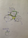
1) apex cordis
2) basis cordis
3) sulcus coronarius
4) sulcus interventricularis anterior
5) sulcus interventricularis posterior
6 - 10
How is #8 called inside?

6) truncus pulmonalis
7) sinus trunci pulmonalis
8) conus arteriosus → inside: infundibulum cordis
9) atrium dextrum
10) ventriculus dexter
11 - 14

11) aorta
12) v. cava superior
13) auricula sinistra
14) auricula dextra
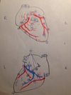
1 - 5

1) aorta ascendens
2) arcus aorticus
3) truncus pulmonalis
4) a. pulmonalis sinistra
5) truncus brachiocephalicus
6 - 10

6) a. carotis communis dextra
7) a. subclavia dextra
8) a. carotis communis sinistra
9) a. subclavia sinistra
10) lig. arteriosum
1 - 5

1) arcus aorticus
2) aorta descendens
3) aorta ascendens
4) truncus brachiocephalicus
5) a. carotis communis sinistra
6 - 10

6) a. subclavia sinistra
7) v. cava superior
8) a. pulmonalis dextra
9) a. pulmonalis sinistra
10) v. cava inferior
11 - 15

11) vv. pulmonales dextrae
12) vv. pulmonales sinistrae
13) atrium dextrum
14) atrium sinistrum
15) sulcus coronarius
16 - 20

16) sinus coronarius
17) sulcus terminalis
18) auricula sinistra
19) sinus transverus pericardii
20) ventriculus sinister
21 - 25

21) ventriculus dexter
22) sulcus interventricularis posterior
23) apex cordis
24) basis cordis
25) truncus pulmonalis
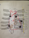
1 - 5

1) v. cava superior
2) v. cava inferior
3) sinus coronarius
4) vv. pulmonales dextrea
5) vv. pulmonales sinistrae
6 - 10
What forms #6?

6) crista terminalis → outside: sulcus terminalis
7) mm. pectinati
8) sinus venarum cavarum
9) valvula Eustachii
10) valvula Thebesii
11 - 15
Give another name for #11.

11) valva artrioventricularis dextra → tricuspid valve
12) chordae tendinae
13) m. pappilaris anterior
14) m. pappilaris septalis
15) m. pappilaris posterior
16 - 20
Give another name for #17.

16) trabeculae carnae
17) trabecula septomarginalis → moderator band
18) septum interventriculare pars muscularis
19) septum interventriculare pars membranacea
20) septum artrioventriculare
22 - 25
Give another name for #25.

22) septum interartriale
23) limbus fossae ovalis
24) fossa ovalis
25) valva artrioventricularis sinistra → bicuspid valve
26 - 28

26) m. pappilaris anterior
27) m. pappilaris posterior
28) thicker wall of right ventricle
Describe the structure of a semilunar valve
Give another name for the smallest part
- pars densa: lower part
- pars flaccida: upper part
- 2 lunuli: thickened edges
- 1 nodulus (=corpus Arantii): closes cusps
The inflow and outflow tract of the right ventricle are divided by.. ?
- crista supraventricularis
- trabecula septomarginalis
The inflow and outflow tract of the left ventricle is divided by.. ?
How is the structure called?
anterior cusp of bicuspid valve
→ vestibulum aortae
Which vessel leaves the left ventricle?
aorta
What are the 3 layers that make up the heart wall?
- endocardium
- myocardium
- epicardium
Which structures make up the myocardium?
- atrial muscle
- ventricular muscle
Differentiate btw atrial muscle layers
- deep layer: surrounding each atrium
- superficial layer: covers both atria