Where do the large intestines run from and to?
From the caecum to the anal canal
What epithelium lines the large intestines?
Mainly columnar epithelium except the final 1/2 of the anal canal which is stratified squamous epithelium
What is the function of the large intestine?
- Removes water from indigestible gut contents
- Turns chyme into a semi solid
- Produces vitamins
- Acts as temporary storage until defecation
Where does colonic mucosa get the majority of its nutrients from?
From short chain fatty acids derived from fermentation of dietary fibre
How do the different parts of the large intestine lie within the peritoneum?
- Ascending and Descending colon are retroperitoneal
- Transverse colon is intraperitoneal and has its own mesentry
- Sigmoid colon has its own mesentery
- Rectum
- Upper 1/3 intraperitoneal
- Middle 1/3 retroperitoneal
- Lower 1/3 no peritoneum
Describe the arterial supply to the midgut
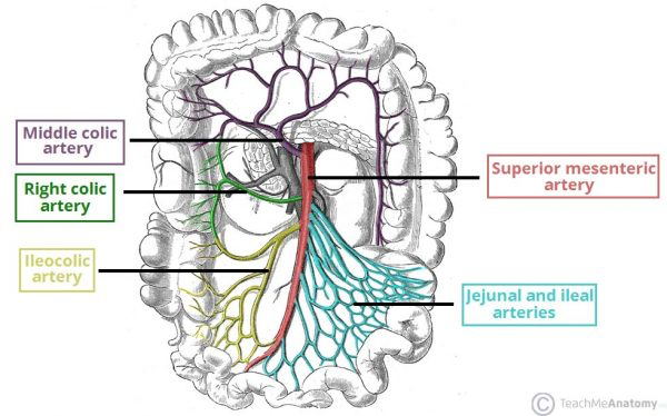
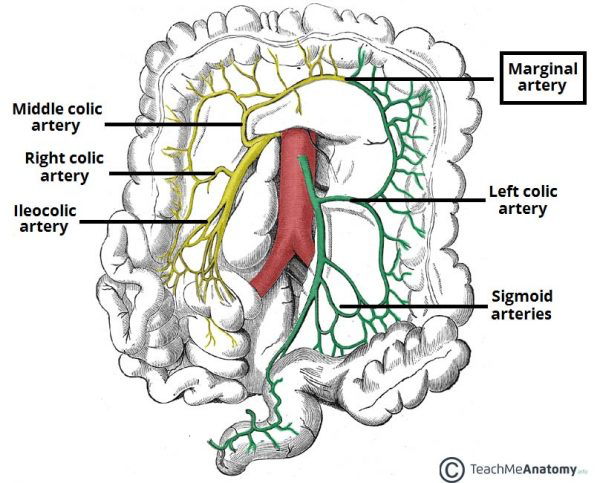
What is the marginal artery of the midgut?
The anastamoses of the distal ends of all the branches of the SMA that supply the midgut

Describe the Arterial supply of the hindgut
Branches off the Inferior Mesenteric Artery
- Left colic - descending colon
- Sigmoid arteries - descending colon and sigmod colon
- Superior rectal artery - upper 1/3 rectum
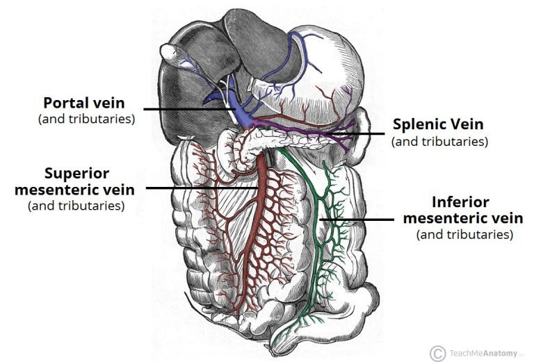
Describe the venous drainage of the abdomen
- Midgut - drains into superior mesenteric vein
- Hindgut - drains into inferior mesenteric vein
- Rectum
- upper 1/3 drains into superior rectal vein into IMC
- Middle and lower 1/3 drains into systemic venous system bypassing the liver
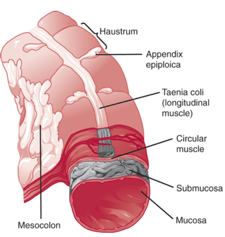
How does the Large intestine differ from the small intestine visually?
- Large intestine is shorter and wider
- Has crypts not villi
- Haustra - incomplete rings instead of plica circulares
How are haustra of the large intestine formed?
Incomplete external longitudinal muscles form 3 distinct bands (teniae coli)
Contraction of the teniae coli forms the sacculations known as haustra
How is water absorbed in the large intestine?
ENaC channels on the apical membrane take water into the colon cells
Water folows the the movement of Na+ and follows through tight junctions
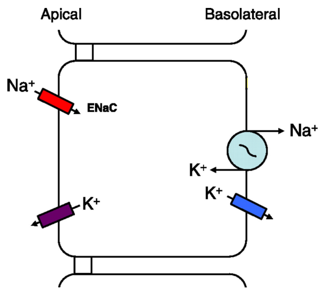
Which hormones adds ENaC channels to the apical surface of the cells of the colon?
Aldosterone
Why are tight junctions in the large intestine much tighter than those in the small intestine?
- Allows a bigger gradient to form
- Don’t get any backflow of ions
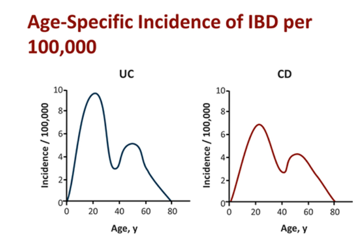
How does the incidence of IBS change with age?
Peaks in young adults ~ 20 years and a smaller peak ~50 years
What are the 2 main types of inflammatory bowel disease?
- Crohn’s Disease
- Ulcerative Colitis
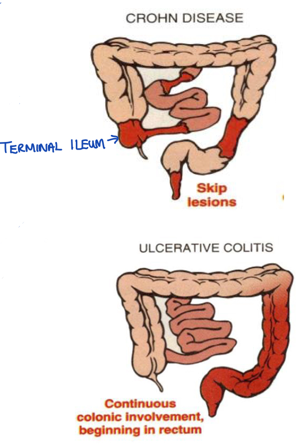
What are the key differences between Crohn’s Disease and Ulcerative Collitis?
Crohn’s Disease:
- Affects anywhere in GI tract
- Mainly involves the Terminal Ileum
- Transmural - through the entire wall of the bowel
- Skip lesions
Ulcerative Colitis:
- Begins in the rectum
- Continuous pattern
- Mucosal inflammation (shallow - not the whole wall)
What extra- intestinal problems are associated with inflammatory bowel disease?
- MSK Pain (Arthritis)
- Skin - Erythema nodosum, pyoderma gangernosum, psoriasis
- Primary Sclerosing Cholangitis of the biliary tree
- Eye problems
What causes inflammatory bowel disease?
- Genetic element
- Unbalance gut colony
- Immune response
- Potentially triggered by; antibiotics, infection, smoking, diet
How would someone with Crohn’s disease present?
- Young patient
- Frequent loose stools
- Non- Bloody stools
- RLQ pain (inflammed terminal ileum)
- Mild perianal inflammation ulceration
- Mildy anaemic
What peri-anal disease would you seen in Crohn’s disease?
(specific to Crohn’s, not seen in UC)
- Haemmoroids
- Skin Tags
- Anal Fissure
- Peri- anal abscess
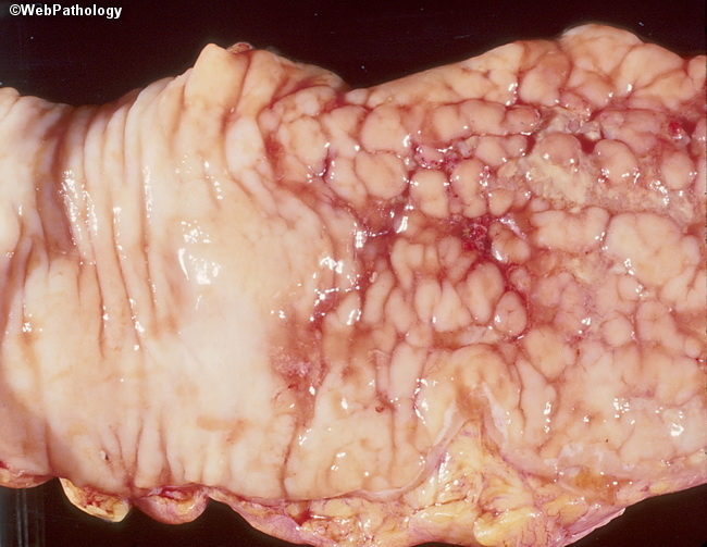
What features would you see on endoscopy in someone with Crohn’s Disease?
- Cobblestone appearance
- Skip lesions
- Fistulae
- Strictures
- Mucosal oedema
- Red and inflammed
- Deep ulcers
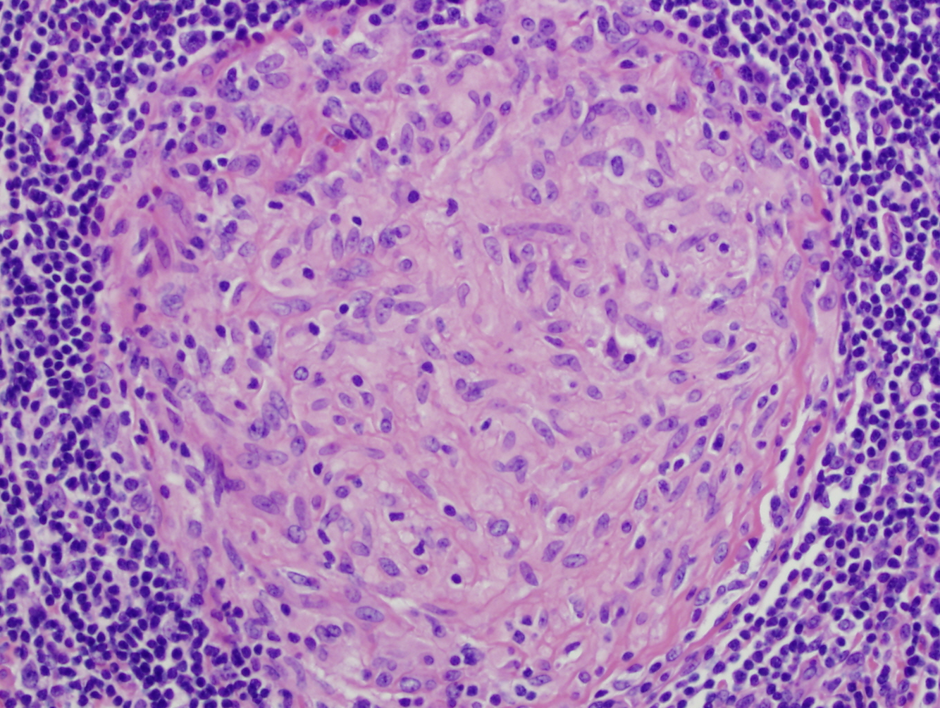
What would you see microscopically in someone with Crohn’s disease?
Granuloma Formation
Central granuloma with epithelioid macrophages
How would you investigate someone with suspected Crohn’s Disease?
- Bloods - check for anaemia
- CT/ MRI - looking for bowel wall thickening, obstruction, extra mural problems
- Barium Enema - identifies strictures / fistulae


-
Purpose of the Gut16
-
Anatomy of the Gut23
-
Abdominal wall and hernias30
-
Anatomy of salivation and swallowing23
-
Intro to Stomach30
-
Pathophysiology of gastric disease37
-
Pancreas and Liver25
-
The Intestines37
-
Liver & The Biliary System41
-
Jaundice and LFTs26
-
Large Intestines / IBS34
-
Distal GI tract pathology46
-
GI Malignancy44
-
Infections of the GI tract40
-
GI Emergencies37
