Portion of heart
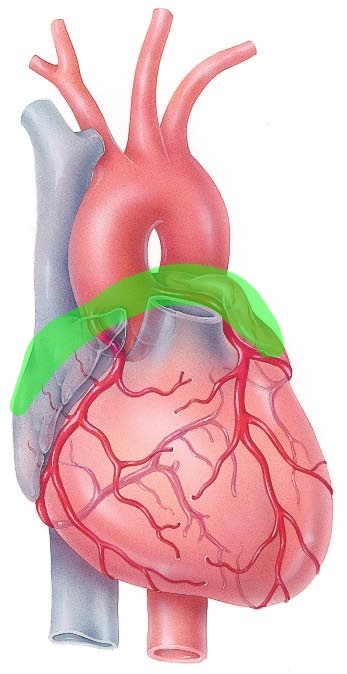
base
Portion of heart
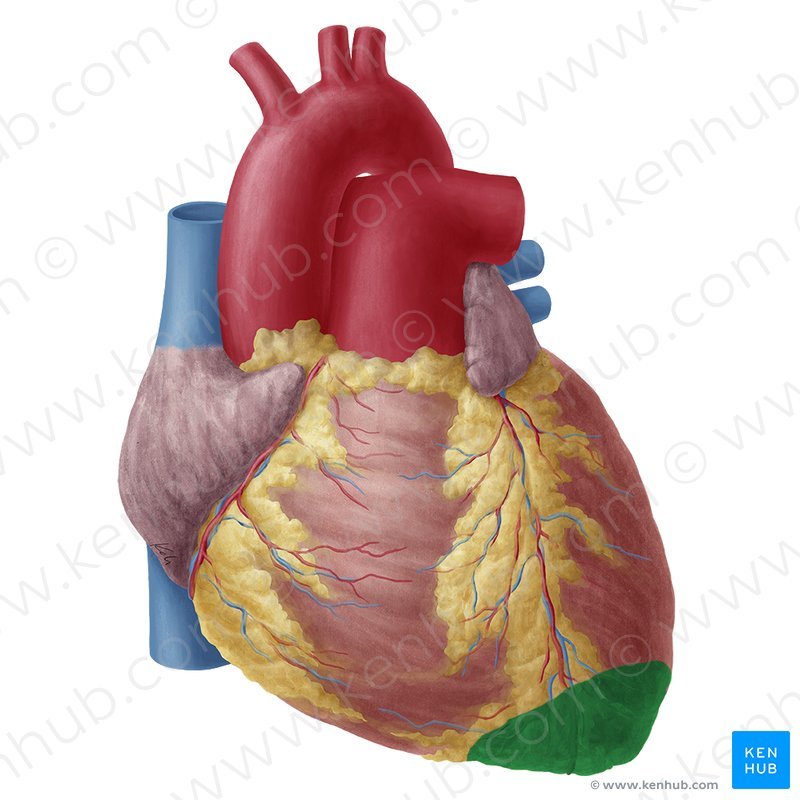
Apex
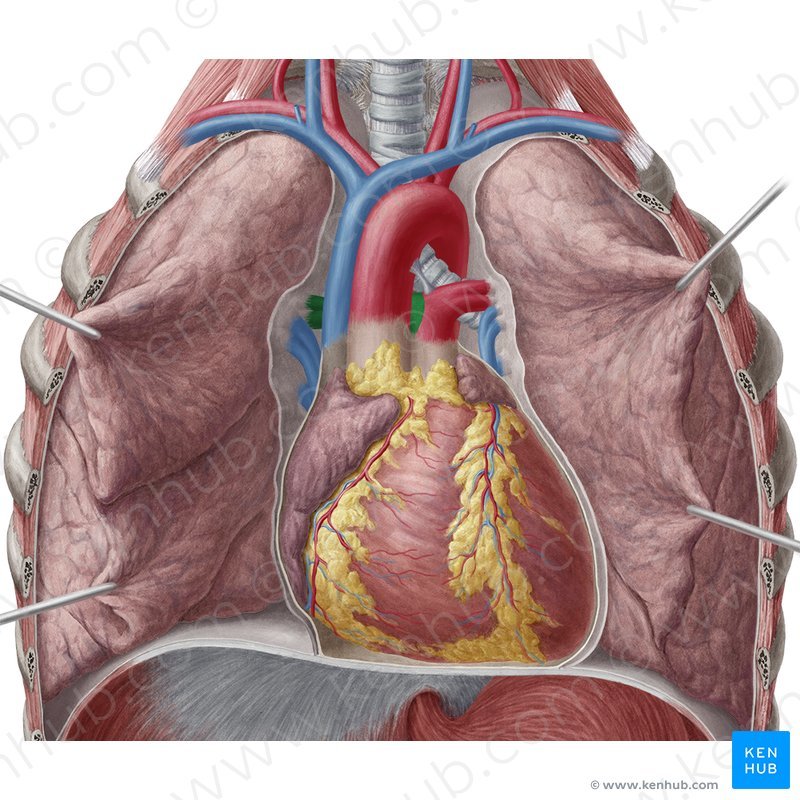
Position of heart
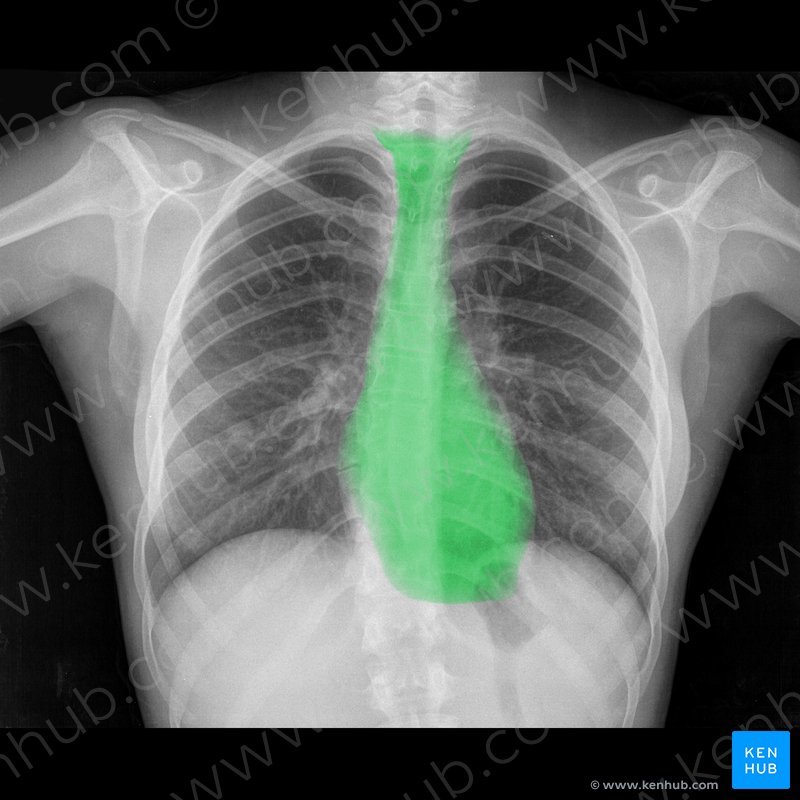
Mediastinum
The heart is on the ___ side of the mediastinum.

Left
A procedure. A probe inserted in the esophagus/stomach, it takes an ultrasound of the heart.
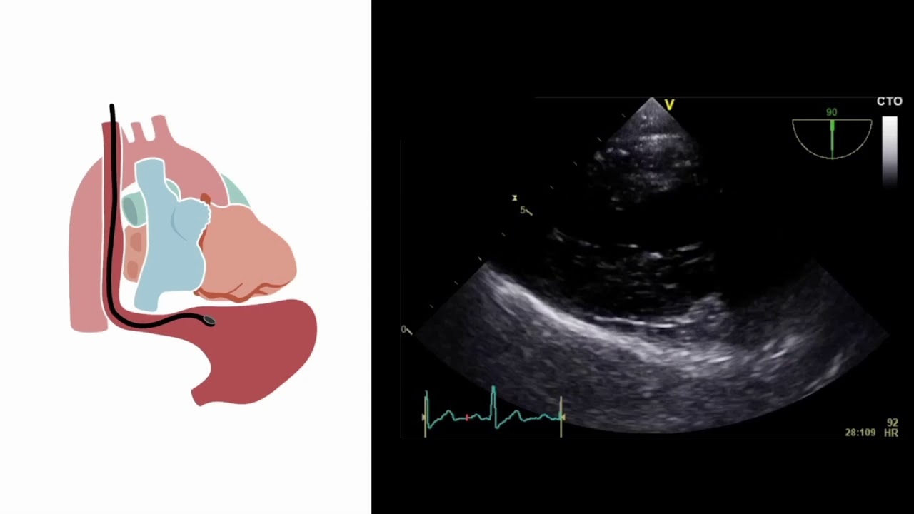
Transesophageal Echocardiography (TEE)
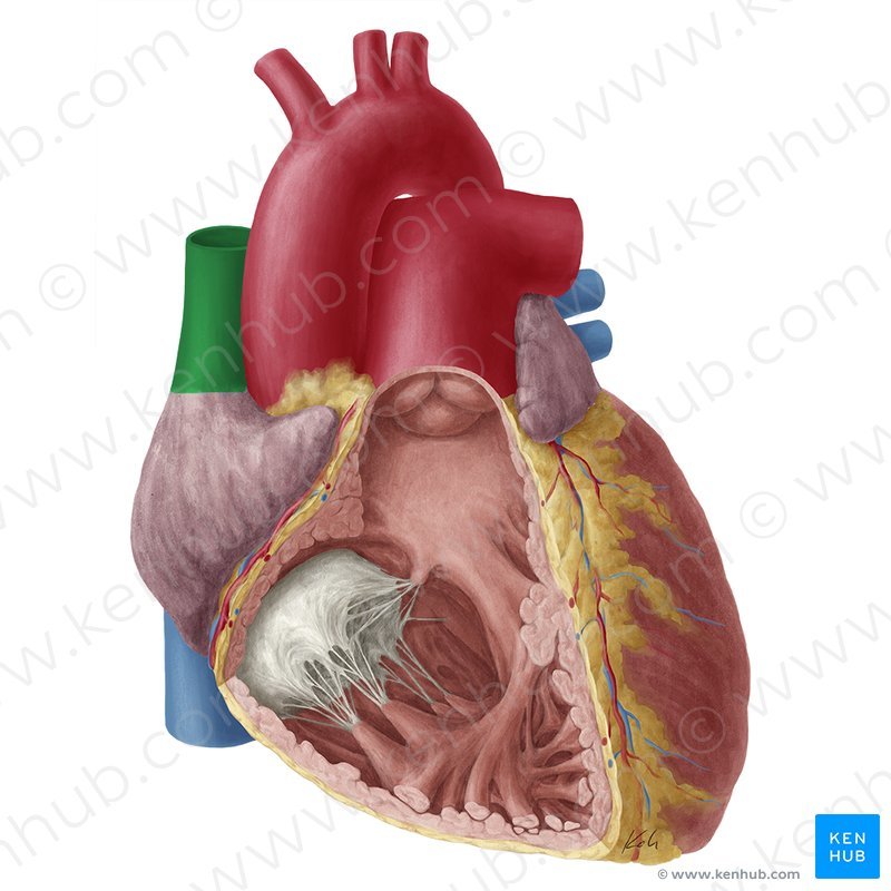
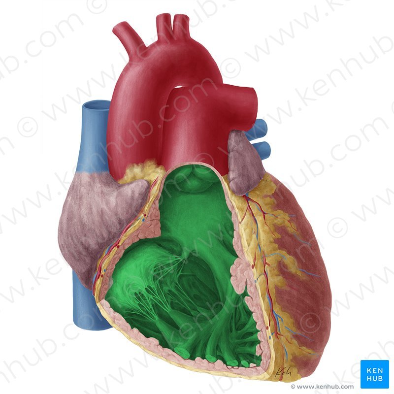
Smooth. Simple squamous epithelium.
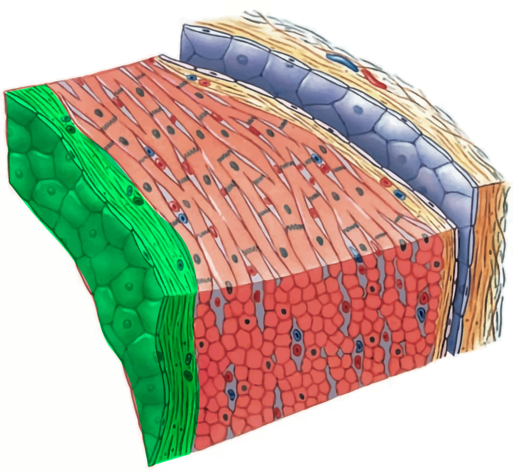
EndoCardium
Heart muscle. Intercalated disks for strength. Gap junctions for synchrony.
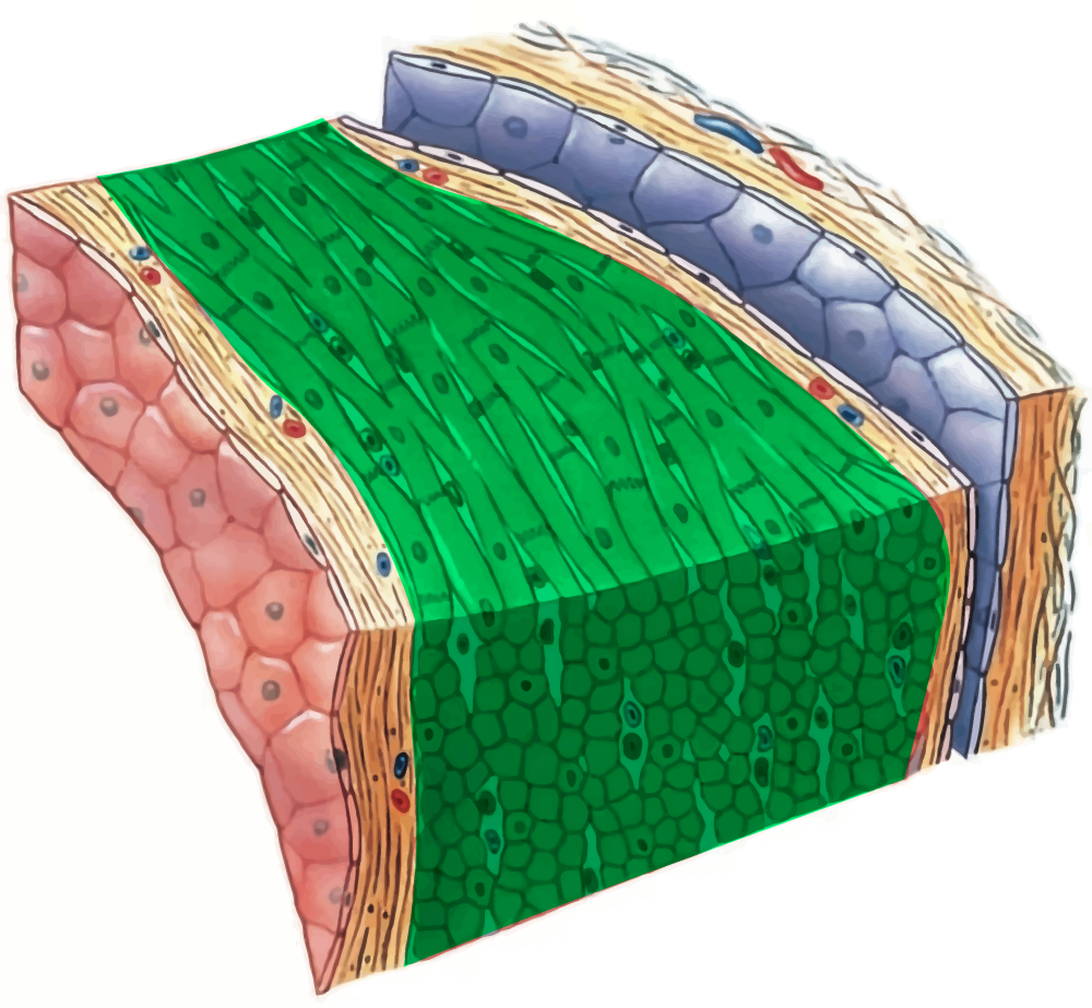
MyoCardium
Above. Visceral Serous.
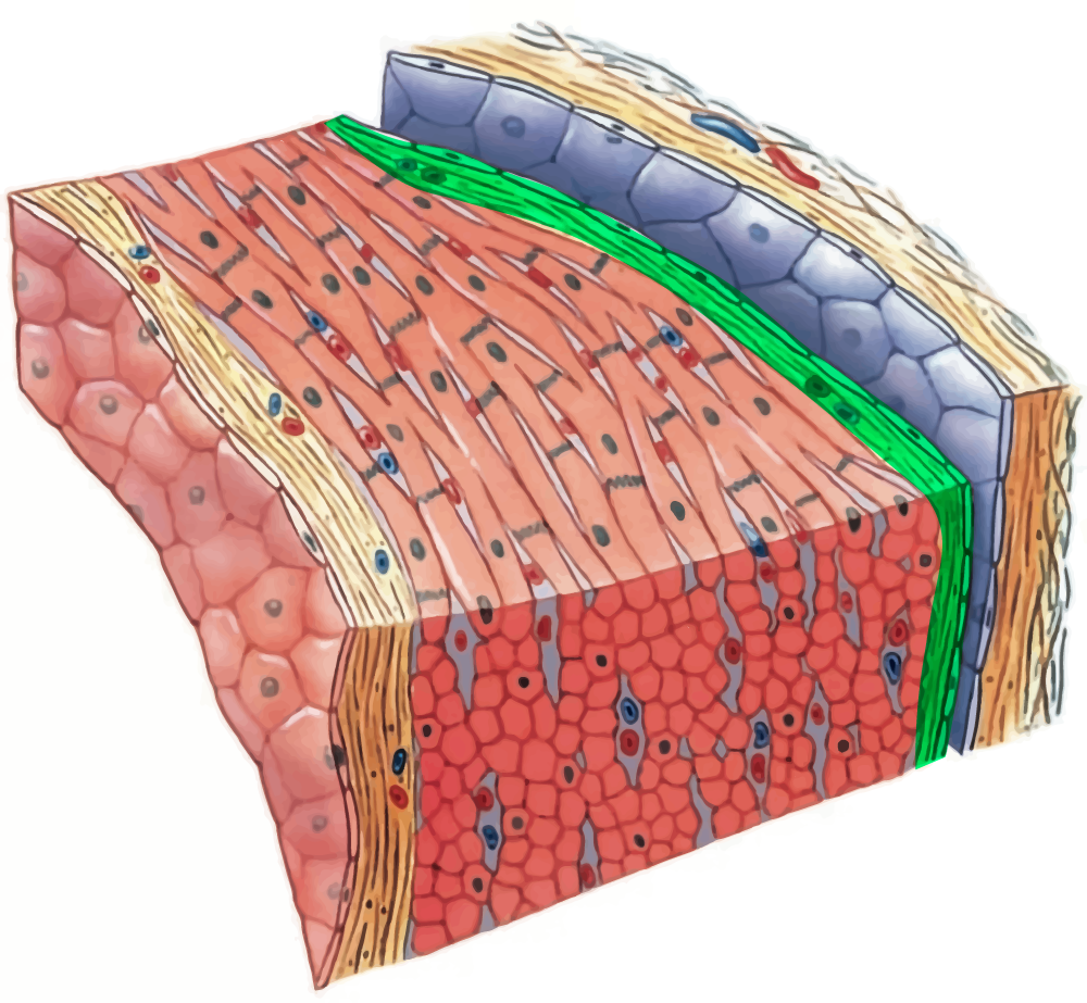
EpiCardium
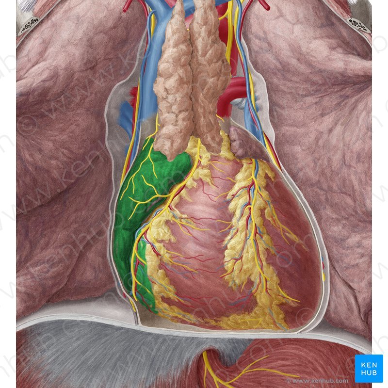
conical sac of fibrous tissue
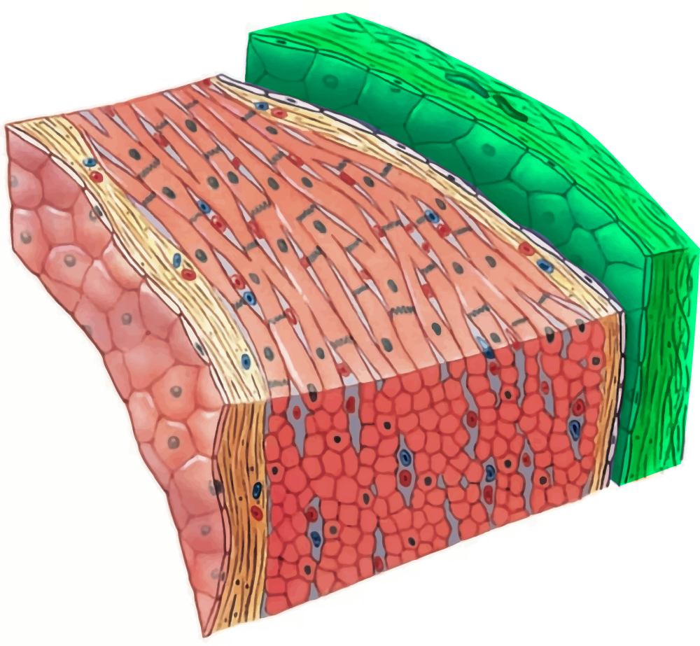
Parietal Pericardium
Infection of the endocardium causes inflammation.
Dental cleanings = antibiotics.
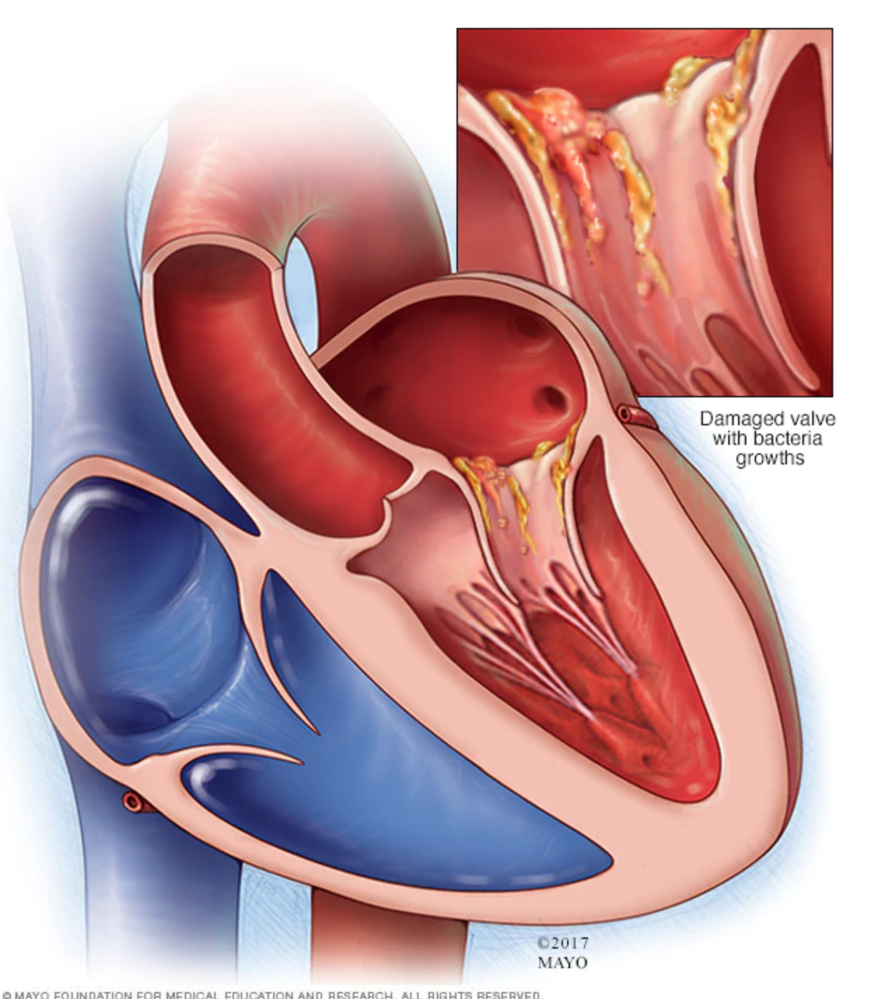
Endocarditis
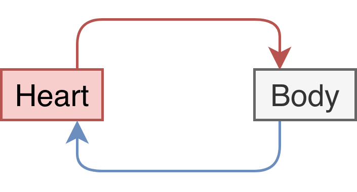
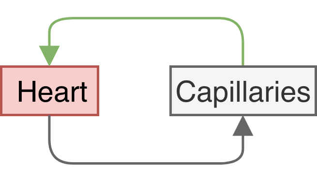
Systemic Circulation
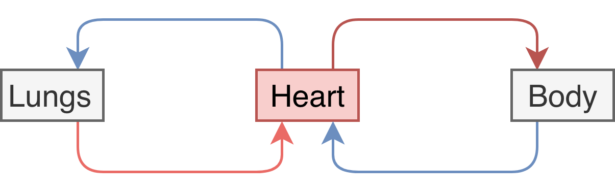
Pulmonary Circulation
Vessels that carry blood away from the heart.
Arteries
Vessels that carry blood toward the heart.
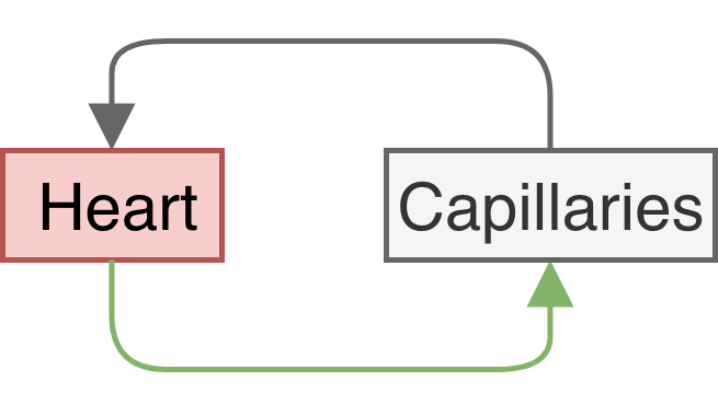
Veins
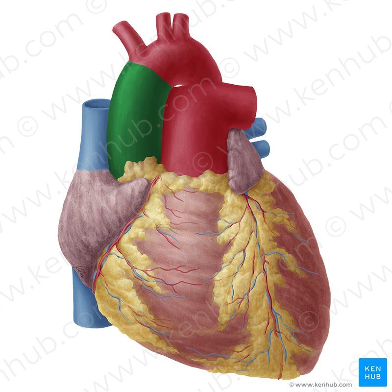
Ascending aorta
Superior vena cava
Right pulmonary artery
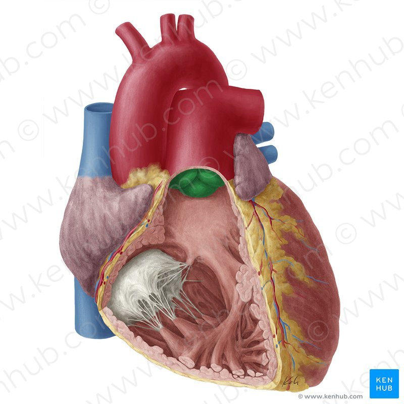
Pulmonary valve
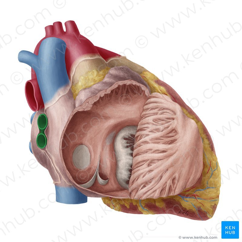
Right pulmonary veins
Right atrium
Right ventricle
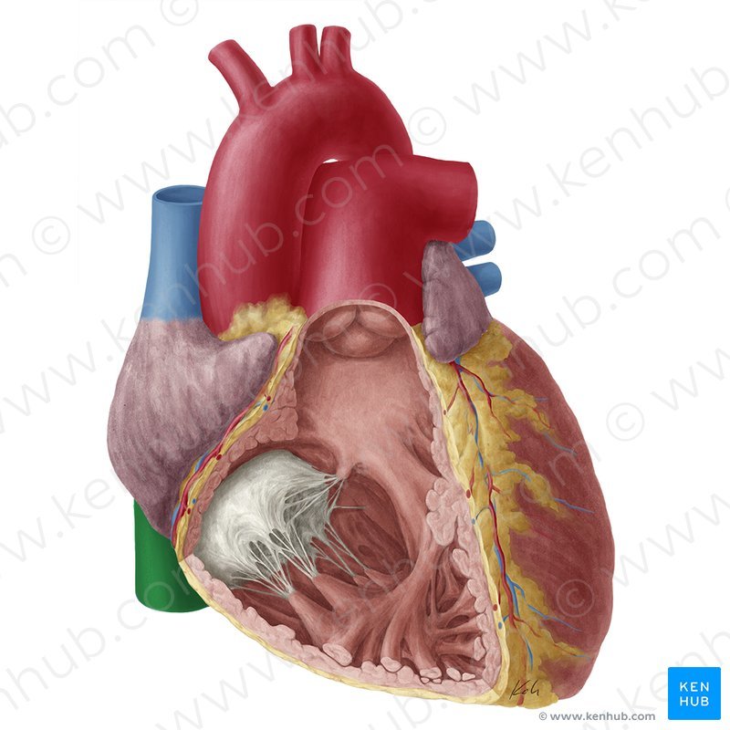
Inferior vena cava
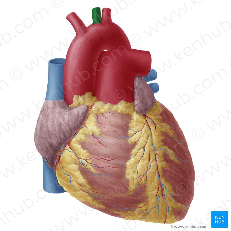
Left common carotid artery
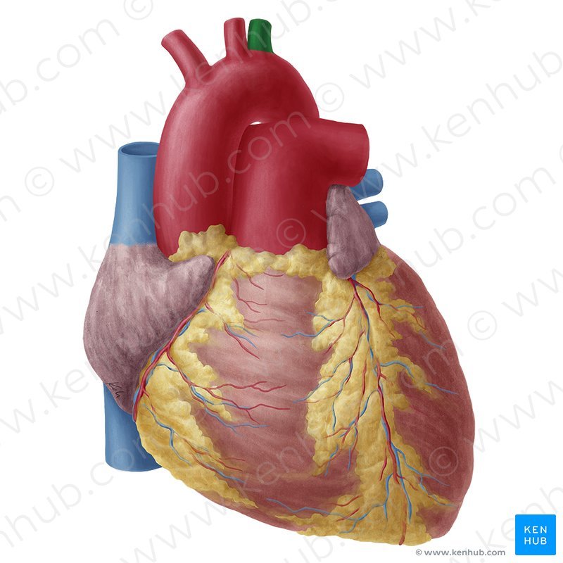
Left subclavian artery


