Indications for Skull X-ray
5
- When they are not otherwise getting a CT Scan (A&O x 3, GCS 15) and: Suspect skull fracture, sinusitis, facial bone tumors, nose pathology, foreign Body
- Evaluation for fractures of the mandible and maxilla
- Evaluation of the skull for lytic lesions such as in multiple myeloma
- Scalp has full thickness laceration or boggy hematoma (looking for skull fracture)
- To evaluate for scalp foreign bodies such as glass
The major drawbacks to skull x-rays
3
1. Lack detail
2. No reassurance if negative in the setting of trauma as unable to evaluate intracranial contents
3. Unable to see fractures in the skull base, if basilar skull fracture suspected a CT scan is indicated
What are lytic lesions called?
punched out lesions because of decreased bone density
What helps differentiate fractures and blood vessels?
When youre wondering if somehting is a fracture or a vessel. If youre branching then its a vessel. If its more straight its a fractire
What could iodine based contrast cause?
ATN
CT Inidcations
4
- Evaluation of the skull and skull base, vertebrae
- Evaluation of the ventricles
- Suspicion of intracranial masses, mass effects
- Looking for acute hemorrhage, ischemia
ischemia is not evident on CT scan until about 24 hours post onset of symptoms
Describe what the following might show us using CT on evaluation:
- Evaluation of the skull and skull base, vertebrae? 2
- Evaluation of the ventricles? 3
- Suspicion of intracranial masses, mass effects? 3
- Looking for acute hemorrhage, ischemia? 2
1.
- trauma
- bone lesions
2.
- hydrocephalus,
- shunt placement
3.
- headache,
- N/V,
- visual symptoms
4.
- stroke,
- mental status change
- Ischemia is not evident on CT scan until when?
- So why is CT our first inital test?
- about 24 hours post onset of symptoms
- CT to rule out hemorrhagic stroke. cant tell us ischemic
IV Contrast in Head CT: Yes or No?
No: 7
Yes: 4
Without contrast:
- Trauma
- R/O stroke
- R/O hemorrhage
- Hydrocephalus
- Dementia
- Epilepsy
- Congenital malformations
With contrast
- Neoplasm (usually very vascular)- usually doing MRI with this anyway
- Infection
- Vascular disease
- Inflammatory disease
Interpreting a CT Scan of the Head for PA’s
4 steps. What are they?
- Look for fluid
- Look for mass
- Look for shift
- Look at each side and compare
- The more dense the tissue, the ______it looks on CT?
- Any calcified structure (like the skull) appears ______?
- New hemorrhage in the brain is _____?
- Water (or CSF) looks _____on CT
- brighter
- bright
- also bright
- dark
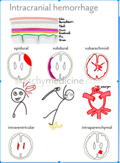
- What is a subarachnoid hemorrhage?
- Where is it at?
- What is a subdural hematoma and where is it at?
- What is an epidural hematoma and where is it at?
- WHat is a epidural hematoma associated with?
Subarachnoid hemorrhage (see picture-layered out on the surface of the brain)
- Arterial bleeding on the surface of the brain
- Between the pia mater and the arachnoid mater
Subdural hematoma
- Venous bleeding between the arachnoid and the dura mater
Epidural hematoma
- Dural artery or venous sinus bleeding between the skull and dura
- Associated with skull fracture
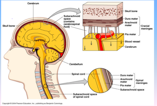
- SAH: What is the injury to?
- Where do the ruptured vessels bleed?
- Injury of small arteries or veins on the surface of the brain
- Ruptured vessel bleeds into the space between the pia and arachnoid mater
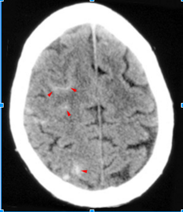
Causes of subarachnoid hemorrhage
2
(most common)?
- Trauma
Most common cause
- Ruptured cerebral aneurysm
What (arrowheads) fills the sulci over the
right cerebral convexity in this subarachnoid hemorrhage?
See Picture
High density blood
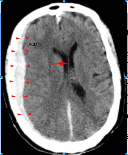
- What is the injury to in a subdural hematoma?
- Where does blood collect?
- Tearing of bridging veins from deceleration and acceleration or rotational forces
- Blood collects in the space between the arachnoid mater and the dura mater
Subdural Hematoma CT has the following characteristics: 3
- Crescent shaped
- Hyperdense, may contain hypodense foci due to serum, CSF or active bleeding
- Does not cross dural reflections
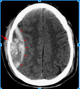
High density, crescent shaped hematoma (arrowheads)
overlying the right cerebral hemisphere. Whats important to note in this image?
Subacute subdural hematoma (arrowheads). Whats important to note about this CT?
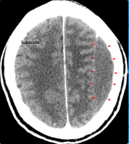
Note the compression of
gray and white matter in the left hemisphere due to the mass effect.
Chronic Subdural hematoma: Whats importnat to notice in this CT?
Crescent shaped chronic subdural hematoma (arrowheads)
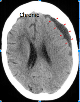
Notice
the low attenuation due to reabsorbtion of the hemorrhage over time.
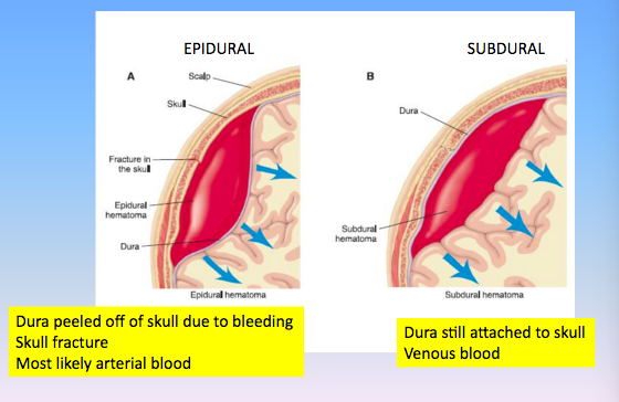
- Epidural hematoma is usually associated with what?
- What is the injury that results from this?
- Where does the blood collect?
- Usually associated with a skull fracture
- The fractured bone lacerates a dural artery or a venous sinus
- The blood from the ruptured vessel collects between the skull and dura
- On the CT, the hematoma forms a what?
- It is usually uniformly ____ _______but may contain hypodense foci due to active bleeding.
- hyperdense biconvex mass
- high density
Describe the differences:
Epidural vs Subdural
2
See picture
Describe the following for Epidural, Subdural and Subarachnoid:
- In relation to the dura
- Respects suture lines?
- Trauma?
- What kind of blood and usually from where?
- Shape on CT?
- Clinical presentation?
Epidural
- Above the dura
- Respects suture lines
- High force trauma
- Arterial blood (commonly the middle meningeal artery)
- Lentiform (lens-shaped) or biconcave on CT
- Acute presentation
Subdural
- Below the dura
- Doesn’t respect suture lines
- Low force trauma
- Venous (from venous plexus)
- Cresent (banana-shaped) on CT
- May be insidious (worsening headache over days)
Subarachnoid
- Below the arachnoid
- No respect for anything
- Aneurysm rupture or high force trauma
- Arterial from the circle of Willis
- Lining surface, going into fissures and sulci and sella (death-star)
- Acute presentation (thunderclap headache)


-
Neurology Anatomy and Physiology50
-
Movement Disorders48
-
Neuroimaging48
-
Neurodiagnostics57
-
Dementia and Delirium83
-
Seizure Disorders91
-
LOC/Coma41
-
Sleep Disorders82
-
Movemnt Disorder Pharm45
-
NEURO-MUSCULAR DDx34
-
Concussions42
-
Neurological Emergencies73
-
Miscellaneous Neurological Disorders39
-
Stroke and TIA80
-
Headaches88
-
Peripheral neuropathy58
-
CNS infections55
-
CNS neoplasms48
