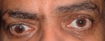
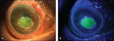
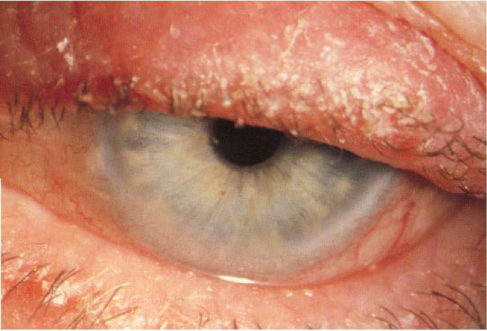
describe abnormal findings with this man’s right eye
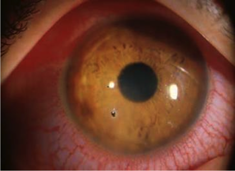
•scleral injection, FB at 7 o’clock
*you would also want to document (if true): PERRLA, EOMI
In an older patient with sudden onset of eye pain, headache and vomiting, what MUST be on your DDx?
acute glaucoma
stroke
What % of ED visits are related to the eyes?
What are the 4 MC complaints?
•3-10% of ED complaints are related to the eyes
- Ocular pain
- Change in vision
- Change in Appearance
- Trauma
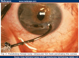
What 3 eye problems are true emergencies and require IMMEDIATE consultation?
Immediate consultation:
- Sudden visual loss
- Globe perforation
- Alkali/Acid burn (alkali is worse than acid burn)
What should you include in taking an initial Hx?
•Chief complaint
•Change in vision
•Change in appearance
•Discomfort (FOB sensation, irritated/scratchy/etc)
•Duration
•Associated symptoms
►Remember to ask about contact use or eye meds
Relevant PMH:
- Surgeries
- Systemic diseases
- Contact lens/vision correction
- Family history
- Ophthamologic medications
List steps in 8 (9) point ER eye exam
- Visual acuity
- Pupils (reactive, symmetric?)
- EOM (LR6SO43)
- Fluoroscein
- Visual fields (specifically test this even if pt reports no deficits→ may be unaware)
- IOP
- Fundoscopic
- Slit Lamp
- (External inspection)
Visual Acuity (single best eye exam)
Uses Snellen chart
•Should be done corrected (with eyeglasses or contacts on or if those aren’t available→ use pinhole card)
•If unable to see chart:
–finger count
–hand motion
–light perception

Pupillary Exam should include:
- Shape
- Size
- Reaction to light
- Accommodation
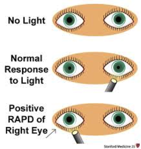
What is Anisocoria?
unequal pupil size
•Physiologic – 20% of cases
- The Large pupil is abnormal in CN III lesion (Adie’s pupil)
- classically young women
- Small pupil abnormal in Horners syndrome
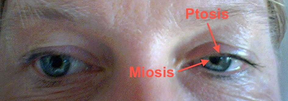
What is Horner’s syndrome?
•Horners Syndrome – ptosis, miosis, anhidrosis (loss of hemifacial sweating)
– many causes – MS, brain tumors, trauma, carotid artery dissection
What can cause an oculomotor nerve palsy?
- ischemia,
- aneurysm,
- trauma,
- brain tumors
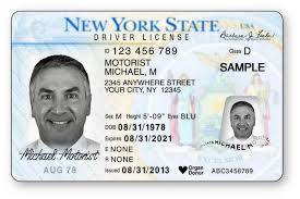
What is a good trick if a patient doesn’t know when you ask them if their current eye presentation is “normal for them”?
ask to see his driver’s license and see how eyes look there
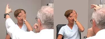
Why is it important to test EOM in all fields, plus convergence?
- Monocular diplopia:
–cornea/lens/malingering
- Binocular diplopia:
–CN source vs EOM source
why is it important to always check visual fields by confrontation?
Visual field defects – glaucoma, stroke, brain tumors, other neuro defects
•Patient may not notice change unless checked
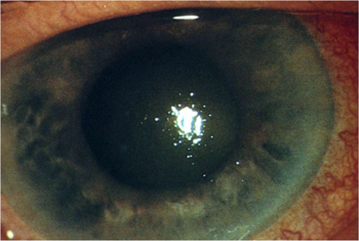
What type of stain is best to evaluate for:
abrasions, dendritic lesions, open globe, ulcers, FB…
Flouroscein
►Have pt pull down lower lid and look medially. Put anesthetic drops in lateral part of globe
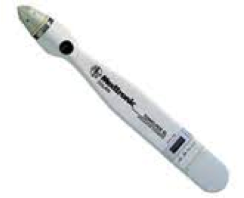
What is normal value for intraocular pressure?
What instrument could be used to measure if you were concerned about increased intraocular pressure?
•IOP: 10-20 is normal
A Tonopen
(do this before you dilate the pupils)
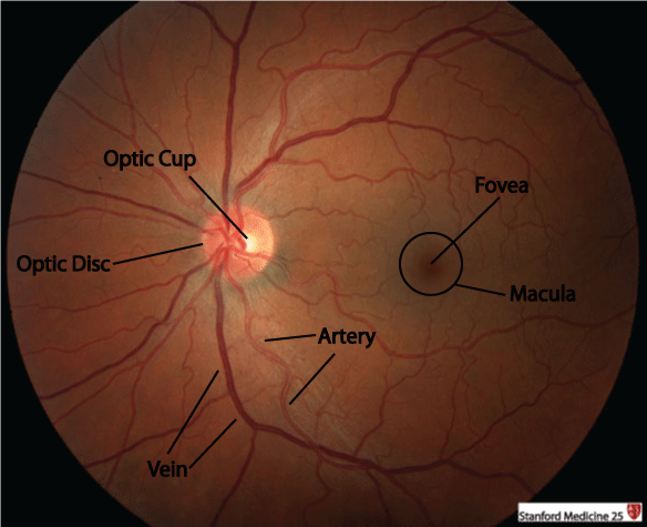
What are we looking at on the fundascopic exam?
(many things…!)
- Red reflex
- Cornea/lens/vitreous/retina/macula/optic nerve/blood vessels
- Retina, optic disc, vessels
Describe steps in Slit lamp exam.
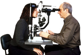
- Start exam anterior to posterior
- Lids/lashes/conjunctiva/tear film/cornea/anterior chamber/iris/lens
- Evert the lid!
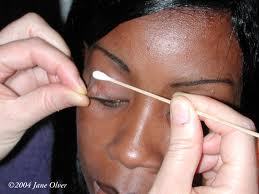
–esp if have foreign body sensation (flip over a q-tip stick if having trouble)
https://youtu.be/https://youtu.be/g0qqwJIKQlY
Describe components of an external eye exam.
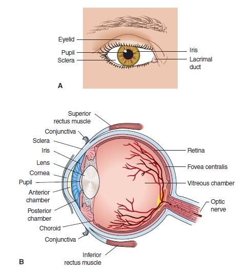
- Systematic
- Orbital rim
- eyelid
- conjunctiva
- mucus membrane that covers the front of the eyeball
- sclera
- the outer wall of the eye, white, fibrous, composed of collagen, and is actually continuous with the clear cornea anteriorly
- At the back of the eye, the sclera forms the optic sheath encircling the optic nerve
- cornea
- iris
*When you examine the “white part” of a patient’s eyes, you’re actually looking through the semi-transparent conjunctiva to the white sclera of the eyeball underneath.
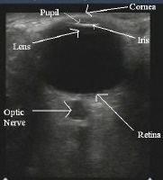
What imaging is quick, done at bedside and is 100% sensitive and 97% specific compared to exam and CT?
•Can Dx scleral and choroidal lacerations, vitreous hemorrhage, retinal detachment, radiolucent and radio-opaque FB, retrobulbar hematomas
When should this imaging NOT be used?
ULTRASOUND
ø Contraindicated in large globe lacerations
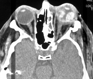
What is the MC way to evaluate:
–fractures of the orbit,
-intraocular foriegn body (>1 mm cuts)
►CT
•Indicated when posterior segment can’t be visualized, suspected occult globe rupture or laceration, and metallic foreign bodies
What is the best imaging to evaluate suspected orbital and periorbital tumors, optic nerve disorders?
MRI
May delineate small wooden and organic FB however must be sure no metallic IOFB present
What is the treatment for Blepharitis?
(Inflammation or infection of lid margin)
Tx: Hygiene; topical antibiotics +/- topical steroids
- Staphylococcal infx common (Abx)
- VZV
- Lid lice (pediculosis) (petroleum jelly)


