The two ways to record action potentials as they occur in an axon.
- intracellular recording. records changes in membrane potential. not feasible to do in lab. amplitude will be constant.
- extracellular recording. do not have to pierce the membrane of the axon to get a reading. look at the timing and frequency of action potentials and not amplitude.
Preamplifier in cockroach experiment.
small voltage is amplified so that it is large enough to be detected and observed. In humans, also has to isolate patient from any source of high voltage.
Plexuses.
Cervical (neck region and contains phrenic nerve), Brachial (shoulder and arm), Lumbosacral (contains femoral and sciatic)
- Plexus. 2. Muscles Innervated. 3. Skin innervated. 4. Location
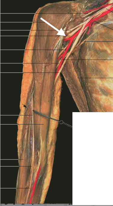
Axillary Nerve. 1. Brachial Plexus. 2. Deltoid. 3. Skin of Shoulder. Branches off with radial nerve and is deep to the M’M nerves.
- Plexus. 2. Muscles Innervated. 3. Skin innervated. 4. Location
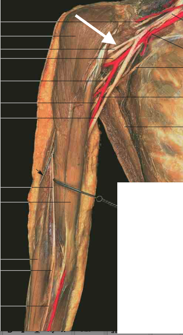
Musculocutaneous Nerve. 1. Brachial Plexus. 2. Brachialis and Biceps Brachii. 3. Lateral Surface of Forearm. 4. paired with Median nerve. anterior to the A & R nerves.
- Plexus. 2. Muscles Innervated. 3. Skin innervated. 4. Location
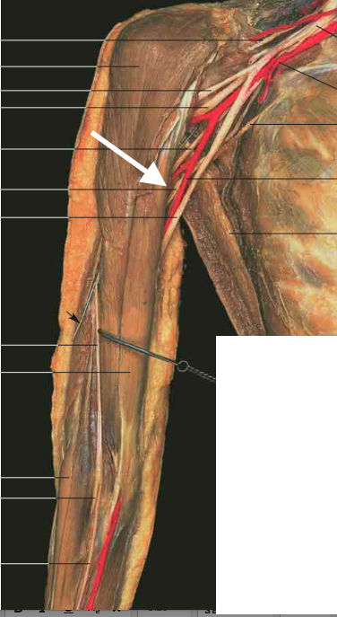
Ulnar Nerve. 1. Brachial Plexus. 2. Flexor/pronator group. 3. skin of medial 1/3 of hand 4. all by itself and most medial.
- Plexus. 2. Muscles Innervated. 3. Skin innervated. 4. Location
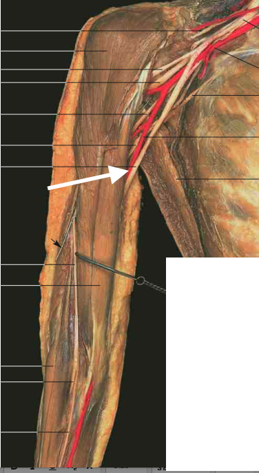
Median Nerve. 1. Brachial Plexus. 2. Flexor/pronator group. 3. Skin of anterior, lateral 2/3 of hand. 4. part of the M’M nerves more medial.
- Plexus. 2. Muscles Innervated. 3. Skin innervated. 4. Location
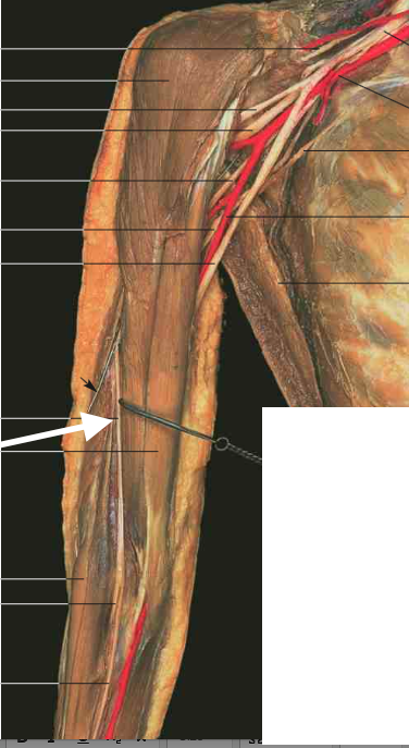
Radial Nerve. 1. Brachial Plexus. 2. triceps, extensor/supinator group. 3. skin of posterior lateral 2/3 of hand. 4. part of the A’R group and bigger than the axillary nerve. runs between the brachialis and brachioradialis.
- Plexus. 2. Muscles Innervated. 3. Skin innervated. 4. Location
Obturator Nerve. 1. lumbosacral plexus 2. adductor magnus and gracilis. 3. medial thigh. 4. only seen on model.
- Plexus. 2. Muscles Innervated. 3. Skin innervated. 4. Location
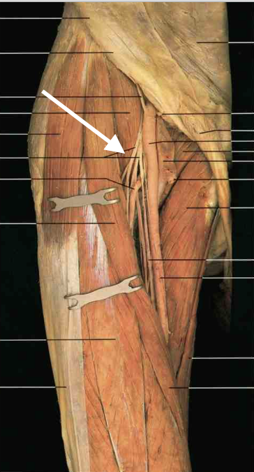
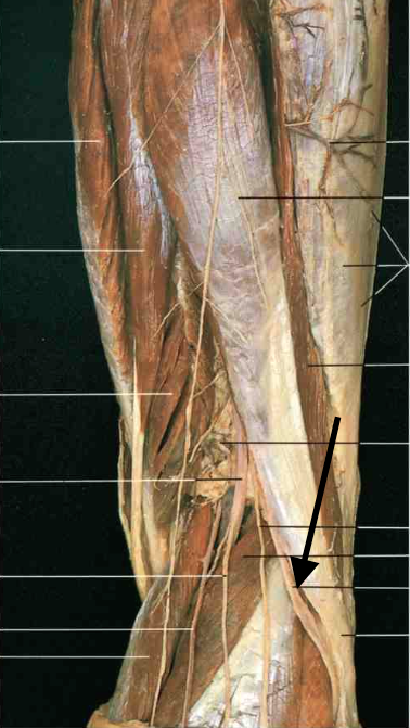
Femoral Nerve. 1. lumbosacral plexus 2. quadriceps. 3. anterior-medial thigh, lower leg. 4. from abdominal region comes out on top of thigh but under fascia.
- Plexus. 2. Muscles Innervated. 3. Skin innervated. 4. Location
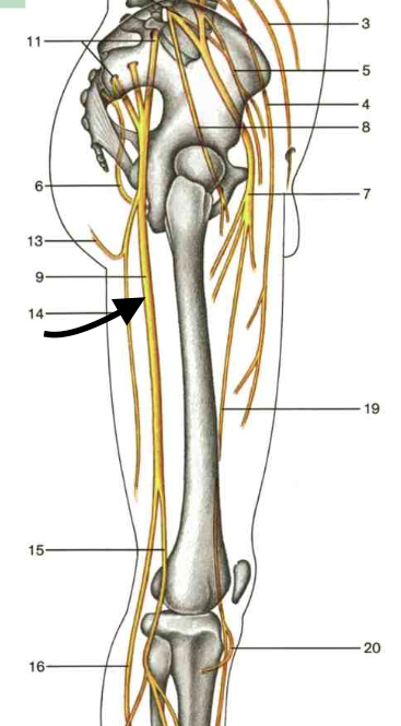
Sciatic Nerve. 1. lumbosacral plexus. 2. hamstrings. 3.? 4. divides into the tibial nerve and common peroneal nerve after hamstring.
- Plexus. 2. Muscles Innervated. 3. Skin innervated. 4. Location
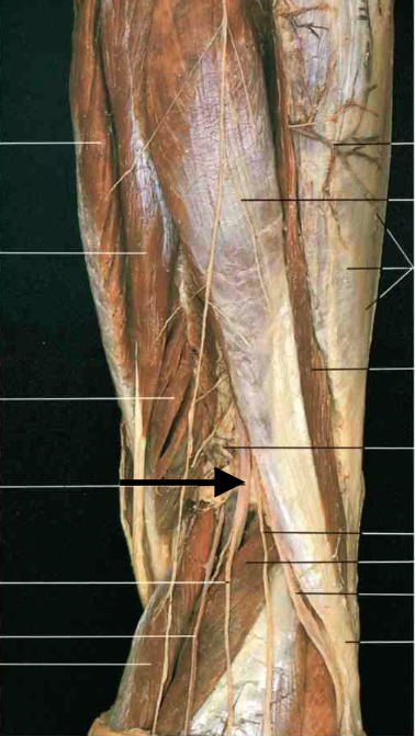
Tibial Nerve. 1. lumbosacral Plexus. 2. gastrocenemius, soleus. 3. posterior lower leg and plantar surface of foot. 4. stays straight after branching from sciatic.
- Plexus. 2. Muscles Innervated. 3. Skin innervated. 4. Location
Common Peroneal Nerve. 1. lumbosacral plexus. 2. peroneal muscles, tibialis anterior, extensors of toes. 3. anterior lower leg and dorsum of foot. 4. goes lateral after splitting from sciatic nerve.
