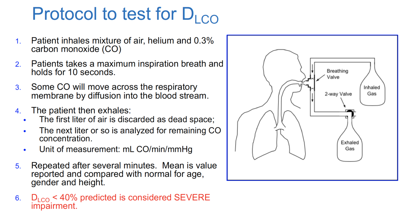
How do you conduct a CO diffusion test?
What are the 3 categories of lung diseases we should be familiar with?

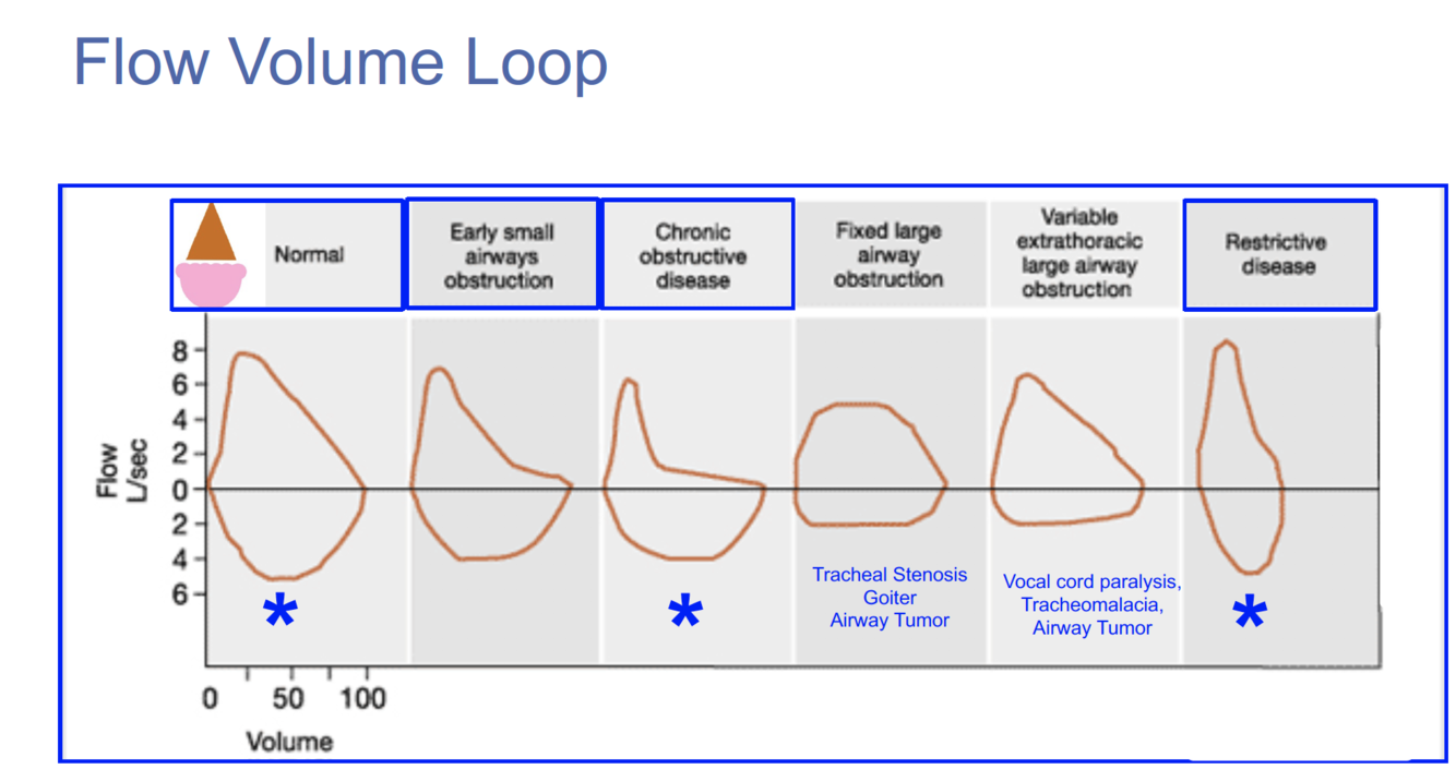
What values on spirometry would be altered in a person with obstructive airway disease?
Increased RV and TLC
Decreased FEV1/FRC ratio
What are all the labels on this diagram?
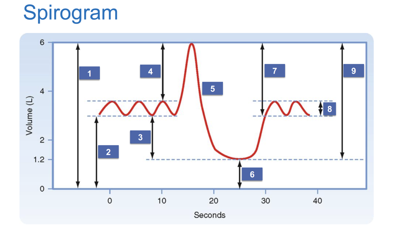
1) Total lung capacity (TLC)
2) Functional residual capacity (FRC)
3) Expiratory reserve volume (ERV)
4) Inspiratory reserve volume (IRV)
5) Forced vital capacity (FVC)
6) Residual volume (RV)
7) Inspiratory capacity (IC)
8) Tidal volume (Vt)
9) Vital capacity (VC)
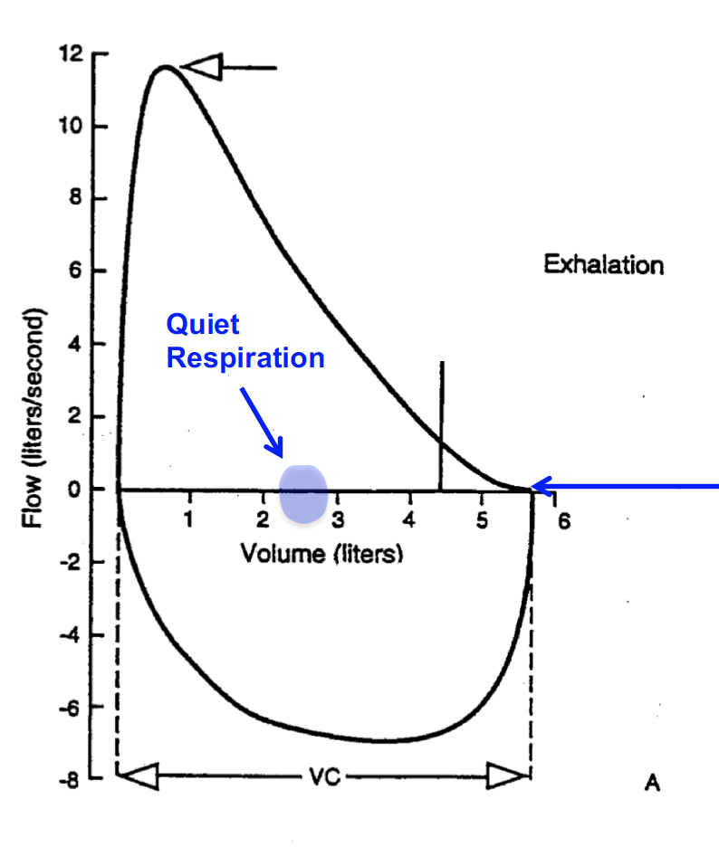
Why is the inhalation flow negative in this graph?
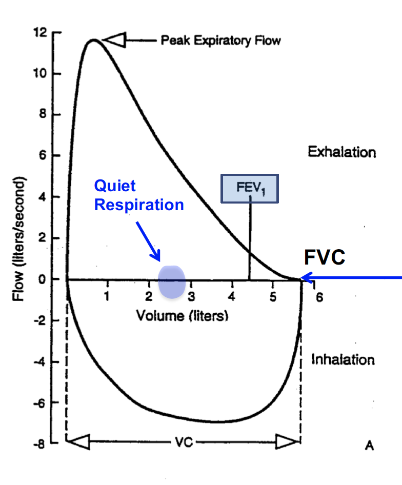
The spirometer measures air taken away from the device as negative (sucking air in during inhalation) and flow that is given back to the device as positive (blowing air out during expiration)
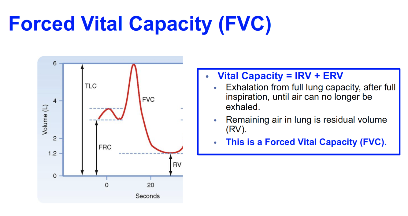
Forced Vital capacity = […] + […]
IRV + ERV
Where is the FEV1 and FVC on this graph?
What is the peak expiratory flow rate?
Rate of change of volume / rate of change of time
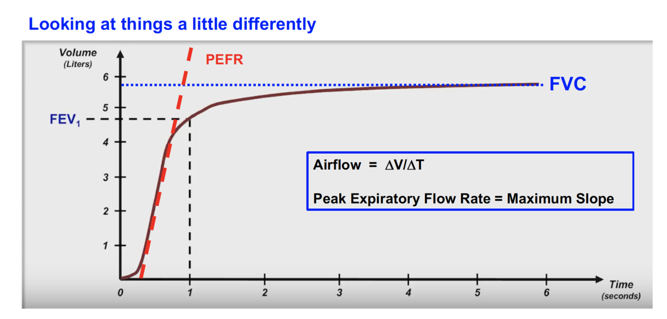
What is FEV1?
What is FVC?
- FEV1 is forced expiratory volume, or the VOLUME of air that a person can force out of their lungs during 1 second
- FVC is forced vital capacity, or the total amount of air that the person can force out of their lungs
What causes an increased diffusion capacity result for CO test?

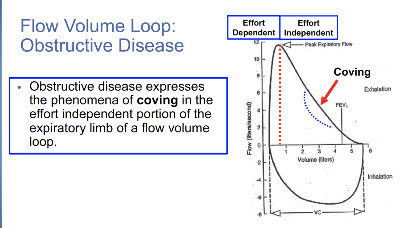
What is coving?
What does this spirometry report tell you?
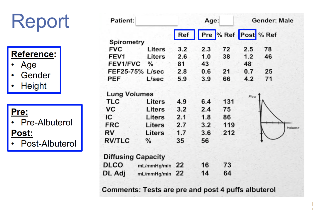
Decreased FVC
Decreased FEV1
Decreased FEV1/FVC –> COPD
Increased TLC –> COPD
Increased RV –> COPD
What values on spirometry would you expect if a person has restrictive airway disease?
All volumes of air decreased
FEV1/FVC is normal to increased
Why would you order a PFT?
- Diagnose symptomatic disease (dyspnea, cough, hypoxemia that is unexplained)
- Screen for early asymptomatic disease
- Prognosis of known disease
- Monitor response to treatment
What could cause a decreased diffusion capacity for CO?
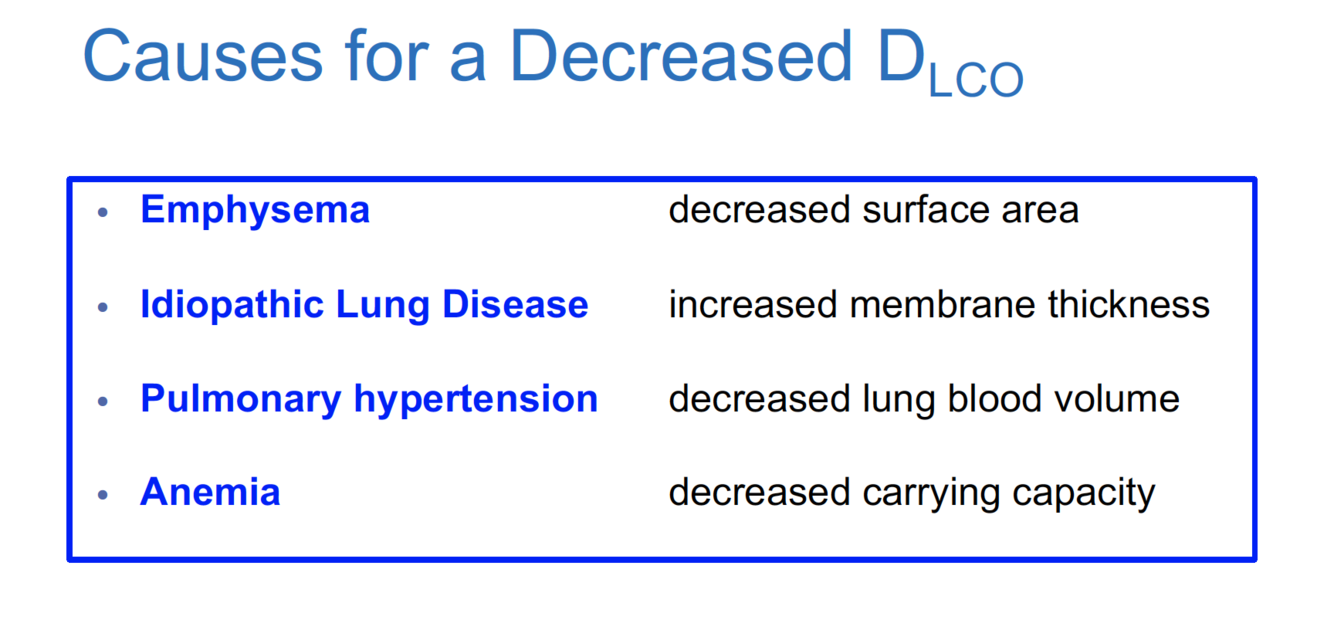
If the diffusion capacity of CO is decreased on spirometry, what does that indicate?
Obstructive disease –> a problem occuring at the level of the respiratory membrane that impairs the ability of CO to diffuse from the blood into the alveoli (fibrosis, surface area, etc.)
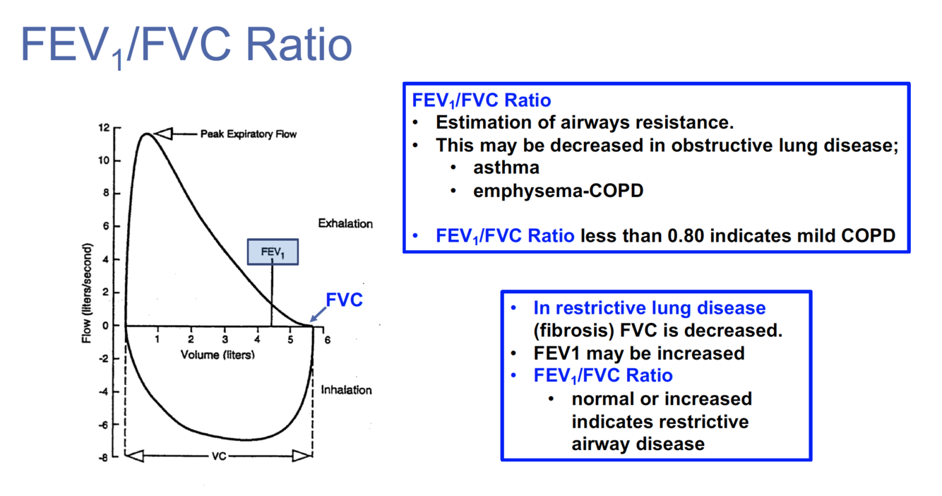
- What does the FEV1 / FVC ratio tell you clinically?
- What is a normal value for this ratio?
- What does a reduced ratio indicate?
- What does an increased ratio indicate
- This ratio is a measure of airway resistance, since the FEV is the volume of air that can be forced out in 1 second and the FVC is the total volume of air that can be forced out overall.
- A normal value for this ratio is around 0.8
- Asthma and emphysema can reduce the FEV1, resulting in a reduction of the overall ratio so ratio < 0.8 may indicate obstructive disease (difficulty getting air out thus decreased FEV1)
- Restrictive lung diseases like fibrosis cause increased restriction of the airways leading to decreased FVC. This will result in an increase in the ratio and thus can indicate restrictive airway disease (difficulty getting air in, thus decreased FVC)
What values on spirometry would you expect if a person had mixed obstructive and restrictive disease?
FEV1/FVC decreased
TLC decreased
-
Lung Anatomy and Histology44
-
Mechanics of Breathing and Ventilation27
-
Epidemiology and GB of Pulm Dis13
-
Pulmonary Circulation21
-
Clinical Correlates13
-
Hb Structure, Function and Gas Transport37
-
Pulmonary Function Test19
-
Control of Respiration25
-
HBCT7
-
Ventilation Perfusion21
-
Pharmacology30
-
Anatomy - Anterior Thoracic Wall22
-
Anatomy - Lungs28
-
Causes and Mechanisms of Hypoxemia17
-
Acid Base 126
-
CVD Epidemiology17
-
Role of Food in CHD Treatment7
-
Atherosclerosis26
-
Dissection - Heart70
-
Anatomy and Histology - Cardiovascular System40
-
Overview of CV System and Cardiac Cycle42
-
Electrical Properties of the Heart42
-
ECG16
-
Contractile Properties of the Heart36
-
Hemodynamics36
-
Control of Cardiac Output16
-
Local and Humoral Control of Blood Flow19
-
Regulation of Blood Pressure18
-
Special Circulations20
-
Posterior Thoracic Wall60
-
CV Physiology and Exercise25
-
Radiology of Heart and Lungs22
-
Surface Anatomy25
-
Anatomy and Histology - Kidney39
-
Overview of Renal System27
-
Dissection - Kidneys11
-
Intro to Renal Physiology27
-
Health Disparities - Renal20
-
Integrated Pharmacology and Drug Clearance17
-
Glomerular Filtration and Renal Clearance23
-
Regulation of ECF Volume and Water Balance35
-
Regulation of K+, Ca2 and PO4-67
-
Regulation of NaCl Balance25
-
Heart valves and sounds45
-
Acid Base 2 Kidneys40
-
HTN Guidelines13
-
Integrated Pharmacology - HTN21
-
ABG event4
-
Integration renal, pulm, CV, hepatic3
