Key discussino topics
- Anatomy of the eye
- Retina: photoreceptors and retinal ganglion cells
- Not horizontal, bipolar and amacrin cells
- Basis of blind spot
- Vitreous and aqeueous humors (increased intraocular pressure and impaired drainage)
- Cranial nerves relevant to visual system, including testing
- Reflexes- accommodation, red, pupilliary
- Occulomotor movements of the eye; innervatio nof muscles, common palsies
- Basics of phototransduction: rhodopsin reacts to photon, hyperpolarises cell, reducing neurotransmitter release (Sensory cells usually release more when stimulated)– cells/ neural pathways are more important than molecular process
- Colour vision: distribution and roles of Rods, Cones− Detailed knowledge of colour blindness is not needed
- Visual acuity, including testing and correcting with lenses
- Visual Fields mapping to Retina and Neural Pathways (stop at cortex)− Know that there is Retinotopic mapping in cortex; do not need details
- Identify and explain common deficits: ptosis, diplopia…
- Discuss how stigma may affect diagnosis and treatment
- Communication across a sensory barrier (e.g. visually impaired people)
What is myopia
and what lens would correct it
- nearsightedness
- the point of focus is in front of the retina because the cornea is too steeply curved
- the axial length of the eye is too long, or both
- Distant objects are blurred, but near objects can be seen clearly
fix with a concave (minus) lens
What is hyperopia
and what lens would correct it
- farsightedness)
- the point of focus is behind the retina because the cornea is too flatly curved, the axial length is too short or both
- In adults both near and distant objects are blurred
- Children and young adults with mild hyperopia may be able to see clearly because of their ability to accommodate
To correct hyperopia, a convex (plus) lens is used
What is astigmatism
what lens would you use to fix it
nonspherical (variable) curvature of the cornea or lens causes light rays of different orientations (eg, vertical, oblique, horizontal) to focus at different points
To correct astigmatism, a cylindrical lens (a segment cut from a cylinder) is used
Cylindrical lenses have no refractive power along one axis and are concave or convex along the other axis
What is presbyopia
when in your life would you develop it
what lens would you use to fix it
loss of the lens’ ability to change shape to focus on near objects due to aging
Typically, presbyopia becomes noticeable by the time a person reaches the early or mid 40s
A convex (plus) lens is used for correction when viewing near objects
What is Anisometropia
significant difference between the refractive errors of the 2 eyes
In adults most double visino is caused by:
▪ Squint (strabismus),
due to:
- Neurological (paresis of III, IV or VI)
- Muscular (weakness, paralysis; e.g. myasthenia gravis) dysfunction
- Mechanical restriction (e.g. tumour)
What causes eye ache
optic neuritis
nflammation affecting nerve function, in this case due to MS episodes
What part of the orbit is the lacrimal gland found
which cranial nerve innervates it
anterolateral aspect of the orbit
innervated by the facial nerve
Ectropion
eyelid turned out
usually due to ageing (chronic scarring/ inflammation) or VII palsy
Entropion
inturned eyelid
Which muscle holds upper eyelid open?
What is it called when the upper eyelid droops?
What are 2 causes
levator muscle, symp innervation by CN III
ptosis
1) CNIII plasy
2) Horner’s syndrome (miosis- constricting of pupil, ptosis and anhidrosis)
What is visual acuity
what does it depend on
what clinical tool do you use to measure it
Visual acuity refers to ability of each eye to ‘see’ fine detail; essentially, the ability to distinguish two points as separate.
Depends on numerous factors:
- Density of photoreceptors
- Degree of convergence of outputs from photoreceptors
- Object illumination
- Pupil diameter
Snellen chart
What is the pinhole test
blocks most light that would be refracted, limiting to centre of cornea/ lens/ retina
If blur reduced, suggests a refractive error
Internuclear opthalmoplegia
in MS, demyelination commonly affects medial longitudinal fasciculus (MLF) in brain stem, which coordinates eye movement
Stops rectus muscles contracting effectively, so eyes do not move in tune with each other, producing double vision (can also occur in stroke and myasthenia gravis)
can also be caused by CN III palsy or CN VI (abducent)
What is RAPD
aka Marcus Gunn pupil
asymmetry of the pupillomotor input between the two eyes
detected by alternating the light from one eye to the other (swinging torch/flashlight test) and comparing the direct light reactions
Can detect unilateral disease of retina/optic nerve(but only anterior to optic chiasm)
Both pupils dilate when light on(affected) eye
Normal: pupil should constrict or stay the same size when the light is transferred to it from the other sideIf the pupil dilates when the light is shone on it, the light reflex is weaker than the consensual reflex, suggesting pathology of the optic nerve
What would you see down a fundoscopy test
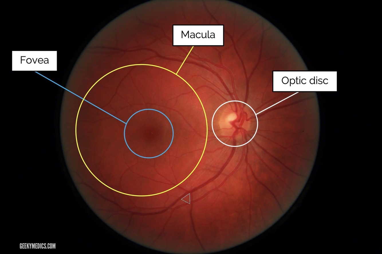
What does the optic disc represent and how does it link to the blind spot
Output from the eye is via the Retinal Ganglion Cells, whose axons form the optic nerve (II).
No photoreceptors present, photons landing on it aren’t detectable- natural blindspot
- What are the 2 main classes of photreceptor?
- What does each facilitate?
- What part of the eye has a high conc of cones?
- How many different kinds of cone are there?
- Cones and rods
- Cones- vision in high illumination and colour vision Rods- vision in low illumination
- Cones are highest in the fovea, where visual acuity is greatest
- 3 types of cone, each responding optimally to different wavelengths of light (red, green and blue)
cataract
loss of lens clarity due to breakdown of proteins in the lens
Affects almost everyone >70
associated with corticosteroid use and diabetes
Retinal detachment
- Neurosensory layer (photoreceptors + ganglion cells) detaches from retinal pigment epithelium as non replenishable vitreous fluid degenerates
- Usually a tear (ageing, trauma) allows liquid in between
- May see ‘floaters’ and ‘flashing lights’ due to vitreous degeneration
- If near macula, surgery is required
Glaucoma
- various disorders, usually with raised intraocular pressure
- Aqueous humour overproduction or impaired drainage can increase pressure on vitreous humour, which presses on retinal nerves and blood vessels
- Can cause pain, loss of vision etc.
