Upper GI Surgery Flashcards

(200 cards)

1
Q

where are the 3 locations of narrowing of the oesophagus?

A
  1. level of cricoid cartilage (C6)
  2. posterior to left main bronchus and aortic arch (T4)
  3. lower oesophageal sphincter (T11)
2
Q

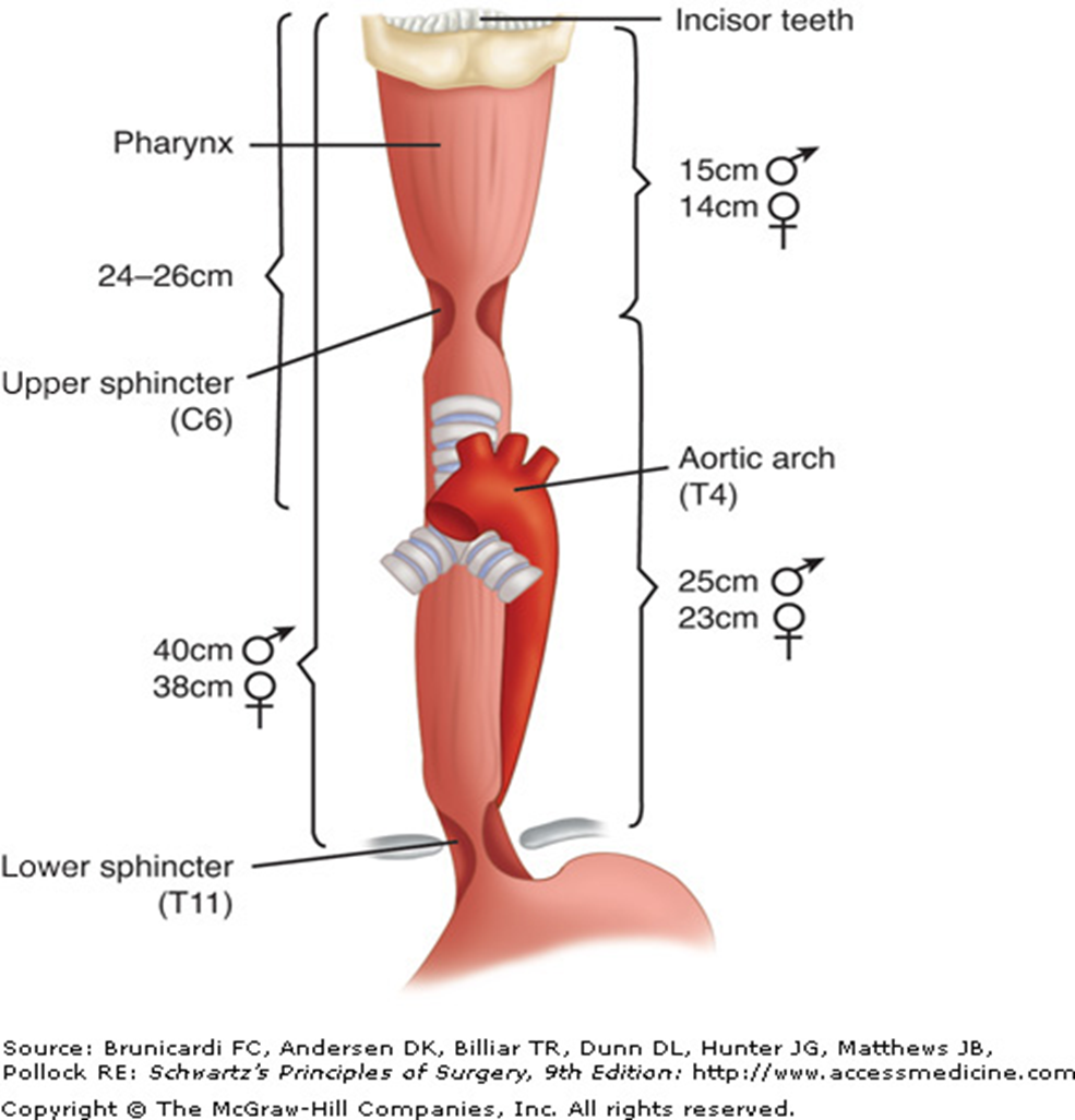
what is the z line in the oesophagus?

A

transition from squamous to gastric columnar epithelium

3
Q

Ix for dysphagia

A

Upper GI endoscopy

Barium Swallow

Manometry (assess LOS fn)

4
Q

projectile vomiting

child hungry after vomiting

failure to gain weight

dehydration/ constipation

Dx?

A

Pyloric stenosis

5
Q

diagnosis of pyloric stenosis?

A

test feed- visible peristalsis

Abdo USS to visualize the hypertrophied pyloric sphincter

abdo xray - may reveal dilated stomach w minimum gas in bowel.

barium meal - reveals the pyloric obstruction w characteristic shouldering of the pyloric antrum

6
Q

what is the metabolic abnormality with pyloric stenosis?

A

hypochloraemic hypokalaemic met alkalosis

7
Q

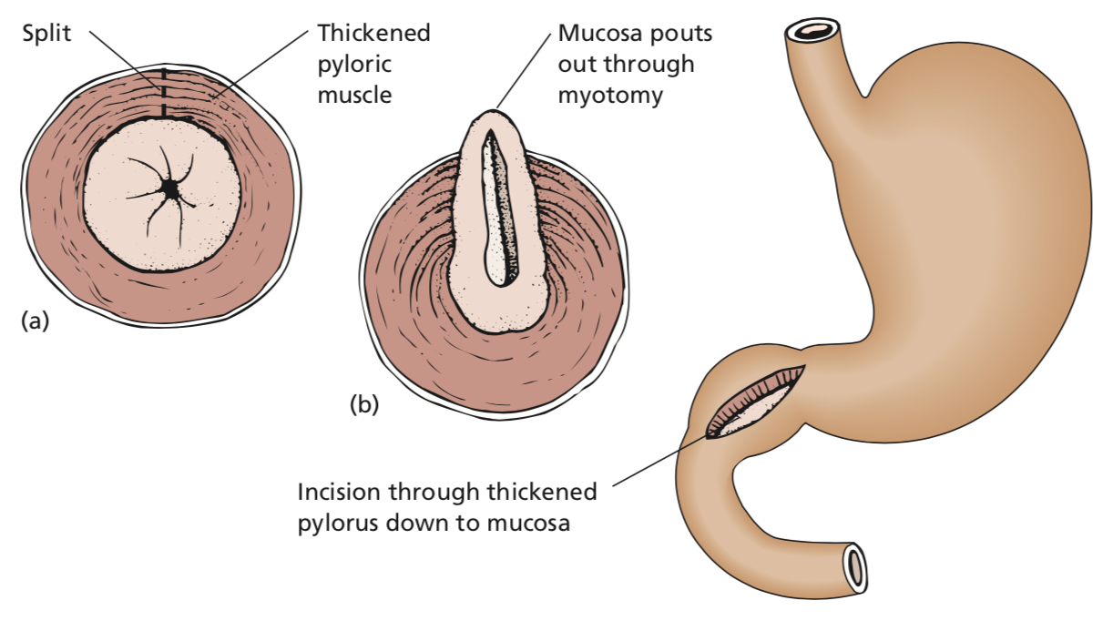
what is Ramstedt’s pyloromyotomy?

A

for pyloric stenosis

  • a longitudinal incision is made through the hypertrophied muscle of the pylorus down to mucosa and the cut edges are separated.

commonly done laparoscopically.

infant is given glucose water 3h after op and followed by 3hrly milk feeds.

8
Q

what medications predispose a pt to peptic ulceration and perforation?

A

steroids

NSAIDs (aspirin, indometacin, ibuprofen etc)

9
Q

examination findings of a pt with perforated peptic ulcer

A

patient in severe pain

cold and sweating w rapid, shallow respirations

abdomen rigid and silent

pneumoperitoneum -> may lead to diminished liver dullness

10
Q

presentation of peptic ulcer perforation on examination

in a delayed (>12h onset) presentation

A

if delayed (>12h) presentation

features of generalized peritonitis with paralytic ileus

distended abdomen

vomiting

pt extremely toxic and in oligaemic shock

11
Q

Ix to order for suspected perforated peptic ulcer

A

CXR: erect.

  • free gas below the diaphragm

CT abdo

  • to detect free intraperitoneal gas and can exclude common differentials e.g. pancreatitis
12
Q

DDx of perforated peptic ulcer

A

perforated appendicitis

acute cholecystitis

acute pancreatitis

myocardial infarction

13
Q

tx of perforated peptic ulcer

A

NG tube: to empty stomach and decrease further leakage

Pain relief: opiates

IV fluid resus

ABx to contend w peritoneal infection

IV H2 blocker or PPI

Immediate operative repair of the perforation

14
Q

what does surgery of perforated peptic ulcer involve?

A

suturing of omental plug to seal the perforation

+

lavage of the peritoneal cavity

+

biopsy of *gastric ulcer to exclude malignancy

15
Q

Postoperative tx for perforated Peptic ulcer

A

H pylori eradication

omeprazole, amoxicillin, clari

16
Q

profuse vomiting, non-bilious

may contain food particles

weight loss, constipation, weakness due to electrolyte disturbance

A

pyloric stenosis

17
Q

Examination findings of pyloric stenosis

A

visible peristalsis seen, from L-R of upper abdomen

grossly dilated, hypertrophied stomach, full of stale food and fluid, can be palpated

gastric splash (succussion splash) can be elicited by shaking pt’s abdomen several hrs after a meal

18
Q

Ix of pyloric stenosis

A

Gastroscopy - following decompression of stomach w NG tube

CT scan

ABG and electrolytes-> hypochloraemic, hypokalaemic alkalosis and uraemia

19
Q

metabolic disturbances of pyloric stenosis

A

dehydrated, Hct raised

serum Cl, Na, K low

plasma bicarb and urea raised

alkalosis

20
Q

DDx of pyloric stenosis

A

ca of pylorus

Other causes of pyloric obstruction are unusual

in the adult:

  • scarring associated with a benign gastric ulcer near the pylorus;
  • carcinoma of the head of the pancreas infiltrating the duodenum and pylorus;
  • chronic pancreatitis;
  • invasion of the pylorus by malignant nodes.
21
Q

differentiating between benign ulcer-> pyloric stenosis vs carcinoma of the pylorus

A
  • Length of history: a history of several years of characteristic peptic ulcer pain is in favour of benign ulcer. Cancer usually has a history of only months and indeed may be painless.
  • Gross dilatation of the stomach favours a benign lesion, as it may take several years for this to develop.
  • The presence of a mass at the pylorus indicates malignant disease, although, rarely, a palpable inflammatory mass in association with a large duodenal ulcer can be detected.
22
Q

tx of pyloric stenosis

A

preop:

IV saline + K to correct dehydration/ electrolyte depletion

daily gastric lavage to remove debris from stomach

Vitamin C

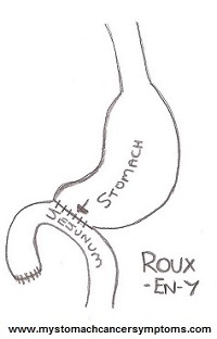
surgical correction:

usually an antrectomy w a Roux-en-Y gastroenterostomy

23
Q

oesophageal causes of GI haemorrhage

A
  • reflux oesophagitis (associated with hiatus
    hernia) ;
  • oesophageal varices (associated with portal

hypertension)

  • peptic ulcer;
  • tumours (benign and malignant).
24
Q

stomach causes of GI haemorrhage

A

gastric ulcer

acute erosions (assoc w aspirin, other NSAIDs, corticosteroids)

gastritis

Mallory-Weiss tear

vascular malformation (e.g. Dieulafoy lesion)

tumours (benign and malignant)

25
Duodenal Causes of GI haemorrhage
duodenitis duodenal ulcer erosion of the duodenum by a pancreatic tumour aortoduodenal fistula, in patients with previous aortic graft
26
small intestine causes of GI haemorrhage
tumours meckel's diverticulum angiodysplasia aortoenteric fistula
27
large bowel causes of GI haemorrhage
Tumours (benign and malignant, commonly adenoCas) diverticulitis Angiodysplasia colitis (UC, Ischaemic colitis and infective colitis)
28
General causes of bleeding?
anticoagulant therapy haemophilia leukaemia thrombocytopenia
29
Ix with GI bleeding
Hb - useful as baseline Serum Urea - raised following upper GI bleed (can distinguish between upper and lower GI bleed) Coagulation screen and Pl count LFTs Cross Match, Group and Save Upper GI fibreoptic endoscopy - will identify the exact site of bleeding in upper GI haemorrhage
30
Tx of Upper GI Bleed
A to E approach-\> stabilize the patient pain relief if shock present: fluid resus/ blood transfusion central venous catheter to measure central venous pressure and assist in fluid replacement urinary catheter to monitor UO treat underlying cause
31
tx of actively bleeding peptic ulcers
treated endoscopically by injection of adrenaline into and around the vessels in the ulcer bed. dual modality tx superior to injection alone: e.g. + coagulation w a heater probe or placement of a clip directly onto the bleeding vessel
32
GI stromal tumours mutation in what gene?
c-kit gene coding for c-kit protein (CD117) on the cell
33
Tx of GIST (GI stromal tumour)
surgical excision chemotx - with c-kit tyrosine kinase inhibitor (imatinib mesilate) -\> can be given to shrin tumour pre-surgery or given post-op to treat metastases or when complete resection was not possible
34
Risk factors for Stomach Ca | (Predisposing conditions)
pernicious anaemia and atrophic gastritis previous gastric resection chronic peptic ulcer
35
Risk factors of stomach ca envt factors
H pylori infection Low SES Smoking Nationality: Japan(?)
36
genetic risk factor for stomach cancer?
HNPCC
37
lymph drainage of carcinoma from cardiac end of stomach?
mediastinal nodes supraclavicular nodes of Virchow
38
lymph drainage from carcinoma of the pylorus?
subpyloric and hepatic nodes
39
type of obstruction when blood supply of the involved segment of intestine is cut off
strangulating obstruction e.g. with strangulated hernia, volvulus, intussusception
40
symptoms of intestinal obstruction
colicky abdo pain distension absolute constipation vomiting
41
Examination of suspected intestinal obstruction should include?
hernias scars - suggests previous operation and adhesions/ band as a cause
42
features of strangulating obstruction vs simple obstruction
change in character of pain from colicky to continuous tachycardia pyrexia peritonism bowel sounds absent/ reduced raised WCC raised CRP
43
causes of intestinal obstruction | (in the lumen)
faecal impaction gallstone ileus food bolus parasites intussusception
44
causes of intestinal obstruction (in the wall)
congenital atresia Crohn's disease tumours diverticulitis
45
causes of intestinal obstruction (outside the wall)
volvulus strangulated hernia obstruction due to adhesions or bands
46
differentiation between small and large bowel obstruction on Abdo Xray?
central vs peripheral position of distented/dilated loops striations that pass completely across width of loop (small bowel) vs haustra of the taenia coli which do not extend across the whole width (large bowel)
47
Mx in acute intestinal obstruction
NG tube for gastric aspiration & to decompress bowel IV fluids + K if K is low ABx if intestinal strangulation is likely
48
signs of non-viability in affected bowel of intestinal obstruction
1. loss of peristalsis 2. loss of normal sheen 3. colour (greenish or black bowel is non-viable; purple bowel may still recover) 4. loss of arterial pulsation in the supplying mesentery
49
closed loop obstruction where is it most commonly seen?
left sided colonic obstruction in the presence of a competent ileocaecal valve. -\> the caecum, the most distensible part of the large bowel, blows up like a balloon and perforation of the caecum, with faecal peritonitis, may occur
50
adhesions from previous surgeries usually lead to what kind of bowel obstruction?
small bowel (about 75%)
51
what are the three main arteries supplying the gut
coeliac super mesenteric inferior mesenteric
52
acute colicky abdo pain, rectal bleeding and shock in an elderly patient who has AF
mesenteric vascular occlusion e.g. embolus, mesenteric arterial/ venous thrombosis, non-occlusive infarction of the intestine
53
neonatal intestinal obstruction ddx?
intestinal atresia volvulus neonatorum meconium ileus necrotizing enterocolitis hirschsprung's disease anorectal atresia
54
Meconium ileus treatment?
instillation of Gastrografin per rectum under X ray control -\> radio opaque and hyperosmolar and contains an emulsifying agent, which facilitates evacuation of the meconium 2nd line: surgery- enterotomy and removal of the inspissated meconium by lavage
55
tx of necrotizing enterocolitis
resus NG tube for gastric aspiration IV fluids TPN and broad spectrum abx
56
Hirschsprungs Disease what pathology?
absence of ganglion cells in the submucosal plexus of Auerbach and intermyenteric plexus of Meissner in the rectum, and sometimes extends into the lower colon.
57
tx of Hirschsprung's
surgery aganglionic segment is resected and an abdominoperineal pull-through anastomosis between normal colon and the anal canal.
58
commonest area of intussusception
ileocolic - through the ileocaecal valve
59
meckel's diverticulum is the remnant of which duct of the embryo?
vitellointestinal duct
60
Crohn's disease - which parts of the bowel is affected
anywhere from mouth to anus
61
Crohns Disease Risk factors
genetic - NOD2 environmental - smoking
62
Macroscopic appearance of affected Crohns' segment
cobblestone appearance of mucous appearance - bowel is bright red and swollen, mucosal ulceration and intervening oedema leads to cobblestone appearance intestine wall is thickened skip lesions fistulae
63
common symptoms of crohns disease
abdo pain diarrhoea palpable mass in the RIF if acute-\> may present like appendicitis intestinal obstruction due to stenotic segments from inflammatory exacerbations-\> fibrosis fistula formation malabsorption with steatorrhoea and multiple vitamin deficiencies perianal disease (fissures to fistulae)
64
what are helful indices of disease activity in Crohns disease?
CRP acute phase proteins
65
complications of Crohns Disease
skin: pyoderma gangrenosum, erythema nodosum anterior uveitis sacroiliitis primary sclerosing cholangitis Renal/ Biliary Calculi
66
medical mx of crohn's disease
elemental diet / nutritional support acute episodes treated with steroids and immunosuppressants such as azathioprine infliximab (anti-TNFa) monoclonal antibody to TNF-a Sulfasalazine/ mesalazine
67
when is surgery indicated for crohns disease?
surgery indicated for severe/ recurrent obstructive symptoms, and for fistulae into bladder/ skin
68
what do carcinoid tumours secrete
5-HT (serotonin)
69
symptoms of carcinoid syndrome
**flushing** with attacks of cyanosis, often precipitated by stress or ingestion of food/ alcohol **diarrhoea** **bronchospasm** **abdo pain**
70
Ix of Carcinoid Syndrome
5-HIAA urinary concentration (will be raised) Chromogranin A serum concentration raised CT liver to seek metastases Radiolabelled octreotide scintigraphy: for screening for tumour. the octreotide binds to somatostatin receptors often expressed on the tumour
71
what medication can be used to control symptoms in carcinoid syndrome?
octreotide, a somatostatin analogue that inhibits 5-HT release
72
diagnostic sequence of acute appendicitis?
colicky central abdo pain followed by vomiting followed by movement of the pain to the RIF + anorexia/ constipation usually
73
symptoms with perforation of the appendix?
temporary remission/ cessation of the pain as tension in the distended organ is relieved followed by more severe and more generalized pain w profuse vomiting as general peritonitis develops
74
ix of suspected appendicitis
WCC: mild raised neutrophils CT abdo US RIF may be diagnostic
75
when is immediate appendicectomy not indicated?
patient moribund w advanced peritonitis -\> 1st aggressive resus w fluids, abx, analgesia attack already resolved -\> can be done electively appendix mass has formed without evidence of general peritonitis -\> immediate sx may be difficult and dangerous w a risk of damage to adjacent bowel loops
76
medical tx of acute appendicitis? apart from appendicectomy
antibiotic prophylaxis - metronidazole and gentamicin drain inserted after appendicectomy
77
what are the muscle types in each 1/3 of the oesophagus?
top 1/3: striated middle: mixed bottom 1/3: smooth muscle
78
how long is the oesophagus?
25 cm long muscular tube | (40 cm from lips to GOJ)
79
where does the oesophagus start?
at the level of the cricoid cartilage C6
80
what is achalasia?
LOS fails to relax during swallowing due to degeneration of myenteric plexus (Auerbach's) -\> decreased peristalsis
81
cause of achalasia?
most commonly idiopathic coule be secondary to Chagas disease (trypanosoma cruzii)
82
dysphagia of liquids AND solids (intermittent) regurgitation esp at night substernal cramps/ pressure relieved by drinking through pain with cold water weight loss dx?
achalasia
83
features of achalasia?
dysphagia of liquids AND solids (intermittent) regurgitation esp at night substernal cramps/ pressure relieved by drinking through pain with cold water weight loss
84
complications of achalasia?
oesophageal SCC in 3-5%
85
Gold standard Ix of Achalasia?
**Oesophageal Manometry** abnormal Lower Oesophageal sphincter pressure during swallow
86
Ix of achalasia?
Manometry: showing abnormal LOS pressure during swallow Barium swallow: Birds beak sign (dilated tapering oesophagus) OGD: exclude malignancy CXR: widened mediastinum, double RH border
87
what sign points to achalasia on barium swallow?
Birds Beak sign
88
Mx of achalasia?
**conservative:** nothing if asymptomatic **medical:** CCB, nitrates (decrease LES pressure) **interventional:** botox injection to LES, endoscopic balloon dilatation **surgical:** heller's cardiomyotomy
89
what is the surgical tx of achalasia?
Heller's cardiomyotomy | (muscles of LES are cut)
90
what is a pharyngeal pouch?
outpouching at the top of the oesophagus between crico and thyropharyngeal components of the inf pharyngeal constrictor at the area of weakness - Killian's dehiscence defect usually occurs posteriorly but swelling usually bulges to L side of neck
91
what is the area of weakness where a pharyngeal pouch usually develops from?
Killian's dehiscence
92
feeling of a lump in your throat, difficulty swallowing (dysphagia), bringing up food after a meal and bad breath. dx?
pharyngeal pouch
93
pharyngeal pouch presentation?
dysphagia regurgitation halitosis feeling of lump in throat gurgling sounds
94
tx of pharyngeal pouch
excision endoscopic stapling
95
Barium swallow showing corkscrew oesophagus?
diffuse oesophageal spasm
96
what is diffuse oesophageal spasm?
condition characterized by uncoordinated contractions of the oesophagus difficulty swallowing (dysphagia) +/- intermittent severe chest pain
97
Ix of diffuse oesophageal spasm?
barium swallow - shows corkscrew oesophagus
98
what is nutcracker oesophagus? or hypertensive peristalsis
normal peristalsis but w raised contraction pressure causes chest pain + dysphagia to liquids and solids (intermittent) oesophageal manometry used to diagnose when pressures \> 180mmHg (like a mechanical nutcracker)
99
what is Plummer-Vinson syndrome?
IDA, dysphagia, oesophageal webs, glossitis, cheilosis pre-malignant: 20% risk of SCC of oesophagus/ pharynx Treatment with iron supplementation and mechanical widening of the esophagus generally provides an excellent outcome
100
IDA, dysphagia, oesophageal webs, glossitis, cheilosis Dx?
Plummer vinson syndrome
101
causes of oesophageal rupture?
Iatrogenic (85-90%): endoscopy, biopsy, dilatation Violent emesis: Boerhaave's syndrome Carcinoma caustic injestion trauma: surgical emphysema +/- pneumothorax
102
features of oesophageal rupture
odynophagia (painful swallowing) mediastinitis: tachypnoea, dyspnoea, fever, shock surgical emphysema (aka subcut emphysema- air in subcut tissues)
103
Mx of oesophageal rupture
resus PPI, NGT, Abx
104
Causes of Squamous Cell Carcinoma of the oesophagus?
1. Toxins: Smoking/ Alcohol 2. Diet: processed/ red meats 3. Obesity, achalasia, Plummer-Vinson syndrome
105
causes of adenocarcinoma of the oesophagus?
GORD-\> Barrett's -\> dysplasia -\> Ca
106
what is the most common type of oesophageal ca in UK?
65% adenoca 35% SCC (commonest worldwide)
107
what regions of the oesophagus are more assoc w adenoca/ and which with SCC?
AdenoCa: lower 3rd SCC: upper and middle 3rds
108
features of Oesophageal Ca?
1. progressive dysphagia and odynophagia: solids then liquids 2. Upper GI signs: voice hoarseness (ca invading recurrent laryngeal n), cough +/- aspiration pneumonia, retrosternal chest pain 3. Red flags: weight loss, anaemia, haematemesis/ melaena, lymphadenopathy
109
Ix of oesophageal ca?
Bloods: FBC (IDA), LFTs (mets), bone profile (mets) Imaging: OGD endoscopy + biopsy Ba swallow: not often used, apple core stricture Staging: TNM e.g. CT
110
gold standard ix for oesophageal ca?
OGD endoscopy + biopsy
111
curative Mx of oesophageal Ca? (25%)
MDT: surgeon, gastroenterologist, specialist nurse, onco, palliative care **surgical:** oesophagectomy **medical**: neoadjuvant chemo to downsize tumour before surgery e.g. cisplatin + 5FU
112
what are the 3 surgical approaches to oesophagectomy?
Ivor-Lewis (2 incisions) McKeown (3 incisions) Trans-hiatal MIT
113
what is the Ivor-Lewis oesophagectomy?
esophageal tumor removed through abdominal incision + right thoracotomy the esophagogastric anastomosis is located in the upper chest.
114
What is the Mckeown oesophagectomy?
like the ivor-lewis, but used for cancers that are higher up in the oesophagus laparotomy + right thoracotomy + L neck incision
115
abdominal incision + right thoracotomy esophagogastric anastomosis located in the upper chest.
Ivor-Lewis oesophagectomy
116
midline abdominal incision + right thoracotomy + left neck incision?
McKeown's oesophagectomy
117
what is transhiatal oesophagectomy?
upper midline incision + oblique incision in neck along lower L border of Sternocleidomastoid patient's diseased esophagus and proximal (top part) stomach is removed.
118
midline abdominal incision + neck incision ?
transhiatal oesophagectomy
119
Palliative Mx of Oesophageal Ca? (75%)
1. palliative care: macmillan nurses 2. Analgesia, palliative chemo 3. intervention: Stenting, Palliative radiotx
120
what is GORD?
LOS dysfunction -\> reflux of gastric contents -\> oesophagitis
121
Risk factors of GORD?
1. anatomical disruption of the gastro-oesophageal junction - hiatus hernia 2. Hypotensive LES/ transient LES relaxations e. g. coffee, alcohol, smoking, obesity, chocolate 3. Delayed oesophageal acid clearance - dysmotility, cigarette smoking (decreased saliva -\> decreased neutralisation -\> increased acid), severe oesophagitis 4. Iatrogenic: hellers myotomy, drugs e.g. nitrates, CCB
122
Features of GORD?
Retrosternal burning - related to meals, worse lying down, relieved by antacids Regurgitation/ acid brash Dysphagia, odynophagia, cough, hoarse voice, asthma \*all symptoms are worse at night
123
complications of GORD?
1. oesophagitis 2. Barrett's oesophagus (metaplasia) 3. Oesophageal AdenoCa 4. strictures causing dysphagia
124
Ix of GORD?
1. Conservative /mx trial -\> diagnosis if responds 2. Imaging: OGD endoscopy +/- biopsy if: **\>55 yo**, persistent systems, anaemia, weight loss, anorexia, recent onset progressive symptoms, melaena, swallowing difficulty 3. Special ix: 24 hr pH testing +/- manometry
125
what findings indicated GORD on 24 hr pH testing?
pH \<4 for \> 4h
126
Conservative Mx of GORD?
Diet: avoid spicy foods, coffee, alcohol, smaller meals, avoid drinking/ eating close to bedtime Weight loss Sleep on side/ bed elevated Stop drugs that may be causing it e.g. NSAIDs, nitrates, CCB, anti-AChM
127
Medical Mx of GORD?
Antacids e.g. gaviscon Full-dose PPI for 1-2 months e.g. Omeprazole 20mg OD / Lansoprazole 30mg if no response -\> double dose PPI BD if no response -\> add H2RA e.g. Ranitidine 300mg nocte
128
surgical mx of GORD?
Nissen's fundoplication - usually laparoscopic - wrap gastric fundus around lower oesophagus - close any diaphragmatic hiatus
129
indications for nissen's fundoplication - surigcal mx of GORD?
all 3: severe symptoms refractory to medical tx confirmed reflux on pH monitoring
130
Complications of Nissen's fundoplication?
dysphagia if wrap too tight gas bloat syndrome: inability to belch/ vomit
131
what are the different types of hiatus hernias?
**Type I: Sliding (80%)** - GOJ in chest -\> GORD common **Type II: rolling** - stomach herniated (paraoesophageal) but GOJ in normal position can -\> strangulation **III: mix of I and II** **IV:** other organs in addition to the stomach (colon, small intestine, spleen) also herniated into chest
132
Ix of hiatus hernia?
CXR: gas bubble and fluid level in chest Ba Swallow: diagnostic OGD: assess for oesophagitis 24 pH + manometry: exclude dysmotility or achalasia
133
what is the diagnostic ix of hiatus hernia?
barium swallow
134
Mx of Hiatus hernia?
Lose weight treat reflux surgery if intractable symptoms despite medical mx -\> should repair rolling hernia even if asymptomatic as it may strangulate
135
what is an ulcer?
a break in the epithelium
136
epigastric pain relieved by eating, worse at nights/ before meals
duodenal ulcer
137
epigastric pain worse on eating -\> weight loss relieved by antacids
gastric ulcer
138
what type of ulcer is most common with peptic ulcer disease?
duodenal 4x more common than gastric
139
Risk factors of Peptic Ulcer disease?
1. Infection: H Pylori 2. Toxins: Smoking, Alcohol 3. Drugs: NSAIDs, steroids 4. Stress: Cushings ulcers, Curling's ulcers Zollinger- Ellison
140
where are gastric ulcers usually found?
lesser curvature of gastric antrum
141
where are duodenal ulcers usually found?
1st part of duodenum
142
Complications of Peptic Ulcer Disease?
**Haemorrhage**: - IDA - haematemesis, melaena **perforation:** peritonitis **Gastric outflow obstruction**: vomiting, colic, distension **malignancy:** gastric ulcers-\> increased risk of gastric ca
143
Features of peptic ulcer disease
1. epigastric pain 2. fullness/ bloating/ belching 3. n/v
144
Ix of peptic ulcer disease? if \>55 +/- RFs +/- no response to Tx
OGD +/- biopsy
145
Ix of Peptic Ulcer disease? \< 55 and No alarm symptoms
h Pylori breath test Stool Antigen test ?gastrin levels if zollinger ellison suspected
146
mx of peptic ulcer disease?
1. stop smoking, decrease alcohol, avoid spicy foods/ drugs e.g. NSAIDS, steroids 2. Medical tx: Triple therapy - Omeprazole + Clarithro + Metro Antacids e.g. gaviscon Acid suppression- PPIs (lansoprazole) or H2RA: ranitidine 3. Surgical mx
147
what is the medical tx of peptic ulcer disease?
Triple therapy - Omeprazole + Clarithro + Metro Antacids e.g. gaviscon Acid suppression- PPIs (lansoprazole) or H2RA: ranitidine
148
what increases acid production in the stomach?
acid secretion is stimulated by gastrin (from antral G cells) and vagus nerve
149
surgical mx of Peptic ulcer disease?
**Vagotomy** **+/-** pyloroplasty +/- antrectomy **Subtotal Gastrectomy w Roux-en-Y:** occasionally performed for Zollinger-Ellison
150
types of vagotomy?
Selective vagotomy or Truncal vagotomy
151
difference between truncal and selective vagotomy?
Selective vagotomy: - vagus nerve only denervated where it supplies lower oesophagus and stomach - Nerves of Laterjet (supplying pylorus) left intact Truncal vagotomy: - decreases acid secretion directly and via decreasing gastrin - prevents pyloric sphincter relaxation (Nerves of Laterjet affected) - must be combined w **pyloroplasty** (widening of the pylorus) or gastroenterostomy
152
what is Antrectomy with Vagotomy?
Truncal vagotomy + Distal half of stomach (antrum) removed -\> gastrin producing cells removed + Billroth I anastamosis: directly to duodenum Billroth II: to small bowel loop w duodenum oversewn
153
Metabolic complications of surgery for PUD?
Weight loss: malabsorption Vitamin deficiency: less parietal cells -\> B12 deficiency, bypassing proximal small bowel -\> Fe + Folate deficiency Blind loop syndrome: overgrowth of bacteria in duodenal stump -\> malabsorption, darrhoea Dumping syndrome: abdominal discomfort, and sometimes abnormally rapid bowel evacuation after meal -\> osmotic hypovolaemia, reactive hypoglycaemia
154
Physical complications of Peptic ulcer disease surgery?
Ca: increase risk gastric ca stump leakage stricture abdominal fullness reflux or bilious vomiting (improves w time)
155
risk factors for Upper GI bleed?
previous bleeds known ulcers oesophageal varices malignancy
156
Signs O/E with upper GI bleed?
Melaena Shock Signs of Chronic liver disease
157
causes of upper GI bleeding?
most commonly peptic ulcer disease (DU): 40% acute erosions/ gastritis: 20% Mallory-Weiss tear: 10% Varices: 5% Oesophagitis: 5% Ca stomach/ oesophagus: \<3%
158
Scoring systems for Upper GI bleed?
Rockall score Glasgow-Blatchford bleeding score
159
what is the Glasgow-Blatchford score?
screening tool to assess the likelihood that a patient with an acute upper GI bleeding will need to have medical intervention such as a blood transfusion or endoscopic intervention. looks at: Hb levels Urea Levels systolic blood pressure tachycardia, melaena, syncope, heart failure, liver failure
160
What is the Rockall score?
risk stratification of patients with upper GI bleed prediction of re-bleeding and mortality **ABCDE** Age Blood pressure fall (Shock) Co-morbidity (e.g. heart, liver, renal failure) Diagnosis Evidence of bleeding: active bleeding, visible vessel, adherent clot D/E is seen on OGD. score \>6 indication for surgery, \>8 high risk of mortality
161
what are oesophageal varices?
portal HTN -\> dilated veins at sites of porto-systemic anastomosis: L gastric and inferior oesophageal veins 30-50% risk of bleeding overall mortality 25%
162
causes of portal HTN
pre-hepatic: portal vein thrombosis hepatic: cirrhosis (most common in UK), schistosomiasis (commonest worldwide) Post-hepatic: Budd-Chiari, Right HF, constrictive pericarditis
163
prevention of Upper GI bleed?
primary prevention: BB, repeat endoscopic banding Secondary: BB, repeat banding, TIPSS (transjugular intrahepatic porto-systemic shunt)
164
What is TIPSS? trans-jugular intrahepatic porto-systemic shunt
artificial channel between inflow portal vein and outflow hepatic vein to decrease portal pressure used prophylactically or acutely if endoscopic therapy fails to control variceal bleeding
165
Mx of acute upper GI bleed?
A to E approach Fluid resuscitation, protect airway, Bloods (Clotting, FBC, U+E, Group and save, x-match) O- blood until cross matched Urgent OGD
166
mx of variceal bleed?
IV terlipressin prophylactic abx e.g. ciprofloxacin oesophageal varices: 1st line band ligation gastric: endoscopic injection of N-butyl-2-cyanoacrylate
167
1st line endoscopic mx of oesophageal varices?
endoscopic banding 2nd line: TIPSS other: balloon-tamponade w Sengstaken-Blakemore tube (only used if exsanguinating haemorrhage/ failure of endoscopic therapy)
168
Mx of non-variceal upper GI bleeding?
urgent endoscopy: * a mechanical method (for example, clips) with or without adrenaline * thermal coagulation with adrenaline * fibrin or thrombin with adrenaline + PPIs
169
Indications of surgery for upper GI bleed?
re-bleeding bleeding despite 6 u transfusion uncontrollable bleeding at endoscopy initial Rockall score 3 or more, or final \>6 open stomach, find bleeder and underrun vessel
170
Mx after endoscopic tx of upper GI bleed?
Omeprazole (decreases re bleeding) H Pylori testing and eradication stop offending drugs e.g. NSAIDs, steroids
171
sudden onset severe pain, beginning in epigastrium then generalised vomiting peritonitis hx of pain after food/ relieved by eating
perforated peptic ulcer most commonly duodenal ulcer
172
perforation of peptic ulcer can create which signs?
ant perforation: air under diaphragm posterior perf: can erode into gastroduodenal artery -\> bleed
173
Ix of suspected perforated peptic ulcer?
Bloods Urine dip Imaging: Erect CXR - air under diaphragm AXR: riglers sign
174
Mx of perforated peptic ulcer
resuscitation: fluid resus, NBM, analgesia, abx (cef and met), NGT conservative: may be considered if pt not peritonitic careful monitoring, fluids, abx omentum may seal perforation spontaneously preventing operation in 50% Surgical: laparotomy repair - send specimen for histology to exclude ca
175
Mx of perforated peptic ulcer after surgery
H Pylori eradication - triple therapy
176
causes of gastric outlet obstruction?
gastric ca late complication of peptic ulcer disease -\> fibrotic stricturing
177
hx of bloating, early satiety and nausea copious projectile, non-bilious vomiting a few hours after meals contains stale food epigastric distension + succussion splash Dx?
Gastric outlet obstruction
178
Ix of gastric outlet obstruction?
ABG: hypochloraemic hypokalaemic metabolic alkalosis AXR: dilated gastric air bubble collapsed distal bowel OGD Contrast meal
179
tx of gastric outflow obstruction?
correct metabolic abnormality: 0.9% Normal saline + KCl Benign: endoscopic balloon dilatation, pyloroplasty or gastroenterostomy Malignant: stenting, resection
180
projectile vomiting minutes after feeding RUQ mass visible peristalsis 6-8wk old infant Dx?
Pyloric stenosis
181
Diagnosis of Pyloric stenosis?
test feed: palpate mass + see peristalsis HypoCl HypoK met alkalosis US to confirm pylorus hypertrophy
182
Mx of pyloric stenosis?
Resus, correct metabolic abnormality NG tube - prevent aspiration ramstedt pyloromyotomy
183
complications of gastric cancer?
perforation upper GI bleed gastric outlet obstruction
184
ix of gastric cancer?
**Bloods** FBC: anaemia LFTs and clotting (mets) **Imaging:** CXR: mets USS: liver mets gastroscopy + biopsy Ba meal **Staging**: CT/MRI diagnostic laparoscopy
185
Mx of gastric cancer?
Palliative care: analgesia, Chemo, PPI, pyloric stenting Curative surgery: resection of tumour partial or total gastrectomy w roux-en-y to prevent bile reflux
186
Risk factors of gastric cancer?
atrophic gastritis h pylori diet: high in nitrates (carcinogenic nitrosamines) smoking
187
pathology of gastric ca?
mainly adenocarcinomas usually located on gastric antrum H Pylori -\> MALToma
188
what is zollinger-ellison syndrome?
gastrin-secreting tumour (gastrinoma) most commonly in small intestine/ pancreas ↑ Gastrin → ↑HCL→ PUD + chronic diarrhoea (diarrhoea due to inactivation of pancreatic enzymes) 25% assoc w MEN1
189
refractory Peptic ulcer disease chronic diarrhoea/ steatorrhoea abdominal pain and dyspepsia dx?
zollinger-ellison syndrome
190
Ix of zollinger-ellison syndrome
Gastrin levels: high pH\<2 in stomach MRI/CT scan Somatostatin receptor scintigraphy- used to find carcinoid, pancreatic neuroendocrine tumors
191
Mx of Zollinger-Ellison Syndrome?
High dose PPI (omeprazole) Surgery: tumour resection may do subtotal gastrectomy w Roux-en-Y
192
what is gastrointestinal stromal tumour? (GIST)
commonest mesenchymal tumour of the GIT \>50% occus in the stomach arise in the smooth muscle pacemaker interstitial cell of Cajal
193
mx of Gastrointestinal stromal tumours?
medical: for unresectable, recurrent or metastatic disease **imatinib**: KIT selective tyrosine kinase inhibitor Surgical resection
194
what is a carcinoid tumour?
neuroendocrine tumour of enterochromaffin cell origin may secrete multiple hormones e.g. serotonin
195
risk factors for gastric carcinoids?
atrophic gastritis -\> low acid production -\> increased gastrin production -\> ECL hyperplasia -\> carcinoid tumour Gastrinomas may also -\> carcinoid
196
Most common cause of Gastric lymphoma (MALToma)?
chronic H pylori
197
Benefits of bariatric surgery?
sustained weight loss symptom improvement: sleep apnoea mobility HTN DM
198
Indications of bariatric surgery?
All: * BMI ≥40 or ≥35 w significant co-morbidities * failure of medical mx to achieve and maintain clinically beneficial weight loss for 6 months * fit for surgery and anaesthesia * integrated program providing guidance on diet, physical acitivity, psychosocial concerns, and lifelong medical monitoring * well-informed and motivated pt if BMI\>50, surgery is 1st line mx
199
what is laparoscopic gastric banding?
inflatable silicone band around proximal stomach -\> small pre-stomach pouch limits food intake, slows digestion at 1 yr: 46% mean excess weight loss
200
what is roux-en-y gastric bypass surgery?
oesophagojejunostomy allows bypass of stomach, duodenum and proximal jejunum alters secretion of hormones influencing glucose regulation and perception of hunger/ satiety greater weight loss and lower reoperation rates