which process causes formation of the gut tube, from which 2 embryonic tissues
lateral folding of embryo causes formation of gut tube from:
- splanchnic mesoderm (from lateral plate layer) - forms visceral layer of peritoneum
- endoderm (from yolk sac) - forms gut epithelium
which structure is initially continuous with midgut
yolk sac, via vitelline duct (regresses wk 5-8) within umbilical cord
describe 3 abnormalities that can arise due to persistence of vitelline duct
- VITELLINE CYST: vitelline duct forms fibrous strands, causing middle portion to form an enterocystoma
- VITELLINE FISTULA: direct communication between umbilicus and GI tract (causes discharge of meconium from umbilicus)
- MECKEL’S DIVERTICULUM: outpocketing of ileum in remnant of vitelline duct. Usually asymptomatic but may contain pancreatic tissue or gastric mucosa, causing ulceration, bleeding or perforation.
by which membranes is the gut tube initially sealed
- foregut temporarily closed by OROPHARYNGEAL MEMBRANE
- hindgut temporarily closed by cloacal membrane
describe the blood supply and mature derivatives of the 3 sections of the gut tube
- FOREGUT
- coeliac trunk
- oesophagus, stomach, duodenum (proximal to bile duct entrance), pancreas, liver and gallbladder - MIDGUT
- SMA
- duodenum (distal to bile duct entrance), jejenum, ileum, caecum, ascending colon and proximal 2/3 transverse colon - HINDGUT
- IMA
- distal 1/3 transverse, descending and sigmoid colon, rectum and upper anal canal (+ internal lining of bladder and urethra)
which process causes formation of the abdominal wall, from which 2 embryonic tissues
lateral embryonic folding causes formation of abdominal cavity with anterior abdominal wall formed from:
- somatic mesoderm (abdominal wall muscles)
- ectoderm (skin of abdomen)
how is the gut tube suspended in the peritoneal cavity
- DORSAL MESENTERY: double layer of peritoneum (visceral + somatic) suspending entire gut tube from posterior body wall
- VENTRAL MESENTERY: double layer of peritoneum connecting foregut only to anterior body wall
describe the visceral innervation of the gut and the consequences for localisation of pain
Visceral peritoneum doesn’t have own nerve supply (insensitive to pain), but visceral afferents accompany sympathetic motor fibres (in retrograde direction): greater lesser and least splanchnic nerves so pain can be referred to dermatomes - is vague.
- foregut: T5-9 - epigastric pain
- midgut: T10-11 - periumbilical pain
- hindgut: T12 - hypogastric pain
why is the location of pain different in early and late appendicitis
- Early appendicitis causes appendix distension/inflammation - affects midgut visceral peritoneum: vague periumbilical pain (T10).
- In late appendicitis, distended appendix compresses parietal peritoneum of abdominal wall. Parietal peritoneum recevies same nerve supply as region of abdominal wall it lines so is sensitive to pain, pressure and temp - can be localised. So pain localised to RIF.
describe the formation of the lesser and greater sacs
formed by rotation of the stomach 90deg clockwise around longitudinal axis:
- space behind stomach bounded by dorsal mesentery = lesser sac
- space anterior and inferior to stomach = greater sac
what happens to the vagus nerves during rotation of the stomach
L vagus n. moves anteriorly… becomes anterior vagal trunk.
R vagus n. moves posteriorly… becomes posterior vagal trunk.
which embryonic structure does the greater omentum arise from
dorsal mesentery, which grows caudally to form sheet hanging from greater curvature of stomach
which structures does the anterior mesentery give rise to
growth of liver divides ventral mesentery into:
- lesser omentum (between liver and lesser curvature of stomach)
- falciform ligament (between liver and anterior abdominal wall)
which structure does elongation of the midgut form in week 6
Primary intestinal loop:
- apex connected to yolk sac via vitelline duct
- has SMA as its axis
which structures do the 2 limbs of the primary intestinal loop develop into
Cranial limb: distal duodenum, jejenum and part of ileum
Caudal limb: distal ileum, caecum, appendix, ascending colon and proximal 2/3 transverse colon
what happens to primary intestinal loop after its formation
i. rapid growth of abdominal organs causes PHYSIOLOGICAL UMBILICAL HERNIATION of loop (wks 6-8)…
ii. during elongation, rotates 90deg counterclockwise about SMA… cranial limb now on R side and caudal limb now on L side…
iii. continued PIL elongation and growth of caecal bud on causal limb…
iv. wk 10: PIL retracts into abdomen, undergoing further 180deg counterclockwise rotation…
v. cranial limb re-enters 1st and settles posteriorly to L side (so small intestines more to L)…
vi. caudal limb with caecum re-enters 2nd and settles more anterior to R side…
vii. ascending colon elongates, placing caecum and appendix in RLQ
what happens to the ascending and descending colon once their final position is attained
become secondarily retroperitoneal via fusion of their mesenteries to posterior abdominal wall
name a common complication of intestinal loop malrotation
VOLVULUS (causing strangulation and ischaemia)
name 4 retroperitoneal organs and 3 secondarily retroperitoneal organs
Retroperitoneal:
- oesophagus
- abdominal aorta
- kidneys
- rectum
Secondarily retroperitoneal:
- ascending and descending colon
- duodenum (exc. proximal part)
- pancreas
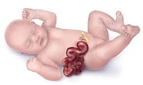
what is this condition and why does it occur

OMPHALOCOELE = umbilical protrusion of intestines with amnion covering caused by persistence of embryological herniation
what is this condition and why does it occur
GASTROSCHISIS: protrusion of abdominal viscera directly into amniotic cavity due to abnormal closure of abdominal wall during embryo folding.
Bowel may be damaged by exposure to amniotic fluid but excellent survival rate.
what are the embryological origins of the anal canal
- upper 2/3 formed from endoderm of hindgut (separation of cloaca by urorectal septum) = simple columnar epithelium
- lower 1/3 formed from ectoderm of proctodeum (which proliferates and invaginates to form anal pit) = stratified squamous epithelium
name the junction between the 2 parts of the anal canal
pectinate line
-
3. GI cellular anatomy17
-
3. GI structural anatomy14
-
8. Abdominal catastrophes14
-
4. The inguinal canal & hernias14
-
5. Function of the stomach15
-
5. Gastric clinical conditions14
-
6. Digestion & absorption17
-
6. Chyme pancreas & liver13
-
7. Anatomy of the liver & biliary tree4
-
7. Liver pathology20
-
7. Pancreatic pathology8
-
2. GI embryonic development23
-
8. IBD13
-
9. GI immune mechanisms7
-
11. GI imaging8
-
11. GI malignancy17
