How many live births have congenital heart disease?
-~1% -most common congenital disorder
3 rationales fro studying CV development
- inherent scientific knowledge 2. understanding the pathogenesis of CHD: dx and prognostic tool development, develop therapies 3. hypothesis: mlc pathways important for CV development are recapitulated during CV disease pathogenesis
5 major processes in cardiac development
- normal cardiac morphogensis 2. left-right patterning 3. valve formation 4. angiogenesis vs vasculogensis 5. contribution of extra-cardiac cells to heart: epicardium and NC
6 parts of normal cardiac morphogenesis
-myocardial specification -formation of linear heart tube -looping -septation -patterning of great vessels -circulatory changes at birth
where is the cardiogenic (heart-forming) region located in embryo? What is this called?
-initially located at the anterior rim of the embryonic disc very early in development -called the cardiac cresent: epithelial layer of cardiac MULTIpotent progenitor cells that make up myocardium, endocardium, pericardium
As the embryo grows, the developing heart assumes a position where? Then what happens?
-ventral to forming forebrain and foregut; as 2 side tubes -heart progenitors migrate ventrally and medially to form a linear heart tube; attached to rest of embryo by dorsal mesocardium
3 events happening at linear heart tube phase
- heart begins to beat: intrinsic pacemaker activity (caudal >rostral) 2. blood flow commences: single circulation in series caud to rostral (remember atria on bottom of linear tube) 3.chamber specification due to molecular determination
Atrium location within linear tube
-caudal end! under LV and RV
Chamber-specific transcriptional programs of linear heart tube
-dHAND is RV and eHAND is LV -hey1 is atria and OFT and hey2 is ventricles
T/F: Advanced cariac morphogenesis does not require midline fusion.
-true
Secondary Heart Field
-large parts of RV and OT derived from secondary field of cardiac precursors -these cells migrate into OT and differentiate into muscle from pharynx -diff gene expressions of these cells within cardiac crescent
Secondary Heart Field is associated with ______ and ______ signaling.
-LiCl (Wnt agonist) -Wnt signalling
Secondary Heart Field defects are attributable to ______ variants in 11-18% cases.
-Islet 1 variants
3 events occurring during cardiac looping
-linear heart tube bends to right and anteriorly, thought to be driven by regional differences in myocardial growth rate–first sign of assymetry in heart!! -begin septation: endocardial cushions (OFT and AV canal) and interventricular septum -myocardial trabeculation
2 end results of cardiac looping
-atria assume a more rostral and posterior orientation -advanced regional specification: bulbis cordis (conus cordis becomes cardiac OFT, truncus arteriosus becomes PA and Ao), AV canal (maturation of AV canal and formation of endocardial cushions)
4 septation events
-AV canal septation -Interventricular septation -Interatrial septation -outflow tract septation (TA and CC)
steps of atrial septation
- septum primum grows from dorsal wall of atrium toward AV cushions 2. initially incomplete (passive): ostium primum= residual connection 3. eventually completely (actively) closed by fushion with cushions 4. new opening in septum primum occurs= ostium secundum (smaller performations up higher) 5. new septum = septum secundum grows from superior atrial wall 6. septum secundum passively closes the ostium secundum
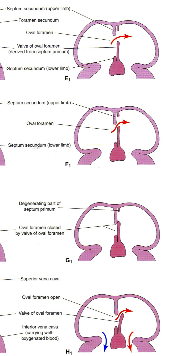
In embryonic circulation, ostium secundum allows for flow from right to left atrium (little pulm circulation). In the adult, septum secundum closes the ostium secundum. Ostium secundum later ______ closes in most, but not all people. What is a variant of this called when the ostium secundum does not close?
-actively -Patent Foramen Ovale: 20-25% of people and potential stroke risk
Ostium Secundum Atrial Septal Defect
-most common type of ASD (larger, constantly lets blood thru) -occurs in center of the septum between LA and RA -due to incomplete formation of septum secundum or incomplete active closure of ostium secundum -variant of this type of ASD is PFO and is very small!
Ostium Primum ASD
-2nd most common ASD -located in lower portion of atrial septum -due to incomplete active closure of ostrium primum -often associated with a cleft or slit-like defect in anterior leaflets in MV
-Ostium Primum cause and association
-due to incomplete active closure of ostrium primum -often assoc with cleft or slit-like defect in anterior leaflet of MV
Sinus Venosus ASD
-least common of ASD -located in upper portion of atrial septum -due to defect in formation of septum primum -often has an abnormal pulmonary vein connection associated with in -one of PVs abnormally connect to the RA instead of the LA (anomalous PV)
-Location of ASDs in atrial septum from top to bottom
- top: sinus venosus ASD 2. middle: ostium secundum ASD 3. bottom: Ostium primum ASD
Rank 3 main ASD in order of how common they are
-Ostium secundum is most common -Ostium primum ASD -Sinus venosus ASD
-
Vasoactive Peptides and Inhibitors: RAAS34
-
Hypertension Overview 9/10/201430
-
Antihypertensive drug therapy60
-
Heart failure38
-
Cardiac Valve Stenosis47
-
Inotropic agents55
-
B agonists20
-
Autonomic ANTAGONISTS25
-
Cholesterol and Lipoprotein Metabolism34
-
Drugs for Lipoprotein issues57
-
Regurgitations50
-
HTN Pathology60
-
Atherosclerosis 138
-
Ischemia pathophysiology41
-
Ischemia Pathology51
-
Unstable ischemic coronary syndromes42
-
Peripheral vascular disease and physiology29
-
Atherosclerosis Meds21
-
Atherosclerosis risk factors: Diet and exercise24
-
Atherosclerosis and Restenosis Mechanisms25
-
Genetic determinants of CVD35
-
Gender issues in CVD37
-
Valvular Disease Patho47
-
Cardiovascular Development50
-
Congenital Heart Disease 1-377
-
Pathology and Pathophys of Myocardial Dz98
-
Antiarrhythmic Drugs42
