Deck 2 Flashcards

(48 cards)

1
Q

Diagnosis?

A

pseudo aneurysm

pepsi-cola sign that is suggestive of pseudoaneurysm

2
Q

Cardiologist notes a femoral bruit a few days after performing transfemoral LHC. He calls you asking how likely you are to accurately diagnose a pseudoaneurysm (PSA) w/ DUS imaging.

What to tell him?

A

DUS is a both sensitive and specific for PSA diagnosis

  • 94% sensitivity
  • 97% specificity
3
Q

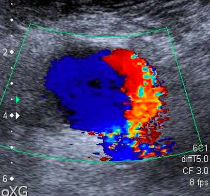
Patient undergoes LE arterial DUS. You come across an isolated image from region near femoral artery (shown below) while reading the study. No further images of the finding are available.

What to do next?

A

Have tech go back and interrogate region w/ pw

Swirling pattern w/ sack off of common femoral artery should make you think of PSA* (but it is not diagnostic)

pw doppler imaging showing bidirectional flow in sack attached to CFA is pathognomonic of PSA

*PSA = pseudoaneurysm

4
Q

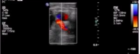
Low-resistance doppler signal in EIA 3 wks after LHC.

*EIA = external iliac artery

A

think of AV fistula

low-resistance doppler signal (LRDS) in the proximal iliofemoral arterial system after LHC→ think of AVF

shown below: LRDS in CFA

5
Q

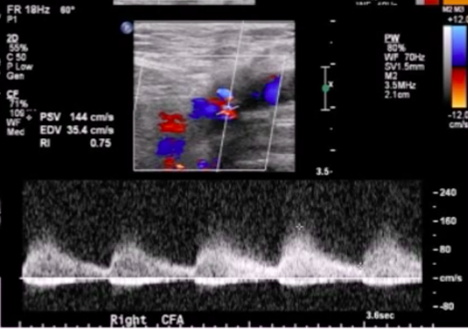
Name five vascular DUS findings that are characteristic of AV fistula.

A
  1. markedly increased arterial PSV
  2. venous pulsatile flow distal to AVF
  3. arterial LRDS* proximal to AVF
  4. color bruit
  5. +/- visualization of fistula tract

*LRDS = low-resistive doppler signal

6
Q

Patient with vascular Ehlers-Danlos syndrome undergoes arterial duplex study. Pertinent findings shown below. Diagnosis?

A

Dissection

7
Q

Frank’s spaghetti is a 60-year-old obese Italian man who comes to your vascular lab for a study prior to initiation of hemodialysis. The ordering physician did not specify what he wants done. What is the most likely reason for patient’s visit to your lab?

A

Patient likely needs superficial venous ultrasound study [vein mapping] Above is performed in order to see if patient is a candidate for 80 fistula surgery, which is preferred over grafting for HT access

8
Q

You are performing a vein map study for a patient with new HD. Your tech wants to know how to do the study. What to tell her?

A

Evaluate the upper extremity veins of the non-dominant arm in the following way: Look at the cephalic vein up to the subclavian vein for compressibility diameter and continuity. May Pl., Turnock it on arm to help bring out Venus structures

9
Q

Patient who is scheduled for vein mapping has small it’s Reddick cephalic vein. What to do next?

A

Evaluate basilic system

10
Q

What is the purpose of evaluating the subclavian an axillary vein’s when doing a vein mapping study?

A

Want to rule out central venous obstruction.

11
Q

What size superficial veins in the upper extremity correlate with higher likelihood of fistula maturation with regard to a vein mapping study?

A

Diameter greater than 2.5 mm

12
Q

Explain to your tech how to perform the arterial portion of a preop 80 fistula exam

A

Evaluate brachial radial and ulnar arteries for of the following: Calcification Waveform characteristics (One triphasic flow) Patency (want lumen diameter greater than 2.0 mm) Anatomic variance [high radial artery take off]

13
Q

Patient with end-stage renal disease on HD has high resistance waveform and left distal subclavian artery. The right subclavian artery has normal flow. Diagnosis?

A

Probably Access site occlusion or stenosis.

14
Q

Important components of post AV access creation vascular ultrasound study?

A

Evaluate inflow artery for stenosis/occlusion Evaluate arterial anastomosis site and beam out and color to look for “swing site” stenosis Evaluate body of access as well as outflow vain for Neil intimal hyperplasia Newly created if you fish Ila should be evaluated for vessel diameter and depth to ensure adequate cannulation

15
Q

How long to 80 fish is usually take to mature?

A

3 to 4 months Balloon assisted maturation is an option to speed along the process.

16
Q

How long before an AV graft can be used after it is placed

A

Usually 2 to 4 weeks

17
Q

What is the preferred flow volume rate for an AV graft compared to a navy fistula?

A

AV graft: greater than 800 mL per minute If you fistula: greater than 600 mL per minute

18
Q

Role of sixes for AV fistula?

A

Fistula diameter greater than or equal to 6.0 mm Fistula depth from skin less than 6.0 mm Low-volume greater than 600 mL per minute

19
Q

Best area to measure flow volume when assessing if you fistula?

A

Brachial artery It is not ideal to measure outflow vessel diameter to use for calculation for volume; because this can give rise to many Errors

20
Q

Major pitfall of assessing AV fistula Doppler flow dynamics?

A

Setting the flow sample size too small and sampling near the wall of the vessel This will lead to underestimation of flow velocity

21
Q

Formula for estimation of AV fistula flow?

A

AVF flow equals brachial artery flow -75 200 mL per minute

22
Q

Patient with 80 fistula has estimated AV fistula flow of 400 mL per minute what to do next?

A

Recommend that they should have assessment for new AV fistula repair of current fistula Can you foundation recommends replacing 80 fistula Winflo is less than 500 mL per minute

23
Q

At what value of AV graft flow does the kidney foundation recommend replacing a graft?

A

600 mL per minute or less

24
Q

Criteria for AV access site stenosis? Venus anastomosis site.

A

Greater than 2 to 1 Peak systolic velocity ratio and Greater than 50% lumen reduction. Above refers to proximal site comparison to distal site

25
Criteria for AV access site stenosis [arterial portion]?
Greater than 3 to 1 pizza Stalock velocity ratio and Greater than 50% lumen reduction
26
Treatment of AV fistula aneurysm?
Stenting or surgical repair
27
What is DH I S?
Distal hypoperfusion ischemic syndrome Occurs when tissue distal to a navy fistula for dialysis becomes ischemic. Thanks
28
Risk factors for DH I S?
Diabetes hypertension elderly patient female gender prefer a vascular disease
29
80 exit site stenosis questioned insert
Insert
30
Describe the four stages of DHI S
Stage one retrograde diastolic flow distal to access without symptoms Stage two: pain on exertion and or during dialysis stage III breast pain stage four: ulceration, necrosis, or gangrene
31
Patient with hand pain as Doppler signal shown below.diagnosis
Distal hypoperfusion ischemic syndrome
32
Pt with HD access site flow that drops 30%.
Think of AVF failure
33
How is ALoD\* in the foot diagnosed (using PAT, physiologic arterial testing)? ALoD = Atherosclerotic Lower extremity arterial Disease
2 methods for diagnosis of foot ALoD\*: 1. **toe/metatarsal PVR** 2. **digit pressures** ALoD = atherosclerotic Lower extremity arterial Disease
34
Patient with abnormal toe pressures in a cold room.
**probably spurious result due to cold-induced vasoconstriction**
35
Ideal size for BP cuff when performing PAT (physiologic arterial testing)?
**width of cuff should be 1.5\*DL** * where DL = Diameter of Limb that you are testing
36
A patient undergoes ABI study with the following data detected: 24 mmHg bradient between right calf and right ankle. Localize the arterial lesion.
**right infrapopliteal disease**
37
73M with multiple bilateral leg wounds has physiologic arterial testing result shown below. Most likely diagnosis?
**bilateral severe arterial disease** ## Footnote falsely elevated ABI values occur due to partially non-compressible (calcified vessels) if clinical data (leg ulcers) and ABI test are inconsistent, go to PVR data
38
This region of the leg should have the highest amplitude on the PVR.
**the calf PVR**
39
This physiologic arterial testing modality is helpful in diagnosis of critical limb ischemia and non-healing ulcers of the foot.
**TCO2 (transcutaneous oximetry)**
40
Pt with ulcer just below knee has the following TCO2 values before and after administration of supplemental 100% O2: pre-O2: 25% post-O2: 30% What to advise the vascular surgeon?
**Amputate proximal to the infrapopliteal ulcer** ## Footnote adequate rise in TCO2 value w/ administration of 100% O2: * ðPaO2 _\<_ 10 mmHg predicts failure of healing post amputation * final PaO2 \> 100 mmHg is considered normal response
41
Patient with critical limb ischemia has TCO2 = 30 mmHg in left foot. o/e the left foot has stage IV ulcer. What to do next?
**Vascular consult for amputation** ## Footnote TCO2 \< 40 ⇒ low likelihood of healing after revascularization
42
18 year-old male with calf pain when he exercises. ADUS is shown below. What to do next?
provocative maneuver to elicit symptoms ## Footnote dx = popliteal artery entrapment disease
43
67 year-old Persian male presents with retinal artery occlusion. Carotid arterial duplex US (ADUS) shows obstructive lesion in proximal left internal carotid artery. You referr him for surgery. Pt says"Doc, don't I need a CAT scan first?". What to tell him ?
There is no need for further testing. Proceed with surgical evaluation ## Footnote no further imaging is necessary prior to CEA for carotid ADUS with the following properties: demonstration of obstructive disease at level of carotid bifurcation symptomatic
44
Your vascular US lab uses photo-plethysmography (FPM) to diagnose VVI\*\*. What would you expect the post-exercise venous filling time (PEVFT) to be for an individual with VVI in the following scenarios? * **after application of the tourniquet** * **without a turniquet** \*\* VVI = venous valvular incompetence
For a pt with VVI undergoing a vein reflux study, PEVFT will be: * \<\< 20 seconds *without a tourniquet* * *\>20 seconds* with tourniquet in place (normalization) note that In the above scenario, a tourniquet simulates calf Muscle Contraction
45
What does the following diagram illustrate with regard to leg vein physiology (i.e. in patients w/out venous insufficiency)?
activation of the calf muscle with exercise →blood is pushed out of the soleal sinuses (into the deep leg veins) → vein valves close, preventing reflux of blood →legs fill with blood via arterial source only (takes \> 20 s)\* \*rapid filling of soleal sinuses ⇒ backflow of venous blood (and thus vein valve incompetence)
46
Patient with "low-pitched, gruff" systolic bruit over the left side of the neck. Most likely diagnosis?
**moderate-range left carotid artery stenosis** ## Footnote The physical exam findings of obstructive carotid artery disease mirror those of AS →→ i.e. later-peaking, higher-pitched bruits are suggestive of more severe carotid disease "low-pitched, gruff" bruit ⇒ likely moderate-range carotid stenosis
47
A carotid bruit cannot be heard until the degree of carotid artery stenosisIs is greater than \_\_\_\_\_.
A carotid bruit cannot be heard until the degree of carotid artery stenosisIs is **greater than 50%**
48
Use the Sexy Lindsy's mnemonic to name the 8 branches of the external carotid artery.
**Sexy Lindsy (Lohan's) Powdered Face often Attracts MD Sperm:** ****_S_**uperior thyroid artery** ****_L_**ingual artery** ****_P_**osterior auricular artery** ****_F_**acial artery** ****_O_**ccipital artery** ****_A_**scending pharyngeal artery** ****_M_**axillary artery** ****_S_**uperficial temporal artery**