Erythrocyte Flashcards

(82 cards)

1
Q

Pathogenesis of Extracellular Hemolytic Anemia

A
  1. Initiating event (immune, metabolic, traumatic, oxidative)
  2. Macrophages recognize and destroy RBCs

——> Anemia

  1. Macrophages degrade Hb to bilirubin

—–> Icterus

——–> Bilirubinuria

2
Q

Pathogenesis of Hemolytic Icterus

A
  1. Macrophage destructin of RBCs results in unconjugated Bilirubin to enter plasma and bind to albumin
  2. Bu/Alb complex transported to liver

—> Hyperbilirubinemia (Bu) (if incomplete removal)

  1. Bu enters hepatocytes and is conjugated (Bc)
  2. Bc excreted in bile
  3. Bc may be regurgitated to plasma if formation > excretion

—> Hyperbilirubinemia (Bc)

  1. Bc passed through kidneys, excrete

—-> Biliruninuria

3
Q

If your patient is icteric, what type of anemia will you suspect (intra- or extravascular)

A

Extravascular

The icterus is caused by macrophages destroying RBCs and then degrading Hb to bilirubin

4
Q

Pathogenesis of intravascular hemolytic anemias

A
  1. Initiating event (immune, metabolic, oxidative)
  2. Marked damage to RBC membrane and lysis of RBCs in blood

—–> Anemia

  1. Hb in plasma

——> Hemoglobinemia

5
Q

Pathogenesis of hemoglobinemia

A

Starts with intravascular hemolysis. Hb is present in plasma and unstable. 3 things can happen next

  1. Hb binds to haptoglobin (Hpt) —> Hb/Hpt complex removed
  2. Hb binds to hemoplexin (Hpx) —> Hb/Hpx complex removed
  3. Too much Hb released so….

—–> Hemoglobinemia

—–> Hemoglobinuria

6
Q

Hemoglobinemia and hemoglobinuria are associated with what type of anemia (intra- or extravascular)

A

Intravascular - RBCs lyse and Hb accumulates in blood/passes through urine
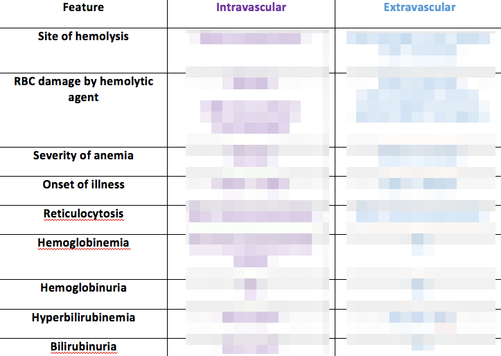
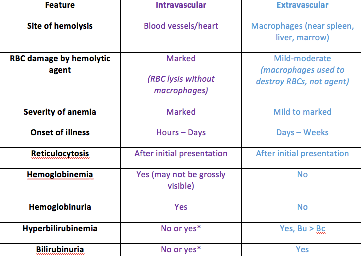
7
Q

Fill out this table

A
8
Q

Antibodies (especially colostral Abs in horses) are a cause of what type of hemolysis

A

Extravascular

9
Q
A
10
Q

Mycoplasma and Anaplasma cause what type of anemia

A

Extravascular hemolytic anemia

11
Q

Penicillin causes what kind of hemolysis

A

Intravascular (and then extravascular)

12
Q

Heinz bodies are found in what type of hemolytic anemia

A

Intravascular (and then extravascular)

13
Q

Your patient has tons of Eccentrocytes. What type of anemia do you suspect

A

Intravascular hemolytic anemia

(extravascular hemolytic anemia will follow)

14
Q

Hypophosphatemia causes which kind of anemia

A

Extravascular

(PO4 needed for major metabolic pathways. Without PO4 —> reduced ATP –> membrane cant repair –> extravascular hemolysis)

15
Q

A dairy cow recently gave birth and you now worry she is anemic. Based on this information, what may be causing this and why type of anemia is she suffering from?

A

She may be suffering from post-parturient hypophosphatemia, which would cause extravascular (??) hemolytic anemia

16
Q

What is a common cause of hypophosphatemia in dogs and how will this effect the dog?

A

Hyperinsulinemia drives PO4 into cells (ie myocytes), resulting in hyphosphatemia and intravascular hemolysis

17
Q

What does L-sorbose intoxication cause?

A

It cause hypophosphatemia

L-sorbose binds to PO4 and inhibits glycolysis —> Reduced ATP —> Reduced membrane repair

—> Intravascular hemolysis

18
Q

Agglutinated RBCs will cause artefact to which indice

A

MCV –> agglutination results in increased MCV

19
Q

What are Wintrobe’s formulas

A
  1. MCV
  2. MCH
  3. MCHC
20
Q

Can you calculate the hematocrit?

A

Yes: MCV x [RBC] / 10

21
Q

Which stain is best for reticulocytes

A

Methylene Blue

22
Q

Whats reticulocyte percentage

A

# reticulocytes / 1000 RBCs

(counted)

23
Q

Whats Corrected Reticulocyte Percentage (CRP)

A

It compensates for the degree of anemia

RP X (Hct/Average Hct for spp)

Tells you what reticulocyte percentage would be if animal were not anemic (as anemia = fewer RBCS = platelets will seem more plentiful)

24
Q

Whats a disadvantage of corrected reticulocyte percentage

A

It assumes a normal hematocrit

–> this may not be normal for all animals. (ie greyhounds have higher PCV than other dog breeds)

25
What is reticulocyte concentration
It is used if reliable [RBC] is available ## Footnote **RP x [RBC}**
26
What is **appropriate rubricytosis**
- Seen in **regenerative anemias** - Regenerative blood cells following blood loss or hemolysis
27
What is inappropriate rubricytosis
Regeneration of RBCs **without anemia** or in **non-regenerative anemia**
28
Causes of inappropriate rubricytosis
- Marrow damage, inflammation, necrosis - Lead poisoning in dogs - Extramedullary hematopoiesis, splenic contraction (horses), splenectomy
29
30
RBC lifespan for cats, dogs, cows, horses
Cats: 70d Dogs: 100d Cows: 280d Horses: 450d
31
Which species have a sinusoidal spleen? What's the clinical significance?
Dogs, horses ## Footnote * - Pools of blood can be released quickly* * - More efficient removal of old, damaged RBCs (by macrophages outside spleen)*
32
What's a morphological sign of regenerative anemia in cows?
Basophillic stripping (Ribosomal RNA that isnt degraded)
33
All acquired anemias are initially what (-chromic, -cytic, -generative)
Normochromic Normocytic Non-regenerative *\*Because it takes 3-4 days for EPO to kick in and for new RBCs to be released*
34
When does reticulocytosis start after onset of anemia? When is peak reticulocytosis
Start: 3-4 days Peak: 7-10 days
35
What is macrocytosis and who has it
Large cells - its seen in **horses** during **regenerative anemia** instead of reticulocytosis
36
How do you classify non-regenerative anemias
**Normocytic** **Normochromic** - No reticulocytosis! - No rubricytosis!
37
Principle causes of non-regenerative anemia
AID Renal failure (chronic)
38
# Fill in the blanks
39
Your patient has normocytic, normochromic anemia. What is cause
- Initial status of regenerative (ie just got HBC) - Non-regenerative
40
Your patient is macrocytic and hypochromic. What type of anemia
Regenerative
41
Your patient is macrocytic and normochromic - what type of anemia
Regenerative
42
Your patient is microcytic and hypochromic - what kind of anemia
Fe deficiency (due to nutrition, AID..)
43
Your patient is microcytic and normochromic. What type of anemia
Fe deficiency Liver failure (microcytes)
44
What does hyperchromic indicate
Usually an artefact!
45
What is this? What is its signficance/cause?
Rouleax formation ## Footnote _Significance_ - Hyperglobinemia - Hyperfibrinogenemia _Cause_ - Inflammation - Dehydration \*normal in horses!
46
You're evaluating a blood stain from your equine patient. Should you be concerned?
No - roleaux is common in horses
47
What's this? Significance? Cause?
**Agglutination** ## Footnote _Significance_ - Formed by antibody bridges between cells _Causes_ - Cold antibody - Heparin induced (horses)
48
What kind of artefacts would you expect from this smear?
Clumps will be seen as large RBCs so: **- Decreased [RBC]** **- Increased MCV** *Could suggest a regenerative anemia (macrocytic..)*
49
You're unsure whether your patient's blood is roleaux or agglutinated. How do you test?
Saline dilution test - Add saline (1 blood: 9 drops saline) **- If clumps disperse ---\> Roleaux** **- If clumps dont disperse --\> Agglutination**
50
How does central palor vary in species (dogs, cats, cows, horses)
Dogs: prominent Cats: mild Cows: mild-moderate Horses: none-mild
51
How will your RBC indices be effected by Fe deficiency
MCV: decreased MCHC: decreased MCH: decreased *Marked central pallor*
52
How will your RBC indices be effected by regenerative anemia
MCV: increased MCHC: decreaed MCH: increased * Reticulocytes* * Polychromatic*
53
What are howell-jolly bodies caused by
Extra chromatic after mitosis of RBC Indicate increased erythropoiesis and decreased splenic function
54
What are heinz bodies caused by
Oxidative damage Defective RBC metabolism
55
Your horse is anemic and has recently been seen eating red maple leaves. What will you seen
Heinz bodies!
56
Your cat has hepatic lipidosis. How will RBCs appear
Elliptocytes
57
RBCs in a patient with intravascular coaguation will be?
Keratocytes
58
Which RBC morphology is indicative of a drying artefact
Echinocyte
59
Which RBC morphologies would you see with microangiopathy
Shizocyte (triangular fragments) Keratocyte (horn projections)
60
How do impedence cell counters identify RBCs
By volume
61
How do optical/laser flow cytometers identify RBCs
By light scatter
62
How do conductivity methods identify RBCs
By electrical resistance
63
How does cetnrigufation analysis identify RBCs
By microhematocrit and buffy coat analysis
64
In which species is it normal to hve orange plasma. Can it indicate pathology?
Healthy in herbivores (plant pigments) Pathology: icterus (bilirubin)
65
Pink/red plasma indicates
Hemoglobin
66
White/hazy plasma indicates
Presence of lipids
67
A thickened buffy coat indicates increase of two types of cells
WBCs Platelets
68
Calculated hematocrit (cHct) and spun hematocrit (sHct) should be within what percentage of each other
3% ## Footnote *If not, something is wrong (usually with cHct)*
69
Whats erythrocytosis
**Increased RBC mass** Increased Hct Increased Hgb Increased [RBC]
70
What is hemoconcentration
Increased concentration of blood components due to decreased plasma volume --\> DEHYDRATION
71
What is polycythemia vera
Clonal myeloproliferative disease causing: - erythrocytosis - leukocytosis - thrombocytosis
72
Common causes of erythrocytosis
1. Hemoconcentration (*dehydration due to diarrhea, voliting, polyuria)* 2. Splenic contractions (*exercise, fright/excitement*) \*dogs, horses\* 3. Hypoxia 4. Neoplasm
73
How can you calculate erythrocytosis using spleen and peripheral Hct
Add peripheral Hct to splenic Hct --\> if higher than 100%, thats how much erythrocytosis you have
74
What are two secondary causes of erythrocytosis (appropriate and inappropriate)
1. Hypoxia --\> stimulates EPO ---\> increased RBC release - **Appropriate** (= due to hypoxia) 2. Neoplasm in liver, kidney stimulating EPO - **Inappropriate**
75
Whats hypoferremia and whats the analyte
Decreased [Fe] in serum/plasma Analyte = Serum Fe
76
Whats hypoferritinemia and what is the analyte
Decreased [ferritin] in serum/plasma Analyte = Serum ferritin
77
What's hyerferritinemia and what is the analyte
Increased ferrtin in serum/plasma Analyte = serum ferriitn
78
What is TIBIC?
Total iron binding capacty -- *total amount that can be bound in serum transferrin*
79
T/F TIBC is an important diagnostic tool
False, it is of little diagnostic value
80
Which assay is useful for Fe storage?
Serum ferritin
81
How can you differentiate if hypoferemia is due to Fe deficiency or inflammation
Stainable Fe test -- *Fe stained in spleen or bone marrowx* - **Inflammation:** will be _increased_ (mac sequestratio) - **Fe deficiency**: will be _decreased_ (less avaialbe and what is available is being used up to make new RBCs)
82
You suspect IMHA. Which two antibdy tests can you use and which antibody are you testing for
Detecting for **ESAIg** (erythrocyte surface associated immunoglobulin) ## Footnote **1) Coomb's test** **2) Flow cytometry for ESAIg**