Choanal Atresia
- Congenital condition characterized by a bony and/or membranous obstruction of the posterior nasal passage.
- Unilateral choanal atresia often presents late with chronic inflammation (e.g., rhinorrhea, congestion) of the affected nasal passage.
- Bilateral choanal atresia manifests as obstructed nasal breathing with intermittent cyanosis immediately after birth; breathing improves when crying, as it allows the infant to breathe through his or her mouth.
VACTERL Association
Mesodermal defects
A group of associated birth defects consisting of
- Vertebral anomaly
- Anal atresia
- Cardiac anomaly
- Tracheoesophageal fistula
- Esophageal atresia
- Renal anomaly
- Limb malformation
Duodenal Atresia
- ∼ 50% of cases are associated with further anomalies, e.g., bile duct and VACTERL association.
- 20–25% of cases are associated with chromosomal abnormalities, especially Down syndrome.
- Occurs when recanalization of the closed duodenum fails to occur or occurs only partially during the embryonic period (usually between the 8th–10th week of gestation).
- Because the development of the duodenum is connected to the growth of the pancreas and the hepatobiliary system, duodenal atresia is commonly associated with anomalies of these organs as well.
Clinical Presentation:
- Intrauterine → polyhydramnios
- Postpartum
- Vomiting, that is typically bilious if the stenosis is distal to the major duodenal papilla
- Atresia or high-grade stenosis → vomiting few hours after birth
- Mild stenosis → vomiting after a few days
- Distended upper abdomen and scaphoid lower abdomen (the lower abdomen is sucked inwards in a concave shape, as there is no gas present in the gastrointestinal tract distal to the obstruction, whereas gas builds up proximal to it)
- Delayed meconium passage (If stenosis (rather than atresia) is present, the newborn may be able to pass meconium; it will, however, take more time because of the obstruction)
X-ray of the abdomen
- Double bubble sign → air and fluid build up proximal to the obstruction and are separated by the pyloric sphincter, which resembles two bubbles on imaging → one in the stomach and one in the duodenum
- Gasless distal bowel
Preoperative management
- Parenteral nutrition via a central catheter shortly after birth
- Fluid replacement and restoration of the electrolyte balance
- Gastric decompression
Surgery → bypass the atresia or stenosis
- The exact procedure depends on the anatomic findings and associated anomalies
- Common procedure → duodenoduodenostomy or duodenojejunostomy with a proximal transverse-to-distal longitudinal (diamond-shaped) anastomosis

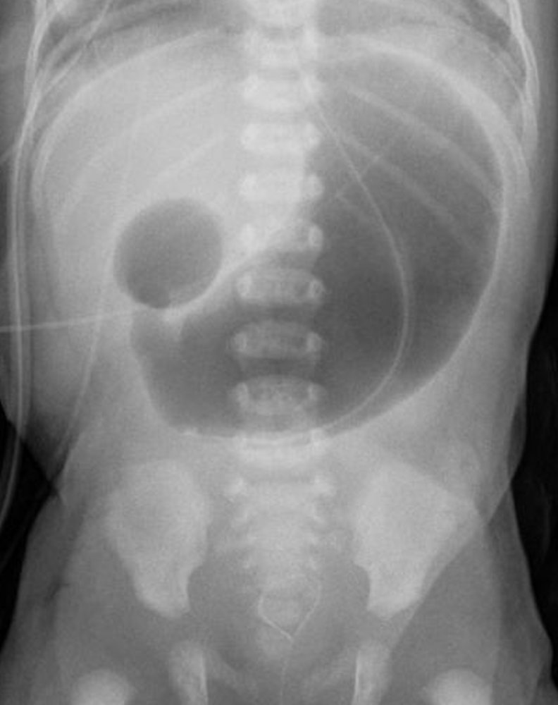
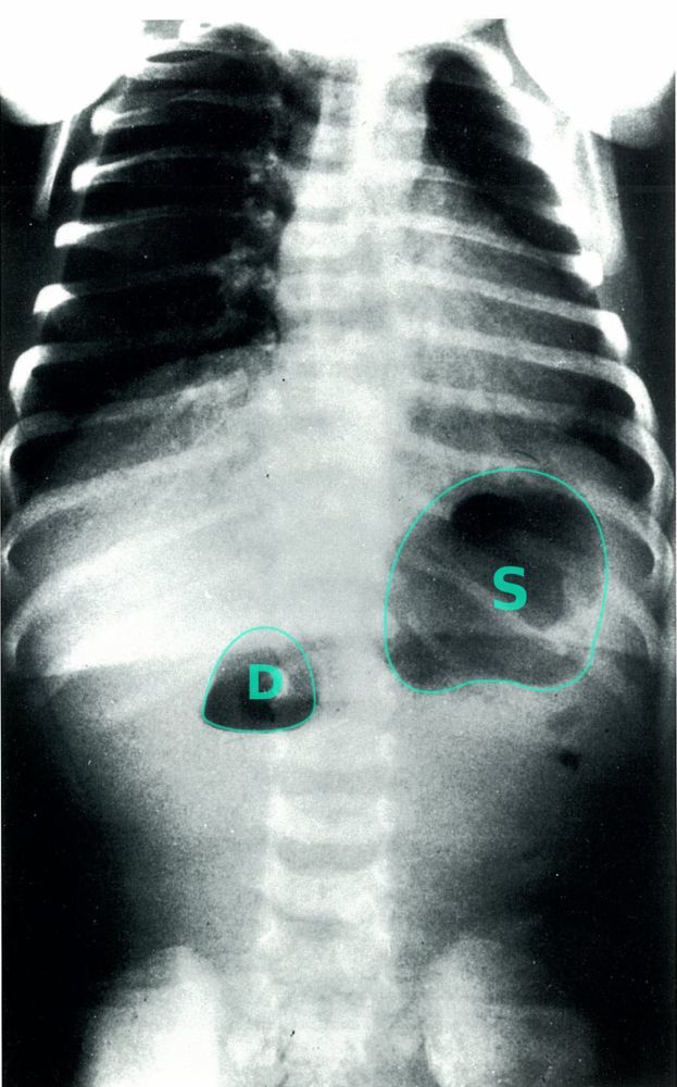
Duodenal Atresia
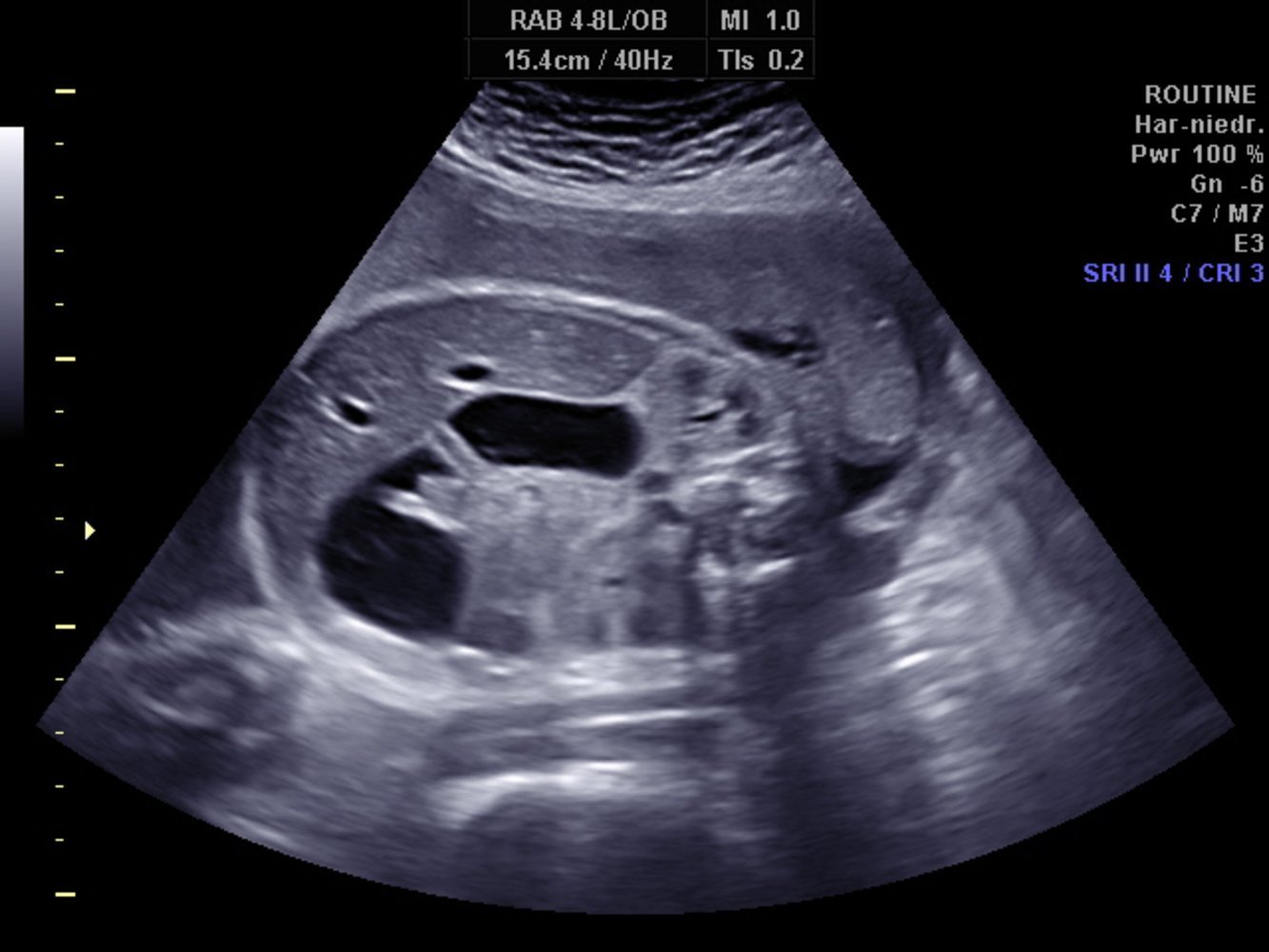
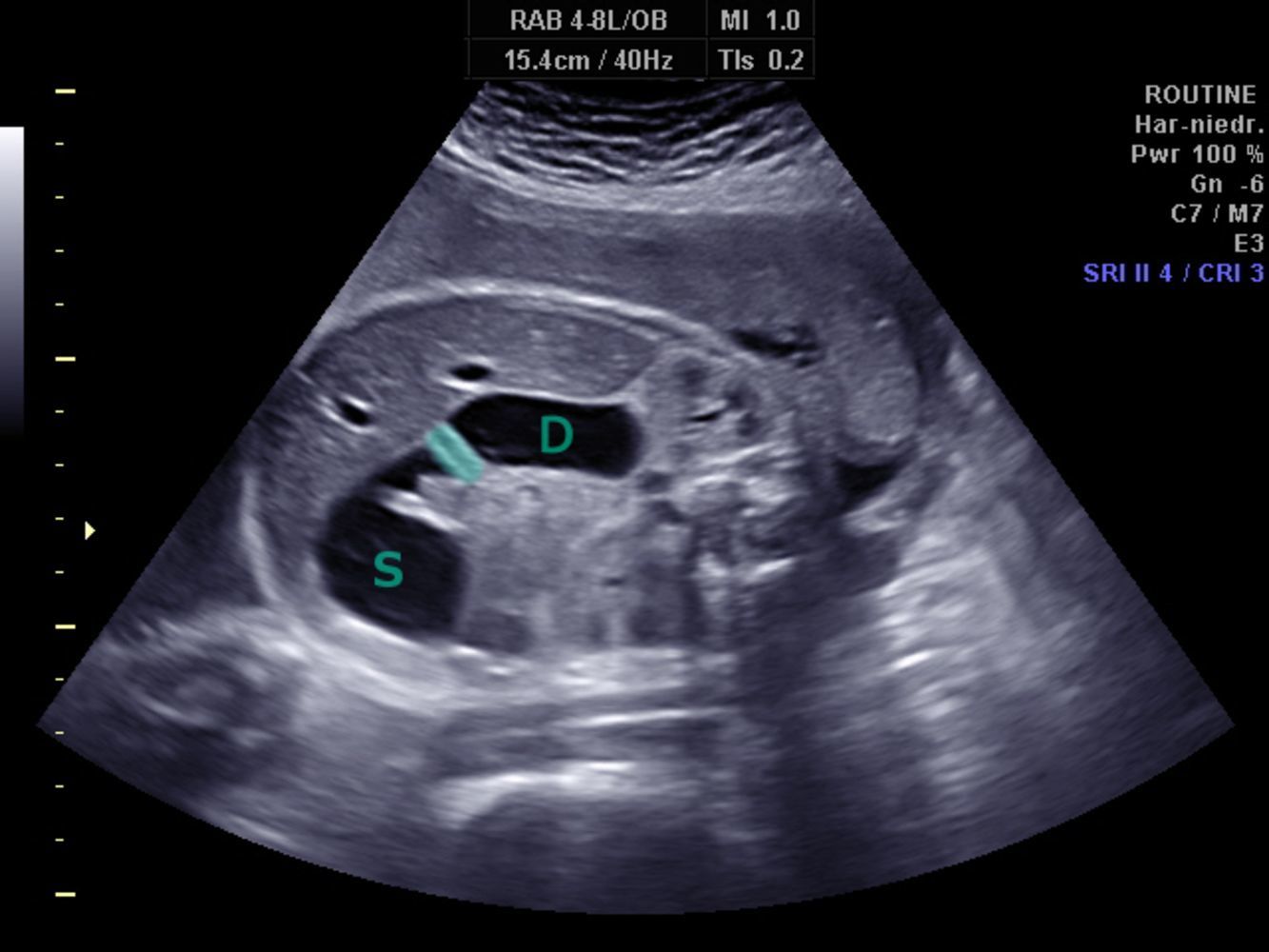
Double Bubble Sign on Ultrasound Seen in Duodenal Atresia
S → stomach
D → proximal duodenum
Shaded area → pyloric sphincter
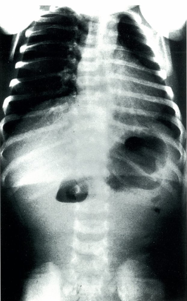
Duodenal Atresia in a Newborn
(dilation of the pre-stenotic bowel segments (duodenum (D) and stomach (S)) with an air-fluid level in each dilated segment (double bubble sign). No air is visible in the post-stenotic bowel loops)
Ileal Atresia
- Absence of the jejunal lumen or ileal lumen
- Less common than duodenal atresia
- Vascular accident in utero (usually a disruption of superior mesenteric artery) → ischemic necrosis and reabsorption of the jejunum/ileum → discontinuous bowel
- Risk factors → vasoconstrictive drugs (e.g., cocaine, MDMA, or cigarettes) during pregnancy
- Clinical features → similar to duodenal atresia
- Polyhydramnios (intrauterine)
- Bilious vomiting and upper abdominal distension (postpartum)
- Diagnostics → abdominal x-ray shows a triple bubble sign (dilated small bowel loops and air-fluid levels) and gasless colon. Distal segment of the ileum assumes a spiral configuration around an ileocolic vessel (apple peel or christmas tree)
- Treatment → surgical correction with bypass of the occluded part of bowel is always required

Jejunal Atresia
- Absence of the jejunal lumen or ileal lumen
- Less common than duodenal atresia
- Vascular accident in utero (usually a disruption of superior mesenteric artery) → ischemic necrosis and reabsorption of the jejunum/ileum → discontinuous bowel
- Risk factors → vasoconstrictive drugs (e.g., cocaine, MDMA, or cigarettes) during pregnancy
- Clinical features → similar to duodenal atresia
- Polyhydramnios (intrauterine)
- Bilious vomiting and upper abdominal distension (postpartum)
- Diagnostics → abdominal x-ray shows a triple bubble sign (dilated small bowel loops and air-fluid levels) and gasless colon. Distal segment of the ileum assumes a spiral configuration around an ileocolic vessel (apple peel or christmas tree)
- Treatment → surgical correction with bypass of the occluded part of bowel is always required

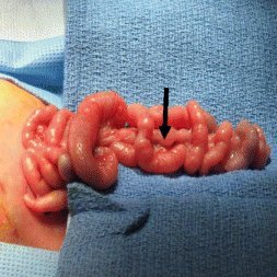
Apple Peel or Christmas Tree Seen in Jejunal Atresia
Hypertrophic Pyloric Stenosis
Etiology:
- Environmental factors
- Exposure to nicotine during pregnancy
- Bottle feeding (bottle-fed infants drink more milk in less time, which may lead to pylorus muscle hypertrophy through overstimulation. Another hypothesis maintains that formula components make it harder to digest and gastric emptying is delayed, which may also burden the pylorus muscle)
- Genetic factors → patients with affected relatives have a higher risk of hypertrophic pyloric stenosis
- Macrolide antibiotics
- Erythromycin and azithromycin are associated with a higher risk of hypertrophic pyloric stenosis, especially when administered within 2 weeks after birth
Clinical Presentation:
- Symptoms usually develop between the 2nd and 7th week of age (rarely after the 12th week).
- Frequent regurgitation progressing to projectile, nonbilious vomiting immediately after feeding
- An enlarged, thickened, “olive-shaped”, non-tender pylorus (diameter of 1–2 cm) should be palpable in the epigastrium
- A peristaltic wave, moving from left to right, may be evident in the epigastrium
- “Hungry vomiter” → demands re-feeding after vomiting, demonstrates a strong rooting and sucking reflex, irritable
- If left untreated → dehydration, weight loss, failure to thrive
Findings:
- Initial imaging → abdominal ultrasound shows an elongated and thickened pylorus
- Barium studies
- Narrow pyloric orifice
- String sign → elongated, thickened pylorus
- Beak sign → The pylorus is only partially open to the stomach because of hypertrophy, resulting in two muscular layers adjacent to one another in an “open beak.”
- Hypochloremic, hypokalemic metabolic alkalosis, a classic result, is now uncommon because infants are typically diagnosed and treated early.
- The loss of gastric hydrochloric acid from emesis results in increased bicarbonate and decreased chloride concentrations in the blood.
- Hypokalemia usually occurs in infants that have been vomiting for many days or even weeks.
- Hyponatremia or hypernatremia may be present (as a result of dehydration)
Conservative measures → before surgery
- Correct electrolyte imbalance (e.g., replace K+)
- IV rehydration
- Frequent administration of small meals (12–24 per day)
- Elevate head
Treatment of choice → pyloromyotomy (a longitudinal muscle-splitting incision of the hypertrophic sphincter)
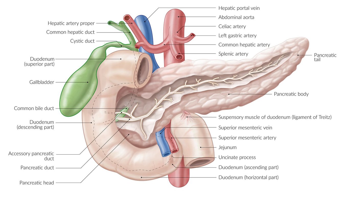
Pancreas Location
- In the abdominal cavity, between the duodenal curvature and the splenic hilum
- Secondary retroperitoneal organ
- Caudal to the omental bursa
- At vertebral level L1/L2
- Head is located within the C-shaped duodenal curvature and contains the pancreatic duct and distal common bile duct
- Uncinate process of pancreas → an extension of the pancreatic head that is located posterior to the superior mesenteric vessels (the remainder of the pancreas is not)
- Neck → lies anterior to the portal vein
- Body → lies anterior to the aorta and extends to the left kidney
- Tail → lies in the splenorenal ligament and extends to the splenic hilum. The distal segment is intraperitoneal.
The uncinate process is _______ to the superior mesenteric vessels. The head, body, and tail of the pancreas lie ______ to the superior mesenteric vessels.
Posterior; anterior
Tumors in the pancreatic ____ often cause bile duct obstruction and can manifest with painless jaundice (Courvoisier sign).
Head
Innervation of the Pancreas
- Innervation → celiac ganglia
- Sympathetic fibers from T6–12
- Parasympathetic fibers from the vagus nerve
Lymphatics of the Pancreas
Celiac, superior mesenteric, and splenic lymph nodes → paraaortic lymph nodes
Vasculature of the Pancreas
Arteries
- Because the pancreas is embryologically derived from the foregut, it mainly receives arterial supply from the celiac trunk and its branches.
- Head and neck
- Inferior pancreaticoduodenal branches (from the superior mesenteric artery)
- Superior pancreaticoduodenal branches (from the gastroduodenal artery)
- Superior and inferior pancreaticoduodenal branches form anastomoses between the celiac trunk and superior mesenteric artery
- Body and tail branches of the splenic artery (itself a branch of the celiac trunk) (the branches of the splenic artery that supply the pancreas include the dorsal pancreatic artery, the great pancreatic artery, and the inferior pancreatic artery)
- Head and neck
Veins
- Head and neck:
- Pancreatic veins → superior mesenteric vein → portal vein
- Body and tail:
- Pancreatic veins → splenic vein → portal vein
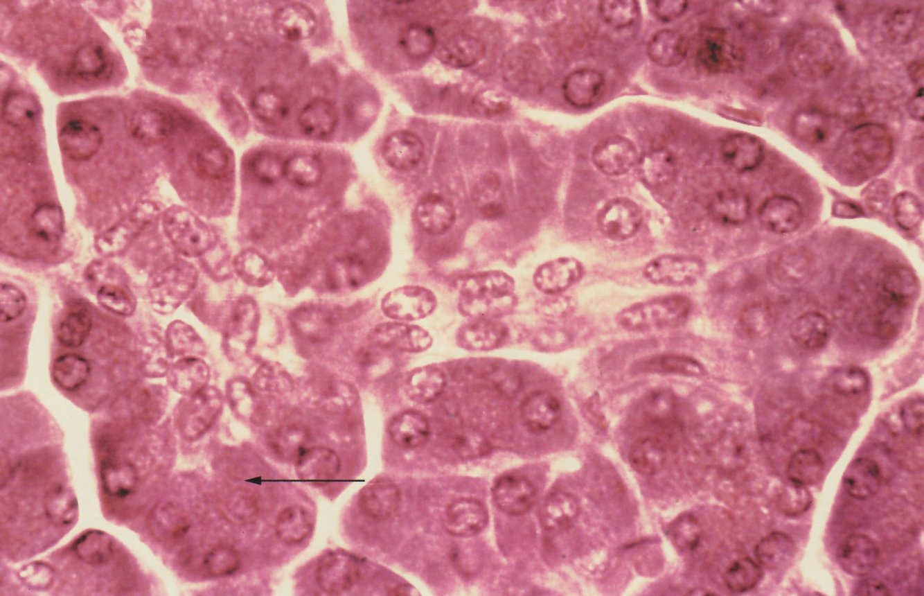
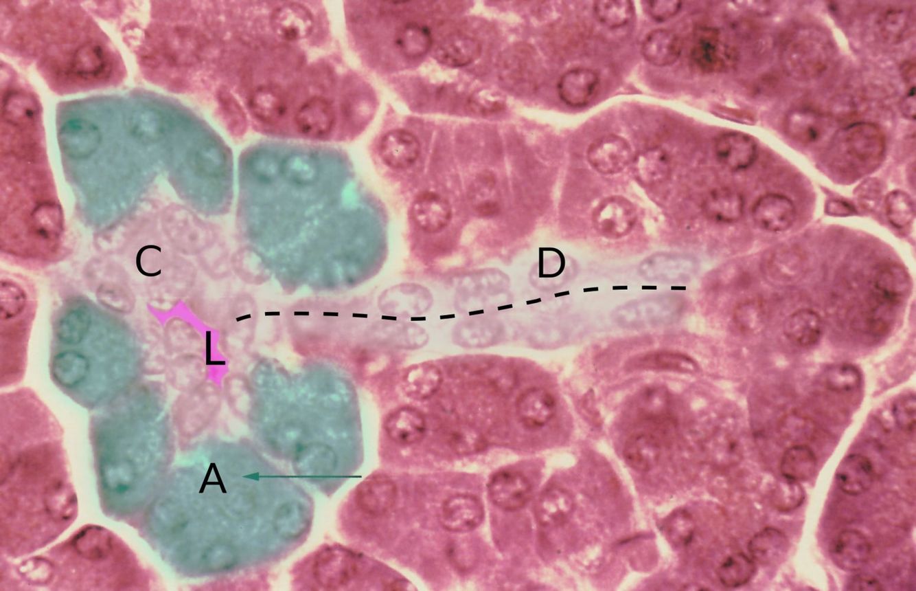
Microstructure of the exocrine pancreas
The functional unit (acinus) of the exocrine pancreas is visible:
- Acinar cells (green overlay; example indicated by A, arrow)
- Centroacinar cells (white overlay; example indicated by C)
- Acinar lumen (pink overlay, L) with the intercalated duct (dashed line, D)
Exocrine Pancreas
- > 90% of the pancreas
- Produces digestive enzymes that are secreted into the gastrointestinal tract
- Composed of serous glandular tissue that is separated into lobules by collagenous septae (septae contain blood and lymphatic vessels, nerves, and excretory ducts)
- Pancreatic acini
- Units of secretory acinar cells surrounding a small lumen
- Secrete proenzymes (e.g., trypsinogen, chymotrypsinogen) into intercalated ducts → ducts eventually merge to form the pancreatic duct → duct carries the enzymes out of the pancreas and to the duodenum
- Centroacinar cells → pale cells in the center of the acini, which secrete bicarbonate ions into the pancreatic fluid
- Electron micrographs of acinar cells show:
- Basophilic rough endoplasmic reticulum at the basal pole
- Eosinophilic proenzyme granules at the apical pole
- Histologically, the exocrine pancreas closely resembles the salivary glands. However, unlike the salivary glands, the pancreatic exocrine glands lack myoepithelial cells in the acini and do not possess striated ducts. Additionally, centroacinar cells are unique to the pancreas.

Endocrine Pancreas
- Produces different hormones that are primarily involved in the regulation of blood glucose levels
- Composed of islets of Langerhans embedded within the exocrine pancreas
- Islet cell types are dispersed throughout the pancreas.
- Alpha cells produce glucagon.
- Beta cells produce insulin.
- Delta cells produce somatostatin.
- Epsilon cells produce ghrelin.
- Pancreatic polypeptide cells (formerly gamma cells) produce pancreatic polypeptide (PP).
- Islet cell types are dispersed throughout the pancreas.
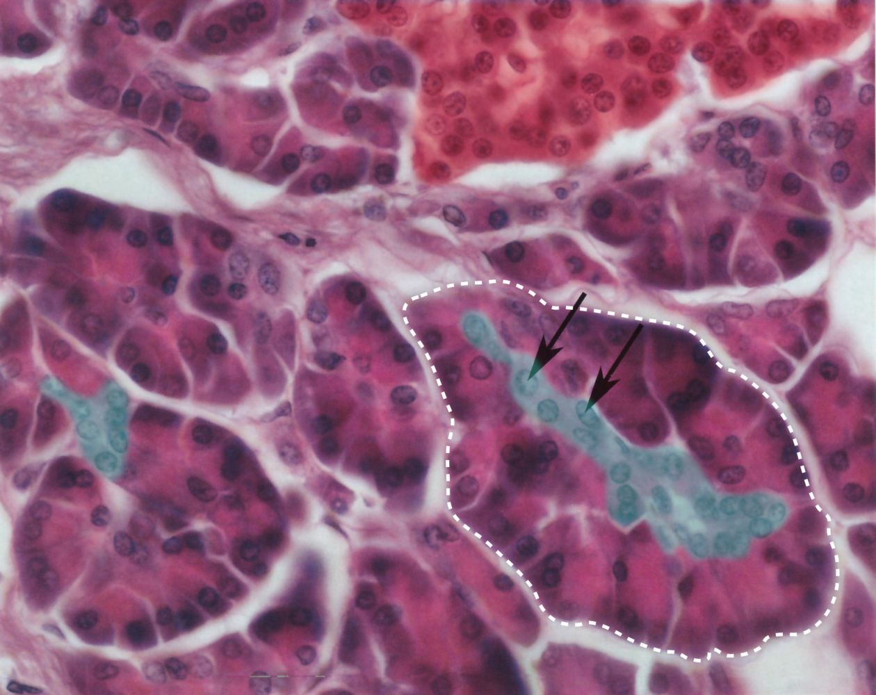
Parenchyma of pancreas
Composed predominantly of deeply staining exocrine acinar cells (example indicated by white dashed outline) and a few scattered islets of Langerhans composed of pale-staining endocrine cells (red overlay). The acinar cells are pyramidal in shape. They have a basophilic base (because of an abundance of rough endoplasmic reticulum) and an eosinophilic apex (because of an abundance of zymogen granules). Centroacinar cells (green overlay; black arrows) are the epithelial cells of the pancreatic ducts and are identifiable as pale-staining cells located centrally within the acini. Centroacinar cells secrete bicarbonate. The Islets of Langerhans are composed of clusters of pale, round endocrine cells that produce insulin, glucagon, and somatostatin.
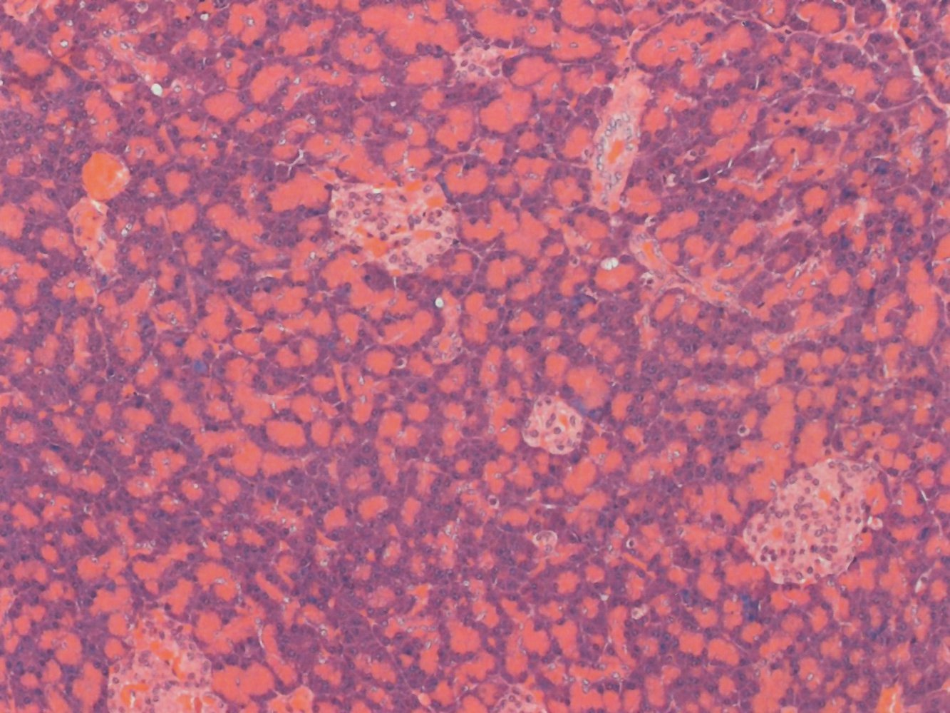
Pancreatic acini and islets of Langerhans
Pancreatic parenchyma is composed predominantly of deeply staining exocrine acinar cells among which are scattered islets of Langerhans composed of pale-staining endocrine cells. Acinar cells are pyramidal in shape. The base of acinar cells is basophilic (examples indicated by white arrows) because of an abundance of rough endoplasmic reticulum and basally located nuclei. The apex of acinar cells is brightly eosinophilic (examples indicated by white arrowheads) because these cells contain abundant zymogen granules. The endocrine cells of the islets of Langerhans (green overlay) are pale and round and produce insulin, glucagon, and somatostatin.
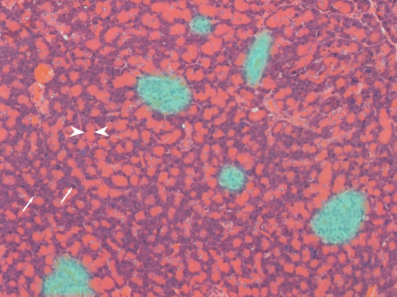
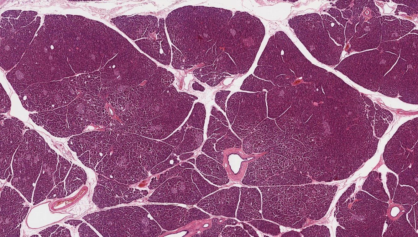
Parenchyma of the pancreas
The pancreas is encapsulated by a thin connective tissue capsule and surrounded by peripancreatic fat. The pancreatic parenchyma is divided into lobules by connective tissue septae in which lie blood vessels, nerves, and branches of the pancreatic ducts. Each lobule of the pancreas is composed of clusters of dark-staining exocrine acinar cells that contain pancreatic zymogens. Scattered amid the acinar cells are the islets of Langerhans composed of pale-staining endocrine cells that secrete pancreatic hormones. Centroacinar cells are the epithelial cells of the pancreatic ducts and are located at the center of the pancreatic acini. Centroacinar cells secrete bicarbonate.
Pancreatic Ducts
- Smaller ducts have cuboidal epithelium.
- Larger interlobular ducts have columnar epithelium.
- Most pancreatic malignancies are adenocarcinomas that originate in the ductal epithelium.
Exocrine Pancreatic Secretions
The pancreatic fluid is isotonic.
It contains the following:
- Digestive pancreatic enzymes
- Pancreatic proteases → digestion of proteins
- Secreted as inactive proenzymes (zymogens) by pancreatic acinar cells into the pancreatic duct (in their active form, proteolytic enzymes would cause autodigestion of the pancreatic tissue. This can occur in acute pancreatitis)
- Trypsin and chymotrypsin
- Proenzymes: trypsinogen and chymotrypsinogen
- Activated in the duodenum → trypsinogen is activated to trypsin by enterokinases, which are located at the brush border of the duodenal and jejunal mucosa.
- Once activated, trypsin activates chymotrypsinogen to form chymotrypsin and, furthermore, converts additional trypsinogen molecules to trypsin (positive feedback loop).
- Elastase (activated by trypsin)
- Digestion of elastin fibers
- Activated by trypsin
- Carboxypeptidase → activated by trypsin
- Nucleases → digestion of RNA/DNA
- Phospholipase A → digestion of phospholipids
- Pancreatic amylase (secreted in active form) → digestion of carbohydrates
- Pancreatic lipase → digestion of lipids
- Pancreatic proteases → digestion of proteins
- Electrolytes (Na+, K+, Cl-, HCO3-) → concentration of Cl- and HCO3- increases with the rate of pancreatic juice secretion
- Water


-
Micro14
-
Public Health Formulas39
-
Public Health262
-
MICRO PHARM198
-
Formulas34
-
IMMUNO PHARM80
-
AUTONOMIC DRUGS131
-
SIDE EFFECTS57
-
PATHOLOGY179
-
CHEMOTHERAPEUTIC DRUGS77
-
PSYCH DRUGS70
-
CARDIO242
-
RESPI2
-
IMAGES207
-
CARDIO PHARM113
-
ENDOCRINE146
-
IMAGES 2230
-
IMAGES 3225
-
ENDOCRINE PHARM89
-
GASTRO-1263
-
GASTRO PHARM83
-
IMAGES 4204
-
HEMATO0
-
MSK0
-
MSK DRUGS0
-
NEURO0
-
NEURO DRUGS0
-
PSYCH0
-
RENAL0
-
RENAL DRUGS0
-
REPRO0
-
REPRO DRUGS0
-
RESPI0
-
RESPI DRUGS0
-
BIOCHEM0
-
GASTRO-2248
-
IMAGES 518
-
PHARM COPY125
