Gastritis Etiology
- Infectious
- Bacterial → e.g., H. pylori, Mycobacterium tuberculosis, Treponema pallidum
- Viral → e.g., CMV, EBV
- Fungal → e.g., Candida spp.
- Parasitic → e.g., Anisakis spp.
- Noninfectious
- Alcohol
- Medications → e.g., NSAIDs, aspirin
- Chemotherapy
- Radiation
- Systemic diseases
- Crohn disease
- Vasculitis → e.g., IgA vasculitis, ANCA-associated vasculitis
- Mesenteric ischemia (e.g., due to external compression or stenosis of the superior mesenteric artery)
- Immune-mediated
- Autoimmune metaplastic atrophic gastritis (AMAG)
- Lymphocytic gastritis, eosinophilic gastritis
- Non-IgE mediated food allergies
- Other
- Ménétrier disease
- Physiological stress → e.g., trauma, burns, critical illness
- Idiopathic
Erosive Gastritis
- Multiple superficial erosions that do not extend beyond the muscularis mucosae and may occasionally cause bleeding
- Stress gastritis → reflecting end-organ damage of the stomach caused by critical illness (e.g., sepsis, shock)
- Chemical gastritis → caused by endogenous secretions (e.g., bile reflux) or exogenous substances. Common causes include NSAIDs, aspirin, alcohol, caffeine, corrosive substances, certain supplements (eg, iron, potassium), and bile reflux.
- May progress to an ulcer
Nonatrophic Gastritis
- Chronic inflammation of the gastric mucosa without atrophic changes
- The mucosa may recover with minimal sequelae or begin to atrophy.
Atrophic Gastritis
- Chronic inflammation of the gastric mucosa, which results in loss of the native glands
- Inflammatory changes are replaced over time by fibrosis or metaplastic changes (i.e., intestinal metaplasia or pseudopyloric gland metaplasia)
Chronic Gastritis
- Inflammation of the gastric mucosa characterized by a predominantly mononuclear infiltrate and loss of the normal architecture of the tissue (e.g., loss of normal mucosal glands)
- Mucosal inflammation, often leading to atrophy (hypochlorhydria → hypergastrinemia) and intestinal metaplasia (↑ risk of gastric cancers)
- Often used to describe long-term or recurring symptoms of suspected gastritis
- H. pylori
- Most common
- ↑ risk of peptic ulcer disease, MALT lymphoma
- Affects antrum first and spreads to body of stomach
- Autoimmune
- Autoantibodies to the H+/K+ ATPase on parietal cells and to intrinsic factor.
- ↑ risk of pernicious anemia
- Affects body/fundus of stomach
- H. pylori
Acute Gastritis
- Inflammation of the gastric mucosa, predominantly by a neutrophilic infiltrate
- Often used to describe self-limiting symptoms of suspected gastritis
- Erosions can be caused by:
- NSAIDs (↓PGE2 → ↓ gastric mucosa protection)
- Burns (Curling ulcer) (hypovolemia → mucosal ischemia)
- Brain injury (Cushing ulcer) (↑ vagal stimulation → ↑ ACh → ↑H+ production)
- Especially common among patients with alcohol use disorder and those taking daily NSAIDs (eg, for rheumatoid arthritis)
Curling ulcer
- Acute gastritis
- Due to burns (hypovolemia → mucosal ischemia)
Cushing ulcer
- Acute gastritis
- Due to brain injury (↑ vagal stimulation → ↑ ACh → ↑H+ production)
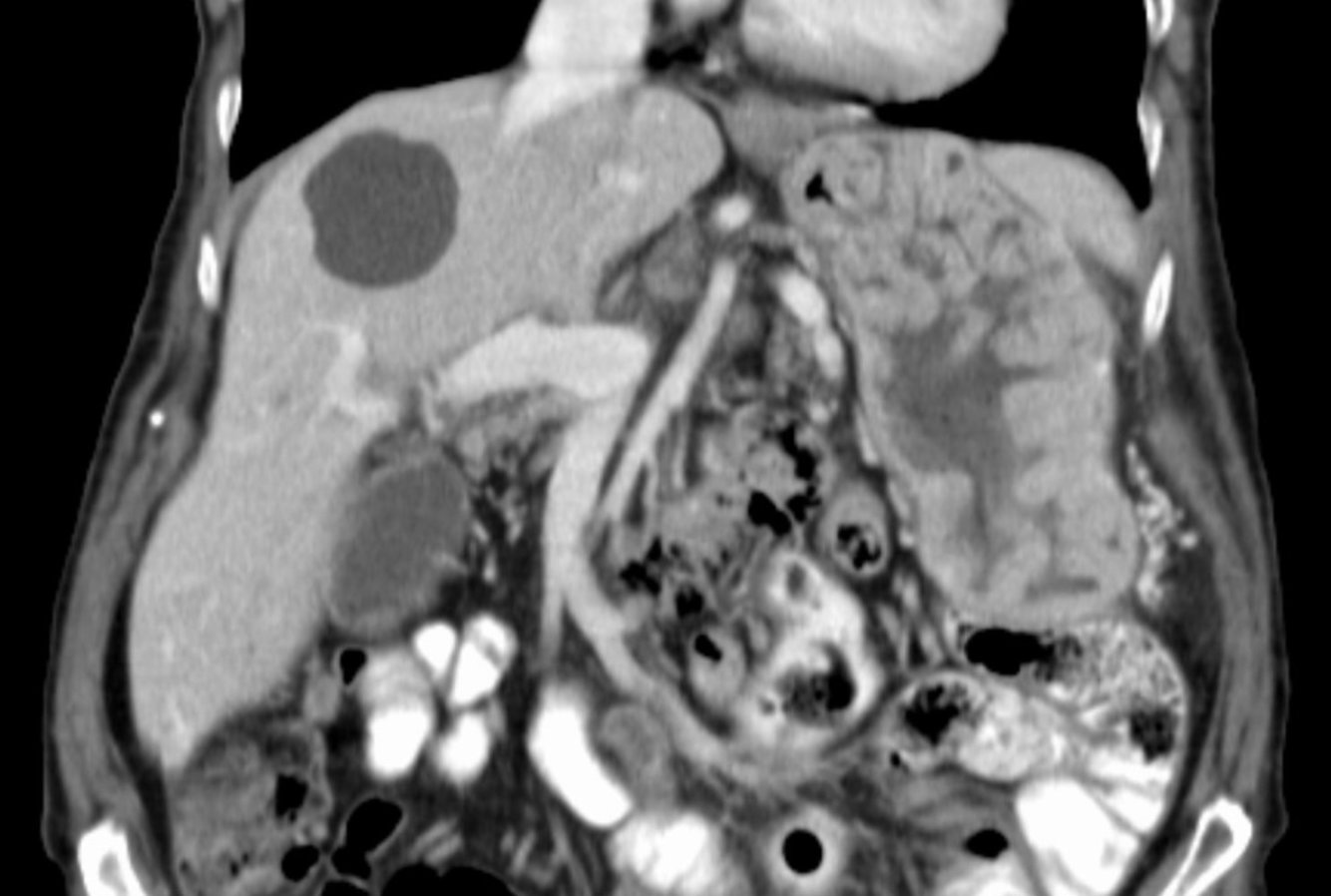
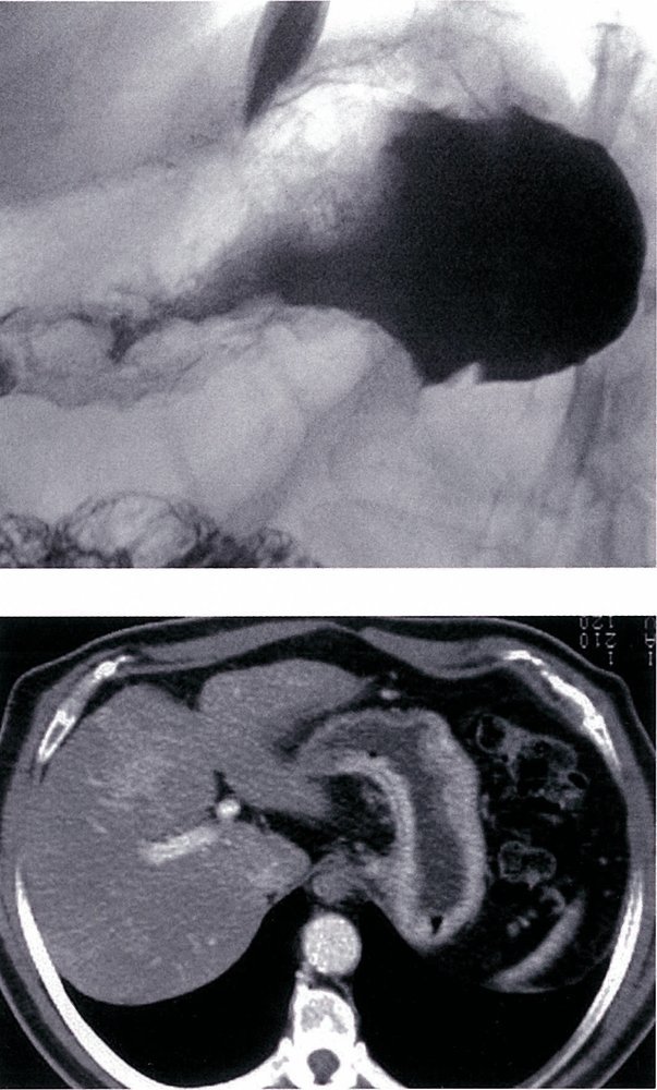
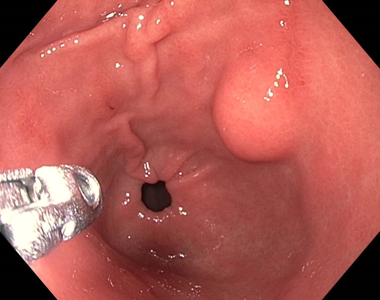
Ménétrier Disease
- Gastritis featuring massive enlargement of the mucosal folds
- Associated with excessive TGF-α
- Foveolar hyperplasia leads to:
- Increased mucus production → loss of protein
- Atrophy of parietal cells → decreased gastric acid production
- Hyperplasia of gastric rugae
- Clinical features
- Dyspeptic symptoms (i.e., abdominal pain, nausea, vomiting, diarrhea, weight loss)
- Protein-loss gastropathy → hypoalbuminemia and peripheral edema
- Consider full-thickness biopsy to exclude malignancy.
- Management
- Most patients
- Supportive care → high-protein diet and possibly IV albumin infusions
- Severe cases with persistent protein loss
- First line → cetuximab (EGF receptor antibody)
- Second line → total gastrectomy
- Most patients
- Complications
- Peripheral edema
- Malignant degeneration

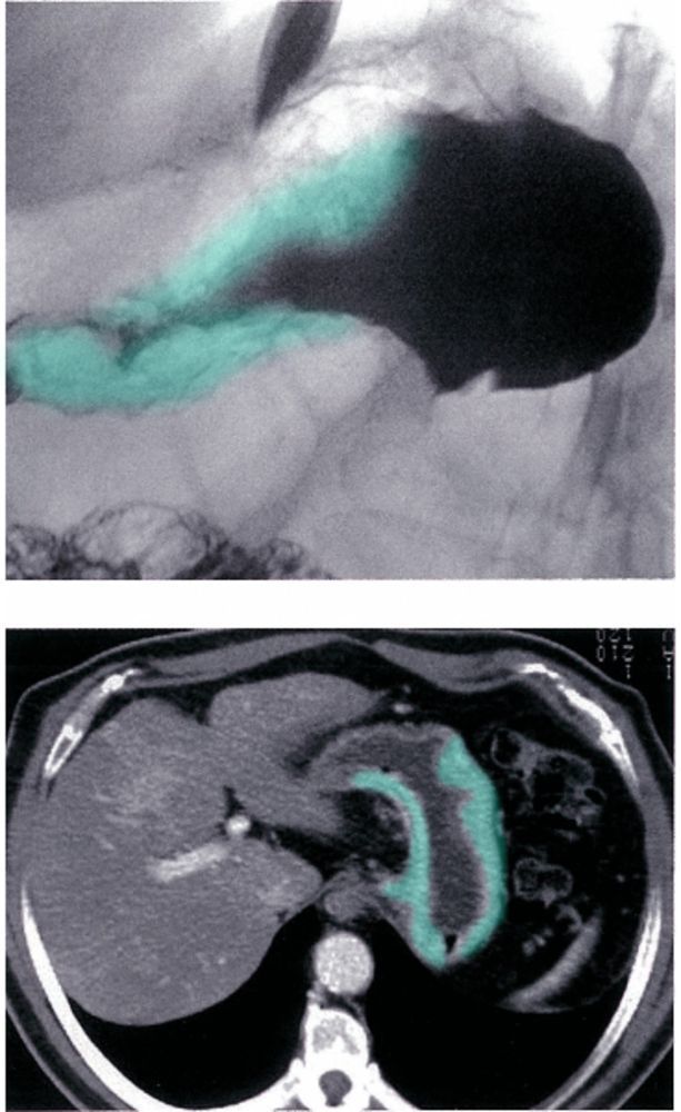
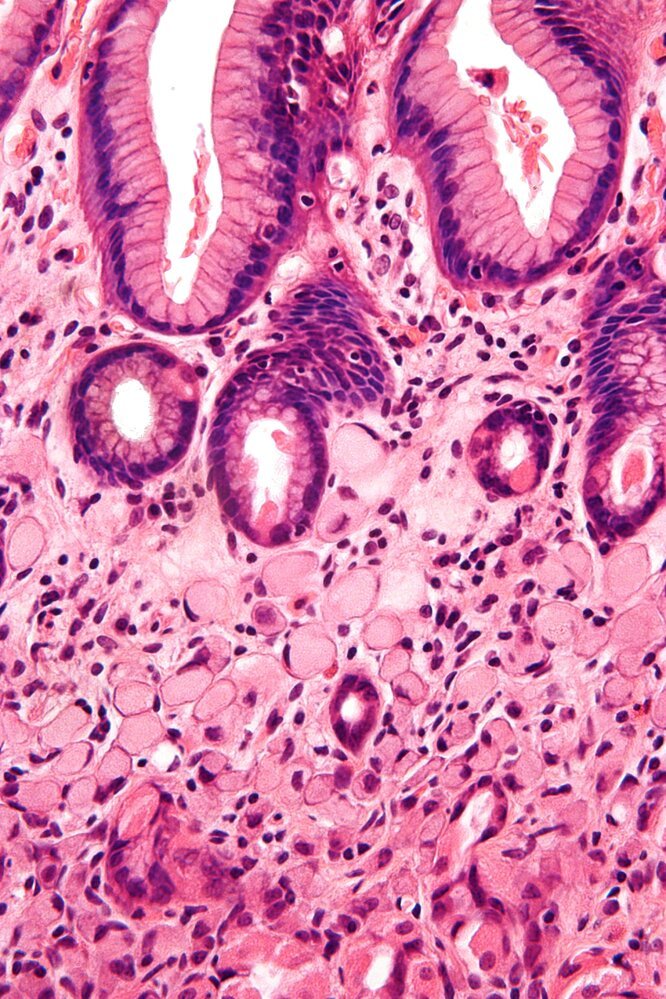
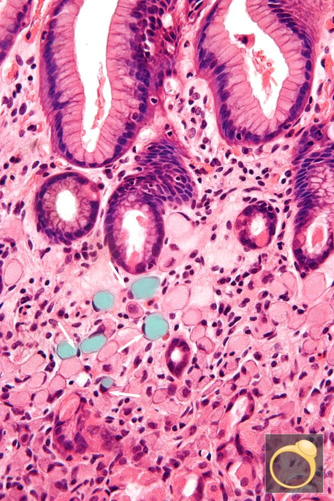
Hypertophic Gastropathy
(Menetrier Disease)
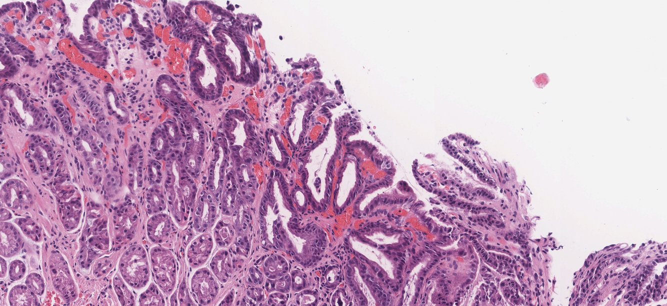
Foveolar Hyperplasia
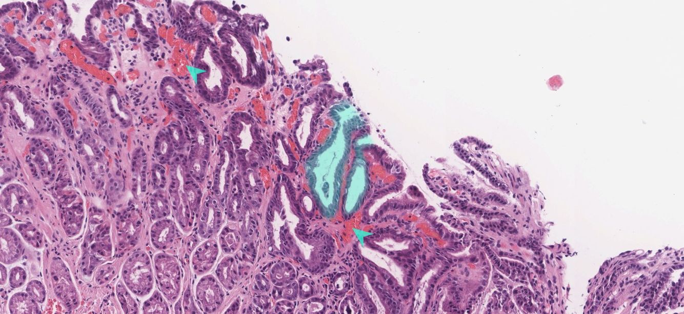
Green overlay → foveolar hyperplasia, i.e. hyperplastic antral glands
Green arrows → hyperemia in the lamina propria
Gastric Cancer Risk Factor
- Exogenous risk factors
- Diet rich in nitrates and/or salts (e.g., dried foods, foods preserved by curing or smoking) (bacteria are believed to convert ingested nitrates into carcinogenic nitrites) and low in fresh vegetables containing antioxidants
- H. pylori infection
- Nicotine use
- Epstein-Barr virus (exact pathomechanism is unknown, but, in rare cases, EBV can cause gastric adenocarcinoma)
- Low socioeconomic status
- Obesity
- Endogenous risk factors
- Gastric conditions
- Chronic atrophic gastritis and associated pernicious anemia
- Achlorhydria (e.g., due to Ménétrier disease)
- Gastric ulcers
- Partial gastrectomy
- Adenomatous gastric polyps
- Gastroesophageal reflux disease
- Hereditary factors
- Positive family history
- Blood type A
- Hereditary nonpolyposis colorectal cancer
- Gastric conditions
Virchow Node
- Left supraclavicular adenopathy
- Sign of gastric metastasis
Sister Mary Joseph Node
- Palpable umbilical nodule
- Sign of gastric metastasis

Blumer Shelf
- Palpable mass on digital rectal examination
- Sign of gastric metastasis
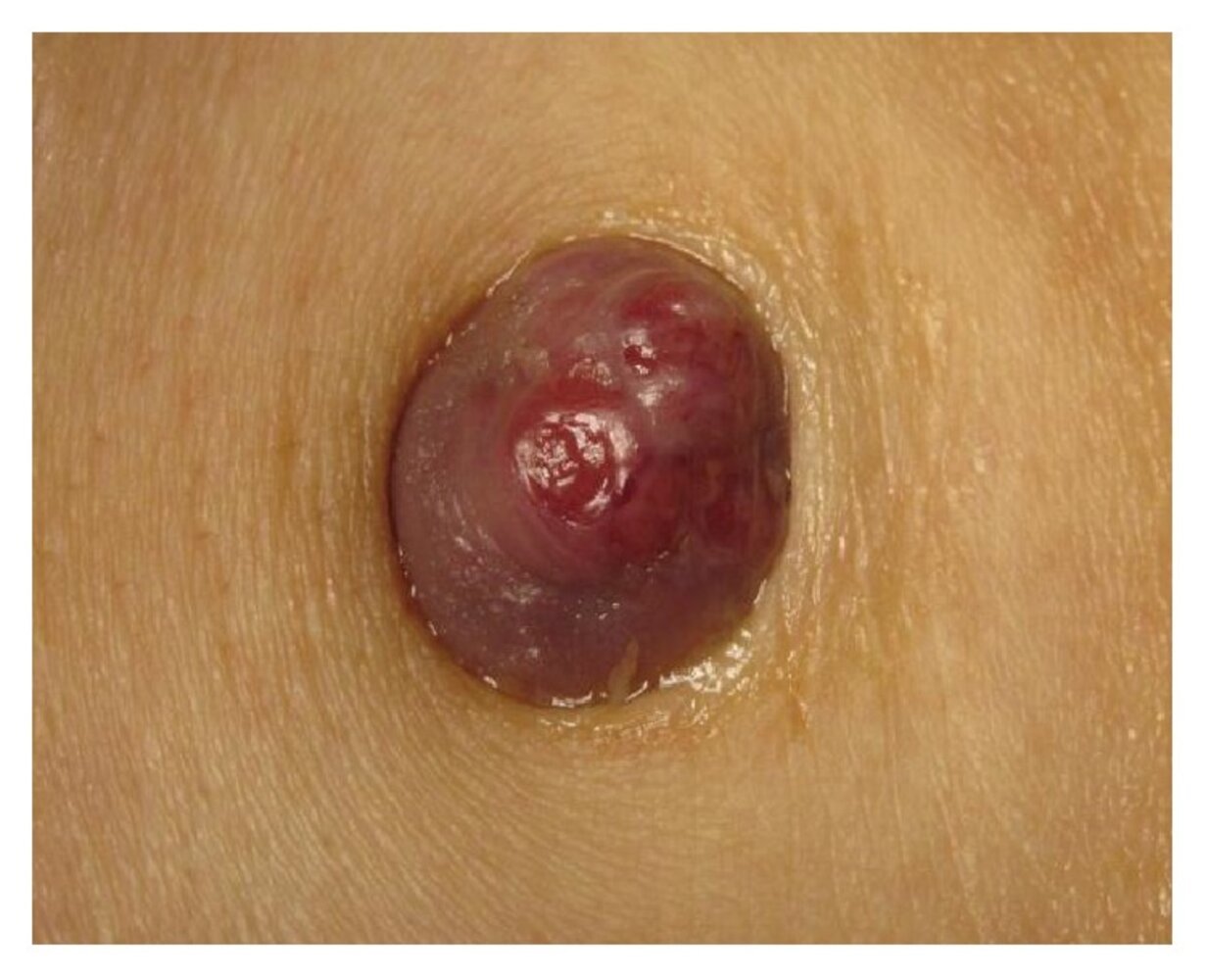
Sister Mary Joseph Nodule
A 2-cm, well-demarcated, protuberant, round, purple nodule is visible in the umbilicus. The lesion shows central ulceration and an accumulation of fluid can be seen in the umbilical crease.
This finding suggests a metastatic lesion of an intraabdominal or pelvic malignancy, referred to as a Sister Mary Joseph nodule. Blood tests, imaging, and histopathological analyses of biopsy materials are required to confirm the diagnosis
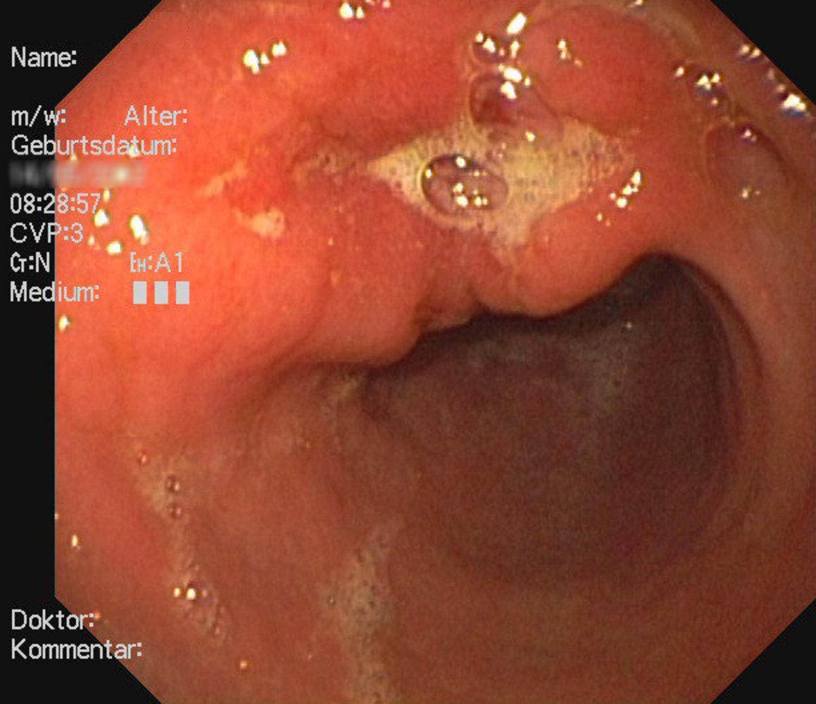
Gastric Cancer
Endoscopy view of the gastric antrum
There is a gastric mass at the level of the lesser curvature with an irregular margin (perimeter marked by green outline) and central ulceration (green overlay).
These findings are consistent with gastric cancer.
Gastric cancer
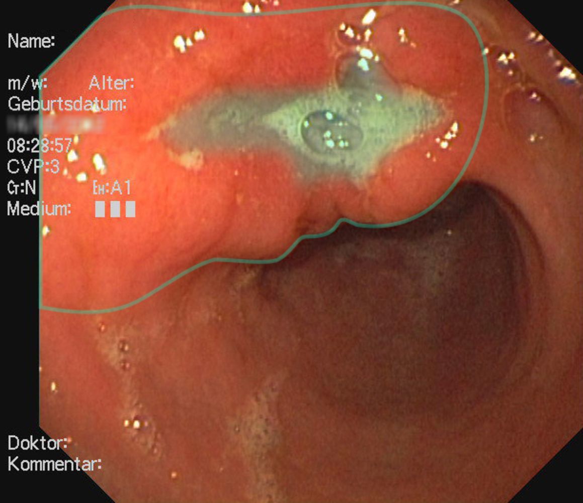
Endoscopy view of the gastric antrum
There is a gastric mass at the level of the lesser curvature with an irregular margin (perimeter marked by green outline) and central ulceration (green overlay).
These findings are consistent with gastric cancer.
Gastric Cancer Markers
- Serologic markers
- Tumor markers → CA 72-4, CA 19-9, CEA
- TNF-α (colonization with H. pylori leads to chronic inflammation, which is caused by increased production of proinflammatory cytokines (e.g., TNF-α). Elevated levels of TNF-α increase the risk of gastric cancer)
- Immunohistochemistry → HER2 testing (HER2/neu gene amplification/overexpression has been associated with the development of gastric cancer. Individuals with HER2/neu-positive status showed decreased survival time, more rapid tumor progression, and higher tumor stages)
Intestinal Type Gastric Carcinoma
- Arises from glandular cells in the stomach
- Typically localized (this means it requires a smaller safety margin for resection compared to the diffuse type)
- Typically seen in older patients
- Polypoid, glandular formation
- Similar to an ulcerative lesion with clear raised margins
- Commonly located on the lesser curvature
Diffuse Type Gastric Carcinoma
- No clear border (potential tissue invasion calls for a wider safety margin during resection to ensure that all malignant cells are removed)
- Spreads earlier in the course of disease
- Infiltrative growth
- Diffuse spread in the gastric wall (even parts of the gastric wall that macroscopically appear to be healthy may have already been infiltrated by diffuse tumor cells)
- Linitis plastica → gastric wall thickening and leather bottle appearance
- Composed of signet ring cells → round cells filled with mucin, with a flat nucleus in the cell periphery
- Associated with E-cadherin mutation (mutations lead to low expression of the E-cadherin protein. Due to its role in cell adhesion and differentiation, E-cadherin protects against tumor formation. Low expression is associated with poorer prognosis (e.g., increased depth of invasion or severe lymph node involvement))

Gastric Signet Ring Cell Carcinoma
Multiple cells with peripheral nuclei (examples indicated by white arrows) and engorged cytoplasm (examples indicated by green overlay) are visible.
These morphological features are characteristic of gastric signet ring cell carcinoma.
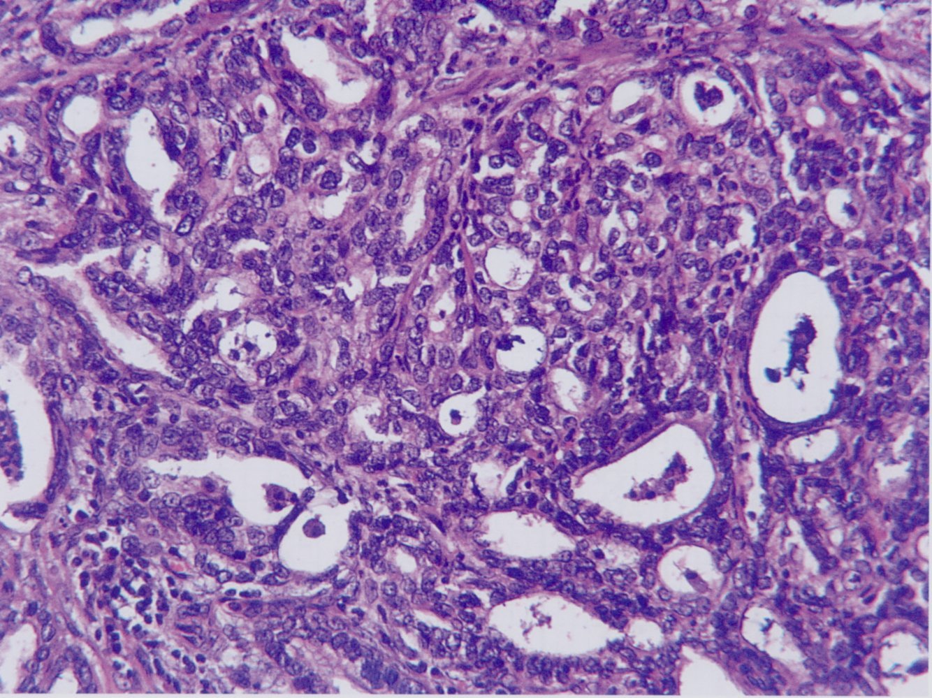
Gastric Adenocarcinoma
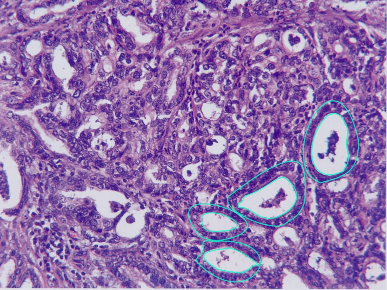
There are atypical cells forming glandular structures (green circles) of various sizes with intraluminal debris.
These findings are consistent with gastric adenocarcinoma.
Gastrointestinal Stromal Tumor (GIST)
- Malignant mesenchymal neoplasm of the gastrointestinal tract that arises from interstitial cells of Cajal or precursor cells
- Associated with c-KIT gene mutations and PDGFRA gene mutations
- Mutations in c-KIT or PDGFRA → phosphorylation of receptor tyrosine kinases → perpetuatal, ligand-independent activation of downstream effectors → ↓ apoptosis and ↑ cellular proliferation → neoplasia
- Localization (localization outside the stomach is considered a clinical sign of malignancy and progression)
- Stomach (60%)
- Small intestine (35%, more frequent in familial forms and syndromes)
- Colon, rectum, esophagus, or omentum (5%)
- Clinical features
- Small tumors (< 2 cm) → often asymptomatic
- Large tumors (> 2 cm)
- Ulceration, bleeding → anemia, melena, and hematemesis
- Obstruction → ileus
- Treatment:
- Involves surgical removal and treatment with tyrosine kinase inhibitors such as imatinib or dasatinib.


-
Micro14
-
Public Health Formulas39
-
Public Health262
-
MICRO PHARM198
-
Formulas34
-
IMMUNO PHARM80
-
AUTONOMIC DRUGS131
-
SIDE EFFECTS57
-
PATHOLOGY179
-
CHEMOTHERAPEUTIC DRUGS77
-
PSYCH DRUGS70
-
CARDIO242
-
RESPI2
-
IMAGES207
-
CARDIO PHARM113
-
ENDOCRINE146
-
IMAGES 2230
-
IMAGES 3225
-
ENDOCRINE PHARM89
-
GASTRO-1263
-
GASTRO PHARM83
-
IMAGES 4204
-
HEMATO0
-
MSK0
-
MSK DRUGS0
-
NEURO0
-
NEURO DRUGS0
-
PSYCH0
-
RENAL0
-
RENAL DRUGS0
-
REPRO0
-
REPRO DRUGS0
-
RESPI0
-
RESPI DRUGS0
-
BIOCHEM0
-
GASTRO-2248
-
IMAGES 518
-
PHARM COPY125
