- Identify the organ
- Name 2 identifiable structures
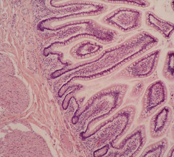
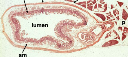
Ileum
Simple columnar epitheium
Goblet cells (secrete mucins)
Absorptive cells
lumen inbetween finger like structures
- Identify this organ
- Name 2 identifiable features
- What notable connective tissue is found here?
1. Espophagus
- Stratified squamous epithelium, not carotinized
Vascular island…
- Lamina proprria (loose connective tissue)
- Identify this organ
- Name 2 identifiable features
- What cells can you see?
- What junctions are in the stratum spinosum?
- THICK skin on sole human foot
- Stratified keratinized squamous
Has all 5 layers… espeically stratum lucidum which means its thick skin
- keratohyalin granules in stratum granulosum
- Desomosomes
- Identify the organ
- Name 2 identifiable features
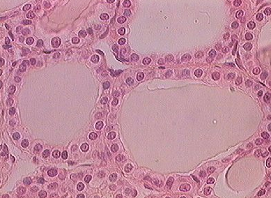
1. Thyroid
- simple cuboidal epitheloum of colloid (thyroid follicles)
spherical nuclei of thyroid cell
Simple squamous epithelium of blood vessels
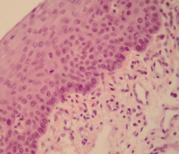
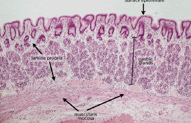
What is this structure? What organ is this slide?
Name 3 cells in this slide
What connective tissue is in this slide?
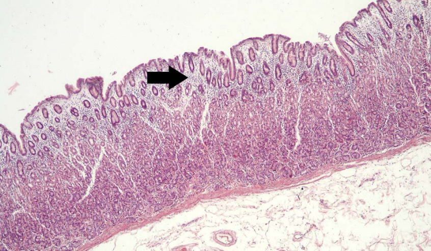
lamina propria in the stomach
- plasma cells
- reticular cells
- eosinophils
below the purple epithelium is LOOSE CONNECTIVE TISSUE
RETICULAR fibers in lamina propria
What organ is this?
- Name 2 identifiable features
- Name 2 types of connective tissue in this cell
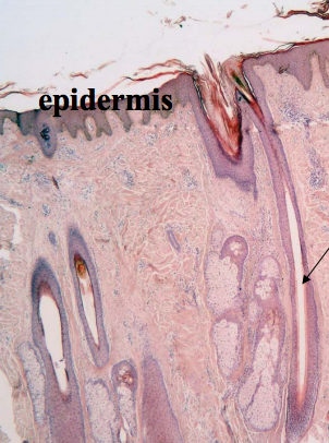
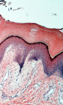
THIN skin of the scalp
- hair follicles, dermal pailla making invaginations into the epidermal layer,
- loose CT in papillary layer, dense irregular tissue in reticular layer
- fibroblasts
What connective is this?
What is the arrow pointing to?
Name 2 identifable features
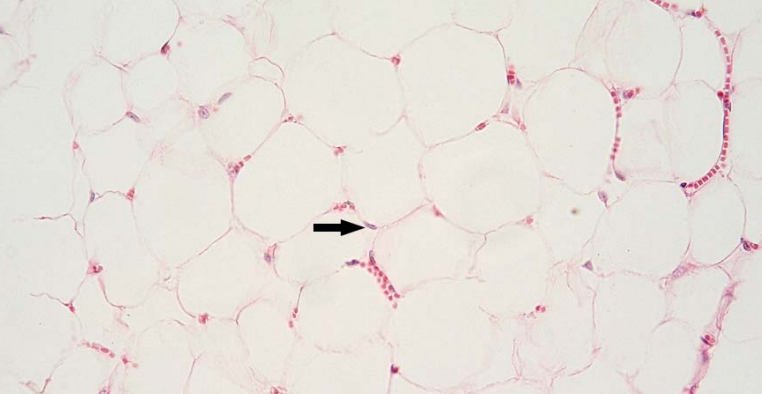
Adipose tissue
Flattened nculei of the addpise tissue
unilocular fat, nuclei pushed ot side of cell
What connective tissue is this?
Name 2 characteristics features
Brown fat adipose tissue
Multilocular fat
centrally located nuclei
high vascular density of brown fat
- What organ is this from?
- Name the connective tissue
- Give 2 identifiable features of CT here?
- where else is this CT found?
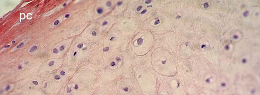
Larynx
- hyaline cartilage in center
- chrondrocytes inside their lacunae (these are often paired!)
hyaline cartiliage beginning to ostify
no vascularture
*this slide is weird because cartialge usually picks up blue stain
- articular surfaces of joints, epiphyseal plate, tracheal rings, ventral ends of ribs
- Classify the epidermis of this slide
- What connective tissue is present?
- what identifiying features are present
- where else in the body is this found?
stratified squamous keratinized epithelium
2 elastic cartilage
- numerous elastin fibers, chrondocytes
- external ear, epiglottis, eustachian tubes
***CLOSE DIAPHRAGM to see elastic fibers to differentiate this between this slide and larynx
What connective tissue is present?
What cells can you see?
What muscle tissue is here?
- dense regular cross tissue in the tendon
- fibroblast nuclei
fragmented muscle tissue… tendon is bright pink
fibroblast nucli appear as small dots
- skeletal muscle
What slide is this from?
What connective tissue is prsent?
2 identifiable features
where else in the body is fibrocartilage found?
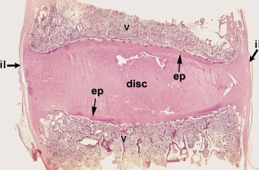
intervertebral disc
dense regular connective tissue in interertebral ligament
hyaline cartilae at the bottom
has fibrocartilage which is uniformly distributed
rounded shape of chrondocytes
pubic symphosis, menisci of knee joint
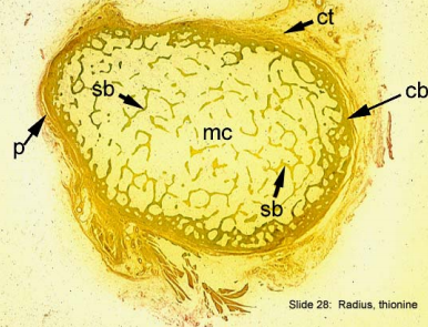
what is this?
waht is it dyed with?
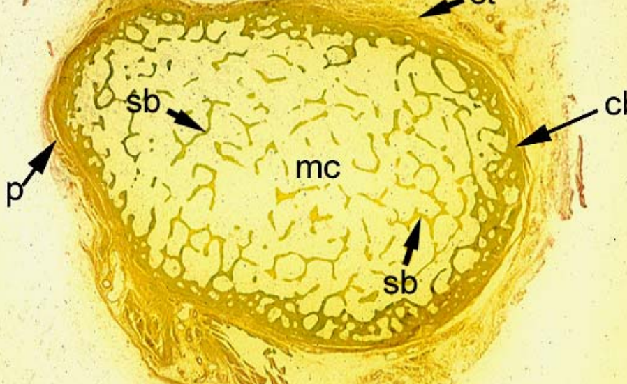
Radius dyed with thionin
e marrow cavity is the large open area
containing trabeculae of spongy bone and bone marrow
spongy bone “spicula” bones.. little lines surrounded by marrow, don’t have haversion canals or volkmans canals
but do have osteocytes

intervertebral disc
dense regular connective tissue in interertebral ligament
has fibrocartilage which is uniformly distributed
rounded shape of chrondocytes
What organ is this?
What connective tissue is present?
Name 2 identifiable features
pinna of ear slide
elastic cartilage
chrondocytes in lacunae
a lot of elastic particles
closed diaphragm and enhanced contrast to make elastic fibers more visible
What slide is this?
Name 2 supporting connective tissues are present?
Name 2 identiying features for each
Cross section of radius
- spongy bone, compact bone
- compact bone: haversion canals (surrounded by concentric lamellae), volksman canal
spongy bone lined by endosteum
Endosteum
Cannaliculi are present
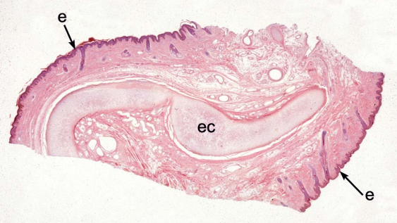
What is the bony structure surrounding teeth?
What cells can you find here?
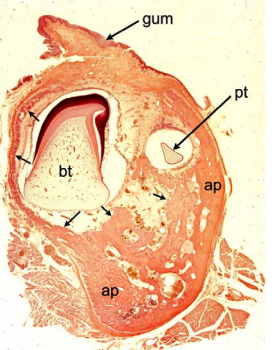
tooth
alveolar process
osteoclasts
multinucleated
osteoblasts
osteoprogenitor
cells.
what organ is this?
what supporting tissue are present
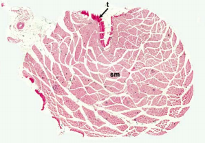
duodenum
smooth muscle in mscularis externa (outer longitudinal and inner circular layers)
less cross striations
cant see borders around fibers
centerally placed nuclei, spindle shaped.
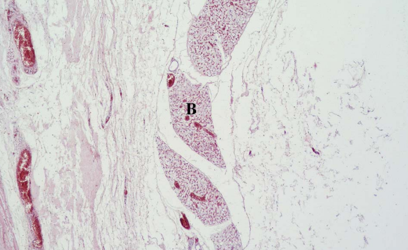
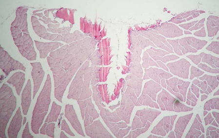
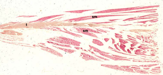
what slide is this?
what connective tissues are present
what muscle tissues are present
what is the endomysin made of? the perimysein?
musculotendon junction
tendon: dense regular CT
skeletal muscle
multinucleated, cross striations
endomysein: reticular fibers
perimyseins: loose CT
what connective tissue present?
what muscle is present?
tendon: dense regular CT
skeletal muscle
multinucleated, cross striations
endomysein: reticular fibers
perimyseins: loose CT
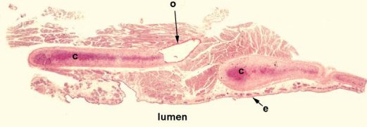
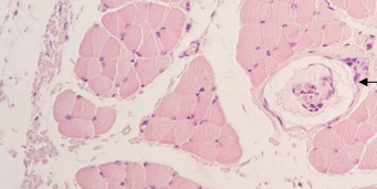
what is this slide?
what muscle is present?
what is covering outside of organ?
what lines the lumen?
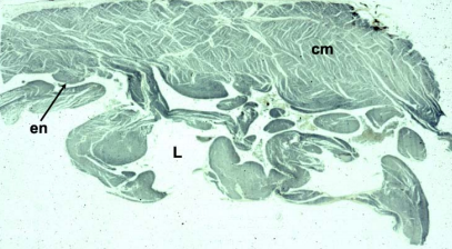
right ventricle
cardiac muscle
nuclei in the center, anastamozeing, see spacees between them (how to tell apart from smooth muscle)
covering out is EPICARDIUM
lining the lumen is the ENDOCARDIUM
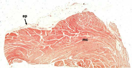
left ventricle
cardiac muscle
centrall placed nuclei, branch and anastamoze
intercalated discs
purkinje fibers at the bottom, responsible for the final distribution fo electrical stimuli to myocardium
what is this tissue?
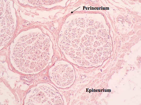
this is nerve tissue
schdmidt lanterman clefts and nodes of ranvier meaning its MYENATED
sciatic nerve
perineurum (made of myofibroblasts and collaen) act as blood nerve barrier
endoneurim: reticular fibers made by schwann cells
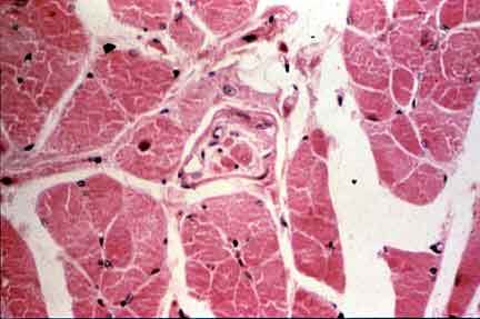
what tissue is this?
what distinguihsing features are here?
skeletal muscle
muscle spindle with intrafusal fibers inside