7.1 In general descriptions, what is the Vestibular System?
Provides sense of balance wrt placement of head in space. Designed to sense motions that arise from head movements as well as the inertial effects due to gravity.
Generally unconscious, but essential for coordination of motor responses, eye movements, posture.
Sensory receptors lie in vestibular labyrinth in inner ear and convey info to primary vestibular sensory neurons.
Primary vestibular sensory neurons relay info via vestibulocochlear nerve (CN VIII) to brainstem and cerebellum where they synapse with projection neurons responsible for eye movements and posture.
Vestibular signals also reach thalamus and cortex, where, with convergence of visual and proprioceptive info, a sense of head position in space is constructed.
7.2 What structures are described by the following?
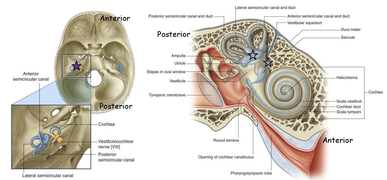
- Convoluted hard walled cave of canals within petrous portion of temporal bone. Filled with perilymph (low K+/high Na+, similar to CSF).
- Convoluted delicate walled sac of ‘ducts’ floating in perilymph and following shape of bony labyrinth canals. Filled with endolymph (high K+/low Na+, similar to intracellular fluid).
- Bony labyrinth (grey star)
- Membranous labyrinth (pale blue star)
NB: petrous portion of temporal bone = purple star
(see attached)
7.3 Differentiate Perilymph from Endolymph
In the vestibular labyrinth:
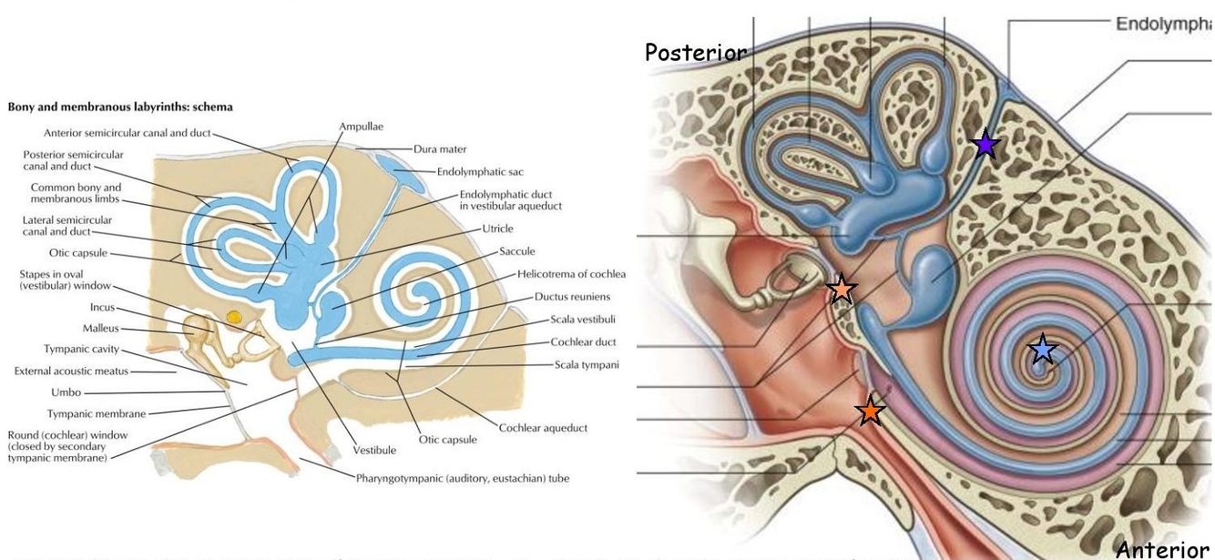
- Perilymph (light orange) secreted by arterioles in periosteum surrounding labyrinth.
- Drains into SA space via perilymphatic duct which runs through cochlear aqueduct (dark orange; see labelled) in temporal bone.
- Endolymph (light blue) secreted by tissue in cochlear duct
- Drains into an extradural sac via endolymphatic duct which runs through vestibular aqueduct (purple) in temporal bone.
Pressure balances in perilymph and endolymph important for functioning of vestibular system. Excess pressure causes vestibular disturbance: Meniere’s disease / Labyrinthitis.
(see attached)
7.4 Label the following sensory organ components of the Vestibular Labyrinth (see attached)
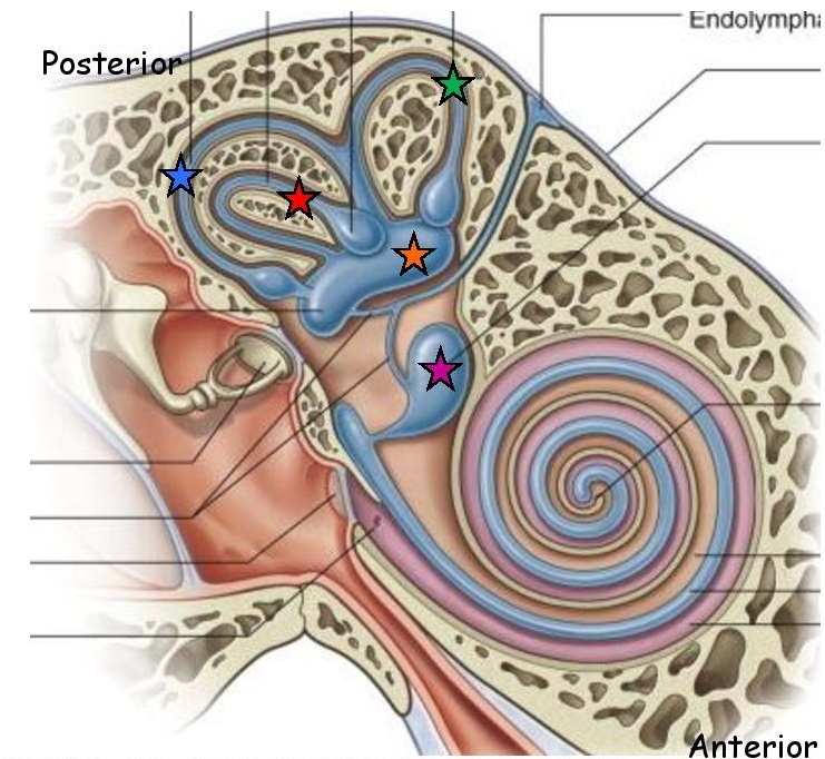
There is 1 vestibular labyrinth on each side of the head. In each inner ear there are:
- 3 semicircular ‘ducts’ in semicircular ‘canals’ (scc):
- Green - anterior scc
- Red - lateral scc
- Blue - posterior scc
- 2 otolith organs in vestibule:
- Orange - utricle
- Magenta - saccule
(To remember positioning of the otolith organs - remember the “tri” in uTRIcle can be likened to the THREE scc’s and thus that the utricle is positioned next to them, as opposed to the saccule, more proximal to the cochlear.)
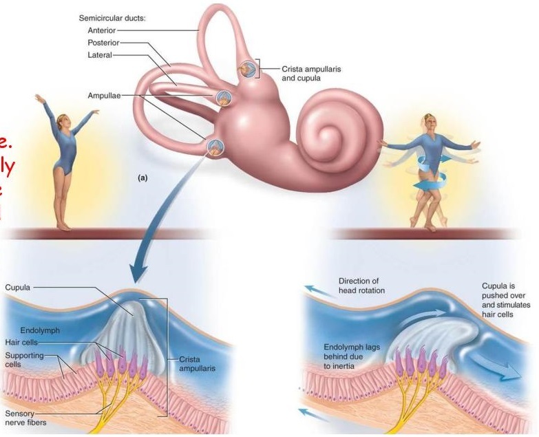
7.5 Describe the semicircular canals in their functional anatomy.
Endolymph-filled ring-like structures that contain sensory receptors that detect endolymph mvmt.
Respond to angular accelerations (rotations) of the head and are maximally sensitive to rotational motions that lie in the plane of the canal:
- Ant - ‘yes’ (nod; sagittal plane)
- Lat - ‘no’ (shake; transverse plane )
- Post - ‘maybe’ (head tilt towards shoulders; coronal plane)
Oriented ‘orthogonally’ - at right angles to each other
Organised into 3 ‘functional’ pairs (based on orientation) - one half of each pair on each side of head:
- Left ant vs right post
- Left lat vs right lat
- Left post vs right ant
‘Ampullae’ house receptive tissue:
- Sensory receptors in scc’s located in the ampullae - dilated ends of each canal near their attachment to utricle inside vestibule
- Endolymph in scc’s and utricle continuous, but at ampulla end - endolymph is partitioned off by flexible septum of receptive tissue - crista ampullaris (see attached) - a ridge that ‘sways’ with the moving flow of endolymph (more detail see 7.6)
For biophysics of scc’s - see 7.7
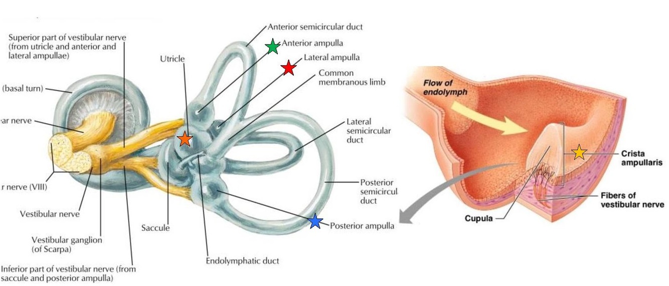
7.6 Describe the purpose of the Crista Ampullaris
Ridge of crista ampullaris (yellow) projects into lumen of ampulla of semicircular canal and is bathed in endolymph (light blue).
Covered by neuroepithelium made of sensory hair cells (green) and supporting cells.
Sensory hair cells’ cilia embedded in gelatinous mass called the cupula (purple).
Hair cells are either excited/inhibited when cupula sways in endolymph and moves wrt neuroepithelium (see hair cell functional anatomy, 7.10, 7.11).
Upon rotational acceleration of the head, the endolymph in the scc’s which are oriented in the plane of the acceleration, is subjected to inertia and bends cupula.
(See attached)
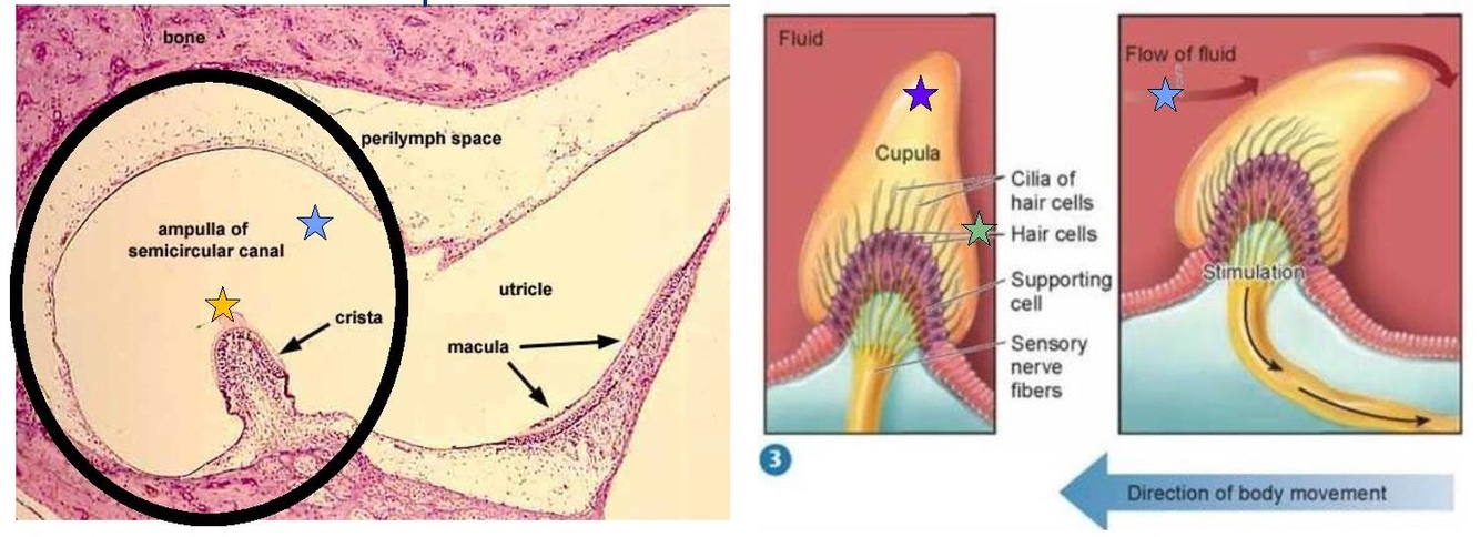
7.7 Describe the biophysics of the semicircular canals.
At the beginning of head rotation within the plane of a semicircular canal (ie spinning = rotational acceleration in transverse plane = lateral canal) endolymph is subjected to inertia and lags behind in canal and so endolymph temporarily moves in opposite direction to head rotation.
Cupula has same specific gravity as endolymph and so ‘sways’ in same direction as endolymph as it experiences inertia. Swaying cupula effectively bends the sensory hair cells embedded in it in the opposite direction to the direction of head rotation.
(see attached)
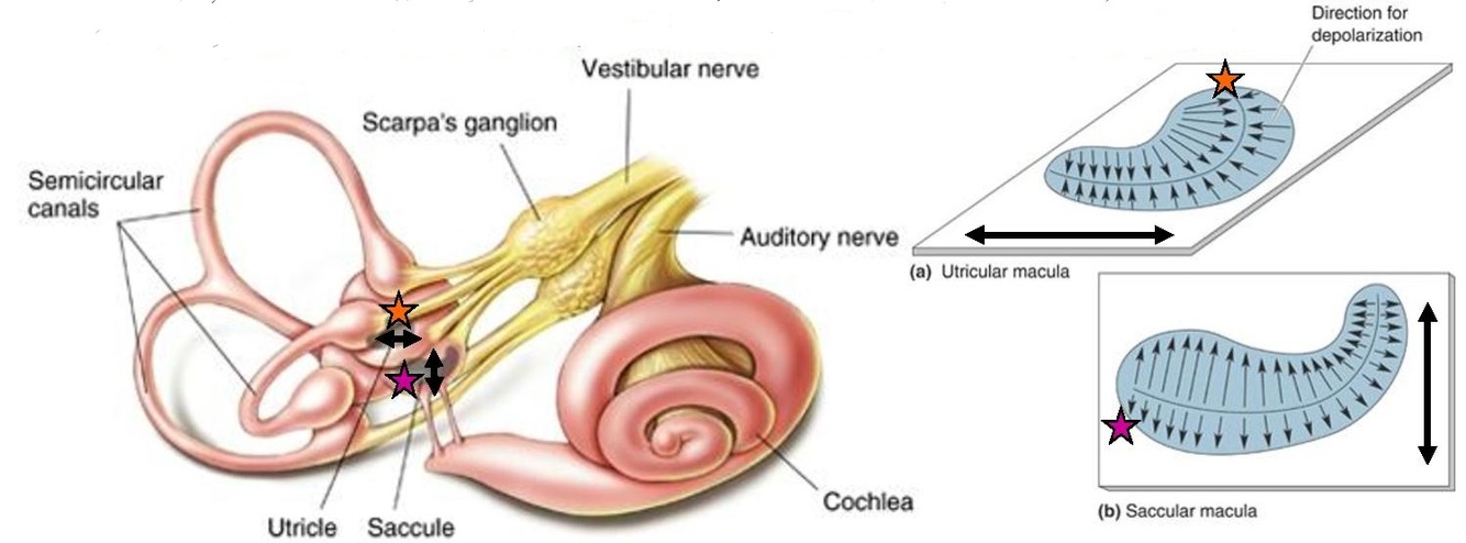
7.8 Discuss how the otolith organs respond to changes in angle (tilt) and linear movements of the head.
The utricle and saccule are endolymph-filled sac-like structures that contain sensory receptors to detect movements dependent on gravity.
They are maximally sensitive to straight line changes in acceleration and direction:
- Utricle (orange) - transverse plane - forward-back, left-right (eg doing a handstand, riding a bike, swaying side to side)
- Saccule (magenta) - sagittal plane - up-down (remember this for saccule by a “sac” - hangs downwards) (eg jumping, riding an elevator)
The floor and wall are lined with receptive tissue. Sensory receptors in otolith organs are maximally sensitive to changes in linear accelerations that occur in the plane along which receptive tissue is orientated.
Receptors are embedded in comma-shaped receptive tissue (neuroepithelial) ‘patches’ - maculae - which are positioned on either the floor or the wall of the otolith organs:
- Macula utriculi - receptors positioned horizontally on floor - oriented to detect linear movements in transverse plane
- Macula sacculi - receptors positioned vertically on medial wall - oriented to detect linear movements in sagittal plane
(see attached)
Although bathed in same endolymph that circulates in semicircular canals, unlike the cristae, maculae are heavier than endolymph and so are more responsive to the pull of gravity rather than changes in endolymph current. (Macula details see 7.9)
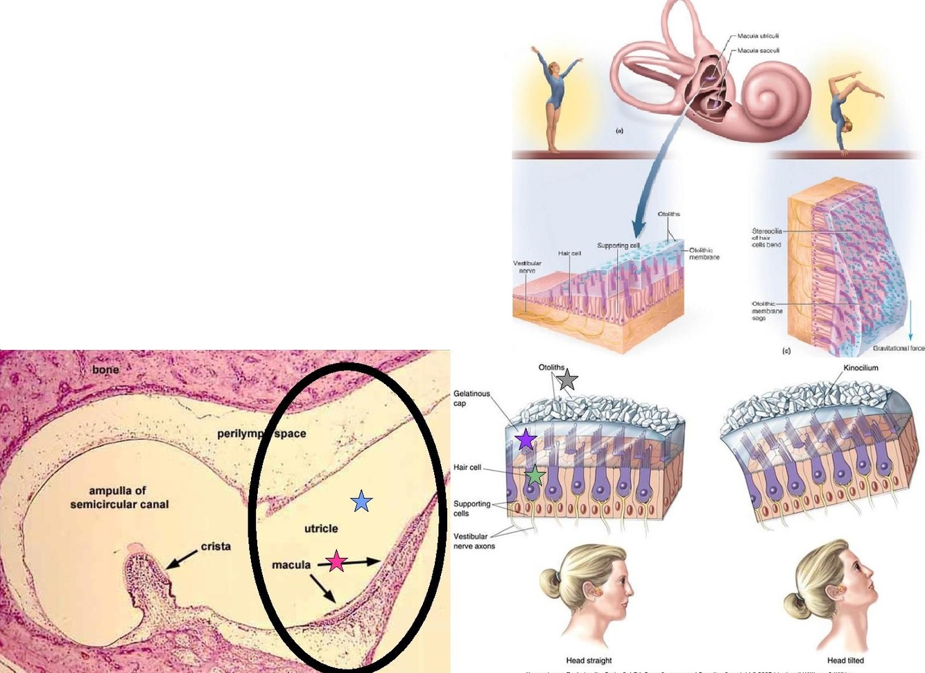
7.9 Describe the purpose of the macula and the biophysics of the otolith organs.
Patches of maculae (pink) project off floor/wall into cavities of utricle/saccule and are bathed in endolymph (blue) (but not moved by endolymph).
Composed of neuroepithelium made of sensory hair cells (green) and supporting cells.
Sensory hair cells’ cilia embedded in gelatinous mass called the otolithic membrane (purple) - this is studded with “weighty” calcium carbonate otoliths (grey)
Hair cells are either excited/inhibited when otolithic membrane moves with respect to neuroepithelium. Because otolithic membrane is weighted, it’s subject to gravitational changes (pulled down when head tilts) as well as to inertia (lags behind when head accelerates)
(see attached)
Biophysics:
- If head is tilted (ie handstand = tilt in transverse plane = wobbles macula utriculi horizontally), the ‘heavy’ gelatinous otolithic membrane is subject to gravity and flops towards the direction of the tilt.
- If head is subjected to linear acceleration (ie jumping up = acceleration in sagittal plane = wobbles macula sacculi vertically), the ‘heavy’ gelatinous otolithic membrane is subjected to inertia and ‘lags’ behind in otolithic organ.
Otolithic membrane is denser than endolymph, so doesn’t ‘sway’ in it.
Upon head tilt or linear acceleration, it’s ‘flop’ and ‘lag’ characteristics effectively bend sensory hair cells embedded in it in same direction as tilt, and in opposite direction to direction of acceleration.
7.10 Describe the sensory hair cells as receptors of the vestibular system.
Sensory hair cells (vestibular hair cells) are accumulated in the ridge of the cristae within the semicircular canals and in the patches of the maculae within the otolith organs.
2 types have been recognised, type…
- (red) Flask shaped, completely surrounded by receptive end of bipolar primary sensory neurons (orange). [use ‘o’ of type ‘one’ to remember rounder shape]
- (blue) Cylinder shaped, contacted directly by receptive end of bipolar primary sensory neurons and motor neurons (light green) [use ‘t’ of type ‘two’ to remember taller shape]
Surrounded by supporting cells and accumulated in sheets of neuroepithelium (cristae or maculae).
Both types have hair tuft made of 30-50 stereocilia (dark green) protruding from surface closest to endolymph.
(Details on kinocilia - see 7.11)
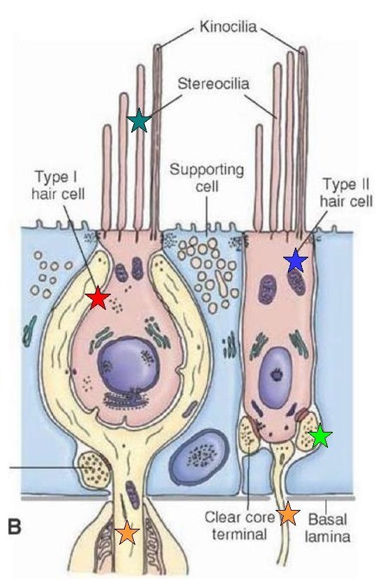
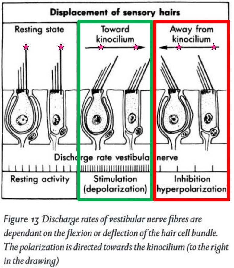
7.11 What is the purpose of the kinocilia of the vestibular hair cells?
Vestibular hair cells similar to auditory hair cells (Organ of Corti) except within each hair tuft of stereocilia there is a single, longer kinocilium projecting on only one side of the cell.
The stereocilia and kinocilium in the hair tuft of a single hair cell all connect via linkages and are embedded in the gelatinous mass that makes up the cupula (semicircular canals) or otolithic membrane (otolith organs)
Movement of gelatinous mass either in response to endolymph flow (scc’s) or gravitational pull (oo’s), bends/displaces the hair tuft
Displacing of hair tuft towards or away from the kinocilium determines whether or not the sensory hair cells release neurotransmitter that stimulates the receptive ends of bipolar primary (vestibular sensory neurons)
- Towards kinocilium - excites hair cell, incr’s nt release, incr’d firing of bipolar primary sensory neurons contacting hair cell
- Away from kinocilium - inhibits hair cell, decr’s nt release, decr’d firing of bipolar primary sensory neurons contacting hair cell
(see attached)
7.12 From where do the vestibular nuclei receive inputs?
- SVN - Cristae (scc’s)
- IVN - Macula in saccule
- LVN - Macula in both otolith organs (saccule, utricle)
- MVN - Cristae (scc’s)
From a different perspective:
- scc’s send info to the SVN and MVN
- maculae send info to the IVN and LVN
- Use the pneumonic: “Can She Melt… Men In Lava”
- C = cristae, M = maculae, the other initial letters represent nucleus position (eg Lateral, ‘Lava’)
- What regions do the following descriptions of projections from the vestibular nuclei represent?
- Fine tune muscle movements
- Controlling eye movements
- Head and body motor control
- Quick reactions of extensor muscles for maintaining balance
- Cerebellum - vestibulocerebellar pathway - (also has some direct connections from 1st-order vestibular sensory neurons!) - fine tune muscle mvmts of head, eyes and those responsible for posture either via direct primary afferent synpase or 2nd-order projection.
- Brain Stem Nuclei - vestibulo-ocular pathway - (occulomotor, trochlear, abducens) - allows eyes to fix on moving object while staying in focus (vestibulo-ocular reflex)
- Thalamus (then to cortex for conscious perception) - vestibulo-thalamo-cortex pathway - allows for head and body motor control and are responsible for conscious awareness of body position
- Spinal Cord - lateral and medial vestibulo-spinal pathways - allow quick reactions of extensor muscles of limbs and trunk necessary for maintaining balance. Influence m. tone and postural adjustments of head and body (vestibulospinal reflex)
-
Mid Sem Miscellaneous (W1-5)29
-
W5 Somatosensory Pathways (Physical Feelings)21
-
L1 Brain Development1
-
L16 Cerebellum16
-
L7 Vestibular System14
-
L8 Proprioception10
-
L6 Auditory Pathways15
-
L15 Motor Pathways19
-
L9 Somatosensory Pathways I10
-
L10 Nociception3
-
L19 Hypothalamus: Feeding2
-
L23 Pain and Suffering1
-
L22 Fear1
-
Spot Test Functions55
-
Brain Stem Sections31
