Functions of renal system
- maintain stable internal environment for optimal cell and tissue metabolism (via solute and water transporting and balance)
- endocrine function - secretes renin (for BP), erythropoietin RBC production), and 1,25- dihydroxy-vitamin D3 (for calcium metabolism)
- release glucose into the circulation by the processes of glycogenolysis and gluconeogenesis
Outer Structure and Location of Kidney
Structure: paired organs in posterior region of abdominal cavity (retroperitoneum). Lie on either side of vertebral column, upper and lower poles go from T12 to L3.
- R kidney slightly lower and displaced downward by overlying lvier
- each kidney ~11cm long, 5-6cm wide, 3-4cm thick
- surrounded by the renal capsule which is embedded in perirenal fat. These layers are further covered with a double layer of renal fascia (fibrous tissue).
- Position of kidney and layer of fat is for protection
- Hilum: indentation in the middle of the kidney, where everything enters/exits (renal blood vessels, nerves, lymphatics, and ureters)

Inner structure of kidney
- Cortex: outer layer of kidney - contains all the glomeruli, most of the proximal tubules and some segments of distal tubule
- Medulla: inner part of kidney and consists of pyramids regions (pyramids extend into renal pelvis and contains loops of Henle and colleting ducts)
- Renal columns: extension of the cortex, extends between the pyramids to the renal pelvis
- Minor and major calyces: chambers receiving urine from collecting ducts and form entry into the renal pelvis (which is an extension of upper ureter)
- Structural unit of kidney: lobe - each composed of a pyramid and overlying cortex (~14-18 lobes in each kidney)

Functional unit of the kidney is the ____________.
nephron
Structural composition of nephron
- tubular structure with subunits (renal corpuscle, proximal convoluted tubule, loops of Henle, distal convoluted tubule, collecting duct)
- ~1.2 million nephrons in each kidney

What are the three kinds of nephrons in the kidney?
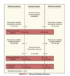
1) superficial cortical nephrons: 85% of nephrons, extend partially into the medulla
2) midcortical nephons: has short or long loops
3) juxtamedullary nephrons: ~12% of nephrons which lie close to and extend deep into the medulla (~40mm); important for concentration of urine
Composition of the renal corpuscle
1) Glomerulus: tuft of capillaries that loop into the Bowman capsule (like fingers pushed into bread dough). Blood supply from afferent arteriole, drained by efferent arteriole.
2) Bowman capsule/space
3) Mesangial cells: shaped like smooth muscle cells; secrete mesangial matrix (type of connective tissue) and lie between and support capillaries. Also have phagocytic abilities simliar to monocyes, release inflammatory cytokines, and can contract to regulate glomerular capillary blood flow.

Glomerular Filtration Membrane - Structure and Function
Structure: 3 layers
- 1) inner capillary endothelium - made of cells in continuous contact with basement membrane and has pores.
- 2) middle basement membrane - selectively permeable network of glycoproteins and mucopolysaccharides
- 3) outer layer capillary epithelium - made of podocytes which are cells that have foto porjections that stick to the basement membrane. Also interlocks with neighbouring podocytes to create filtration slits
Function: filters blood components through its three layers. Separates the blood of glomerulr capillaries from fluid in Bowman space to allow filtration to occur (with the exception of blood cells and plasma proteins with a high MW). Filtrates passes through and finally becomes primary urine.
Structure and function of the juxtaglomerular apparatus
Structure:
- composed of juxtaglomerular cells (which release renin)
- and macula densa (sodium-sending cells) of the distal tubule which is between the afferent and efferent arterioles
Function: control of renal blood flow, glomerular filtration, renin and secretion

Structure of proximal convoluted tubule
- starts from the Bowman space and has an initial voncoluted segment (pars convoluta) and then straight segment (pars recta) that descends towards medulla
- wall made of one layer of cuboidal epithelial cells with surface layer of microvilli (brush border) that increases reabsorptive SA
- then joins Loop of Henle
- brush border and high number of mitochondria in cells of PCT promote reabsorption of 50% of glomerular filtrate

Structure of loop of Henle
- extends into medulla
- cells of the thick segment part of the loop are cuboidal and actively transport several solutes (not water)
- thin ascending segment of the loop narrows and made of thin squamous cells with no active transport function

Structure of distal tubule
- has straight and convoluted segments
- extends from macula densa to collecting duct which is a large tubule that descends down the cortex and through the renal pyramids in the inner and outer medullae (which draines urine into minor calyx)
- composed of:
- principal cells: reabsorb sodium and secrete potassium
- intercalated cells: secrete H+ and reabsorb K+ and bicarbonate

What are the major blood vessels of the kidney?
1) Renal arteries
2) Interlobar artery
3) Arcuate arteries
4) Glomerular capillaries
5) Peritubular capillaries
6) Vasa recta
7) Renal veins

Where do renal arteries originate and end?
Originate: as 5th branches of abdominal aorta, dividing into anterior and posterior branches at renal hilum, subdivide into lobar arteries
End: lower, middle, and upper thirds of kidney
Structure of interlobar artery
subdivisions travel down renal colummns and between pyramids, form afferent glomerular arteries
Structure of arcuate arteries
made of branches of interlobar arteries at cortical-medullary junction and arches over the base of pyramids and run parallel to the surface
Structure of glomerular capillaries
- made of 4-8 vessels, arranged in a fistlike structure
- arise from afferent arteriole, empty into efferent arteriole which carries blood to the peritubular capillaries
- major vessels that regulate intrarenal blood flow
Structure of peritubular capillaries
- surround convoluted portions of the proximal and distal tubules and loop of Henle
- adapted for cortical and juxtamedullary nephrons
Structure of vasa recta
- network of capillaries that forms loops, closely follows loop of Henle
- the only blood supply to the medulla (important for formation of concentrated urine)
Structure of renal veins
- goes in the reverse direction of the arteries
- eventually empties into IVC
Flow of urine
formed by nephrons & flows from distal tubules and collecting ducts through papillary ducts to the renal papillae (projections of the ducts) into the calyces, where it is collected in the renal pelvis and then funneled into the ureters
Describe the structure & innervation of the ureters, and flow of urine through the ureters
Structures:
- ~30cm long, made of long intertwining smooth muscle bundles
- lower ends of ureters pass obliquely through the posterior aspect of the bladder wall
- Sympathetic imput at upper part of ureter (T10 nerve roots) with referred pain to umbilicus
- Parasympathetic sacral nerves innervate lower part of ureter with referred pain to vulva/penis
- Primary arteries for blood supply from kidney, some from lumbar/superior vesical arteries
Flow of urine mechanism:
- smooth muscle cells are nearby to allow electrical stimulation from one cell to another and cause downward peristalsis contraction of urine into the bladder
- Contraction of bladder during micturition compresses lower end of ureter to prevent reflux
- Peristalsis maintained even is ureter is denervated

Structure of bladder
Structure: bag of smooth muscle that forms the destrusor muscle and smooth uroepithelium lining. Uroepithelium forms the interface between urinary space and underlying vasculature and connective/nervous/muscle tissue.
- when bladder fills with urine, distension occurs and the layers of uroepithelium slide past each other to become thinner and bladder volume increases
- contains an smooth triangular area (aka trigone) that’s between the openings of the two ureters and urethra
- has a profuse blood supply; innervated by parasympathetic fibers
- position varies with age and sex

Function of uroepithelium
- allows for the bladder wall to expand when it fills
- lines the urinary tract from renal pelvis to urethra
- acts as a barrier to prevent movement of water and solutes between urine and blood
- communicates information about urine pressure and composition to surrounding nerve and muscle cells


-
PCTH - ALS Intro (Auxiliary Directives)48
-
PCTH - Clinical Procedures5
-
Additional Directives36
-
Neonatal Resuscitation & Emergency Childbirth Medical Directives21
-
PCTH - Trauma Cardiac Arrest Medical Directive48
-
PCTH - Medical Cardiac Arrest Medical Directive22
-
PCTH - Cardiac Arrest Special Considerations34
-
PCTH - Opioid/Hypoglycemia/Adrenal Crisis Directives44
-
PCTH - Analgesia & Allergic Reaction43
-
PCTH - Cardiac Ischemia and STEMI Bypass63
-
PCTH - ACPE/CPAP37
-
PCTH - Croup/Bronchoconstriction36
-
Patho - Neurological System568
-
PATHO - Endocrine System153
-
PATHO - Urinary System144
-
PATHO - Digestive System217
-
PSYCH - Stress, PTSD, & Crisis Intervention59
-
PSYCH - Panic, Anxiety, Mood Disorders80
-
Differential Diagnosis - Altered LOA59
-
Differential Diagnosis - Trauma & Non-Traumatic Pain70
-
Differential Diagnosis - Allergies, Non-Traumatic Pain, Respiratory50
-
HALO - Term Test 1 Normal Childbirth112
-
HALO Term Test 2 - Birthing Complications77
-
PATHO - Reproductive System174
-
HALO - Term Test 3 (Pediatric & Neonatal Resuscitation, Bariatric, Geriatric)107
