What structures embryologically form the diaphragm?
- Septum transversum (ventrally)
- Mesentery of foregut (dorsally)
- 2 pleuroperitoneal folds (dorsally)
During queit respiration, what % of change in thoracic volume is achieved by diaphragmatic movement vs intercostal mm?
- 75% diaphragm
- 25% intercostal
What reason is given for potenital spread of abdominal disease to mediastinum / pleural space?
- unidirectional drainage of lymph nodes -> final destination thoracic trunks
Detail the anatomical portions of the diaphragm?
- Central tendinous part
- Peripheral muscular part (3 areas)
Pars sternalis => attaches to xiphoid cartilage
Pars costalis => attaches to 8-13th ribs
Pars lumbalis => 2 crura. R attaches to craionventral border L4, L attaches to body of L3
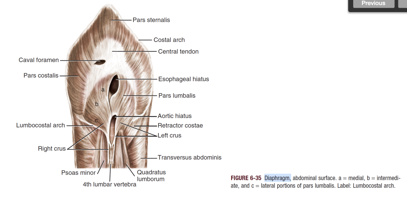
Which 2 recesses are formed by the diaphragm?
- Phrenicocostalis (costodiaphragmatic) recess
=> formed between layesrs of pleura lining diaphragm and ribs
- Phrenicolumbalis (lumbodiaphragmatic) recess
=> formed similarly, but region dorsal to crura and ventral to vertebra (bilateral)
List the 3 openings within the diaphragm, and what they contain
Aortic hiatus
- Aorta, hemiazygous, azygous, lumbar cistern of thoracic duct
Oesophageal hiatus
- Oesophagus, vagus trunks
Caval hiatus
- CaVC
Which portions of the diaphragm are not visible radiographically?
- Visibility dependent on adjcanet opacity.
- Accordingly majority of thoracic portion visible
EXCEPT recesses, as lung not contacting
- Ventral portion of abdominal diaphragm may be visible if falciform fat present
Where is the cupula?
= the body
Most cranial convex portion on both DV and laterals
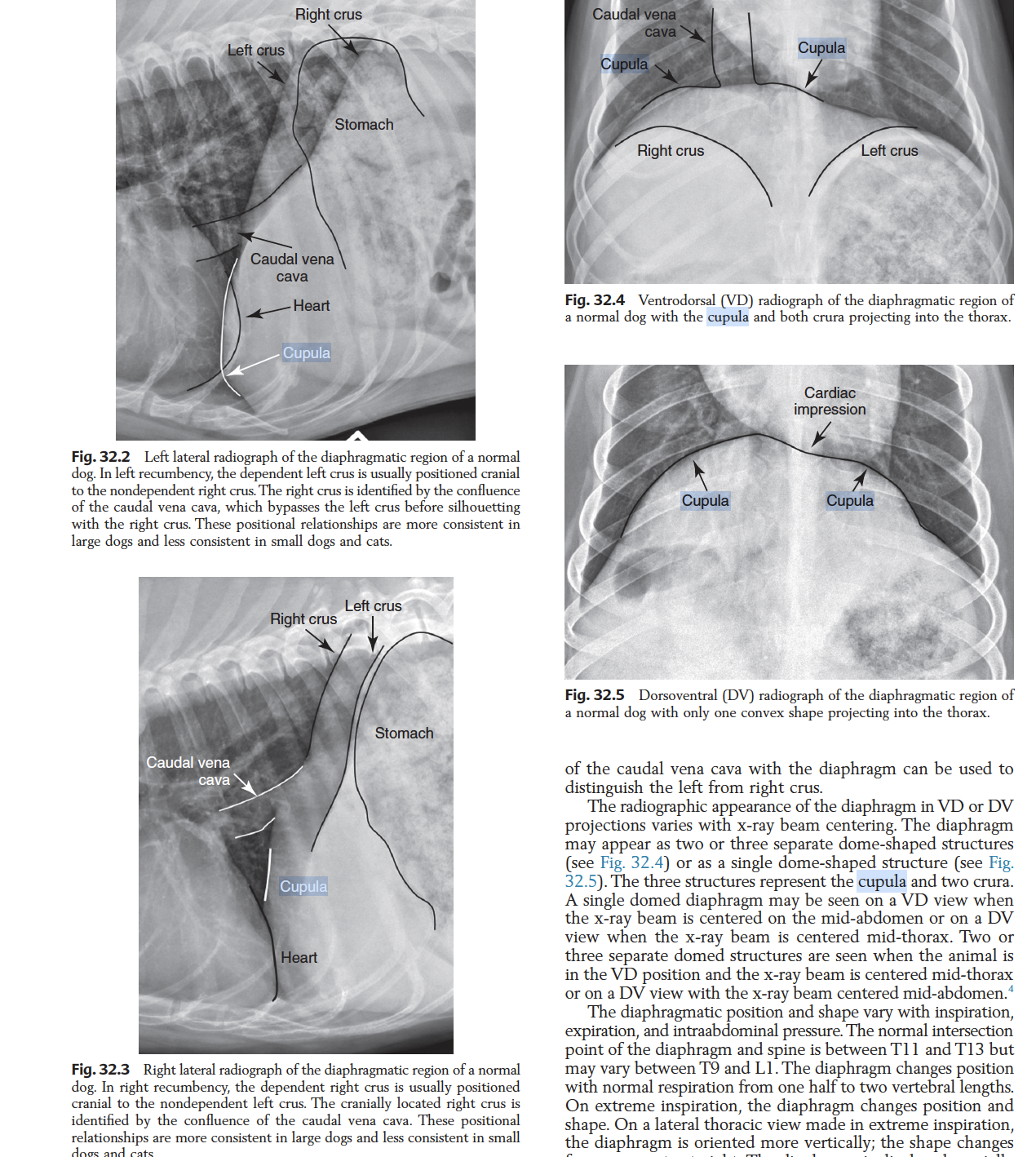
What effect can poor radiographic technique (cranial centring, rotation) have on the appearance of the diaphragm in the lateral projections?
- INcreased seperation of the crura (up to 2.5 vertebral lengths)
In what views does the diaphragm have a dome / mickey mouse shape?
Dome: DV thorax, VD mid abdomen
Mickey: VD thorax, DV mid abdomen
Where does the diaphragm extend caudally to (intersection with spine)? How does it change with extreme resp?
- Normal: T11-13
- May vary between T9-L1
- Extreme: More verteical, flattened / straight, tenting in the cat
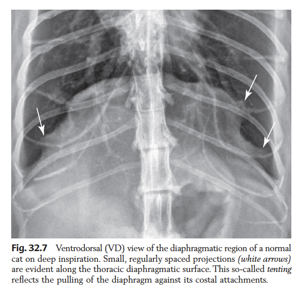
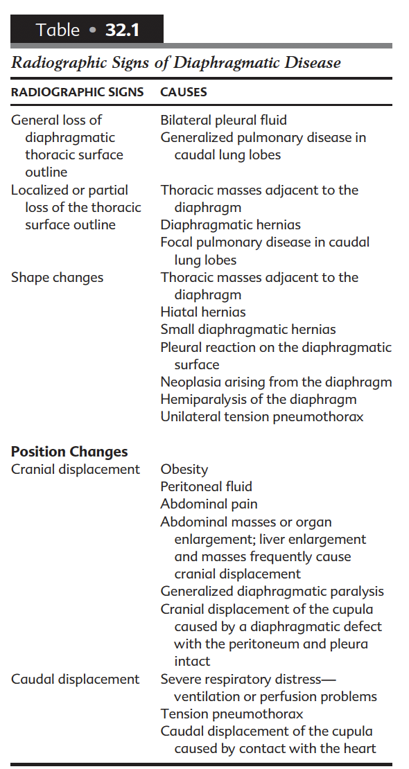
Table - Rx signs of diaphragmatic disease
Where are the most common changes to diaphragmatic shape seen? why?
- Cupula -> heart contact, patient postiioning, large breed dogs
What are the possible causes of asymmetry of the diaphragm? Name one way to confirm your suspicions for more unusual dx….
- Unilateral tension pneumo
- Hemiparalysis -> FLURO
List 5 broad types of diaphragmatic hernia
- Traumatic
- Peritoneopericardial
- Hiatal
- Peritoneopleural
- Other congenital diaphragmatic defects
Describe the pathophys of traumatic hernia
- Increased abdominal pressure with OPEN glottis
=> leads to large peritoneo-pleural pressure gradient
- Subsequent rent formation
What radiographic techniques are described to better characterise diaphragmatic herniation?
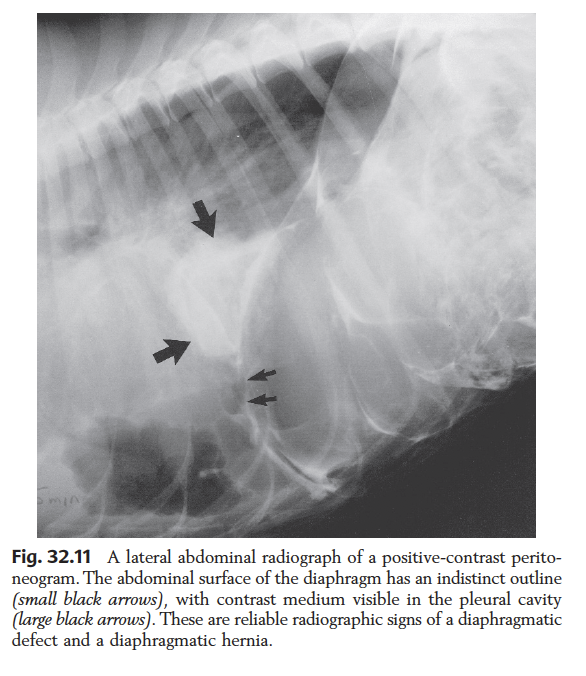
- Positional radiographs
- Removal of pleural fluid + repeat
- Barium study (0.5ml / kg, 30%w/v)
- +ve contrast peritneography (2ml/kg, iodinated), other selective +ve contrast techniques
- Horizontal beam
=>LAST RESORT, position animal so accumulates cranially
- other modalities
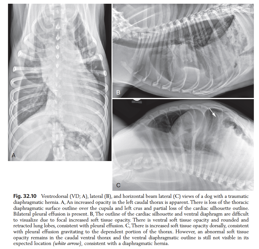
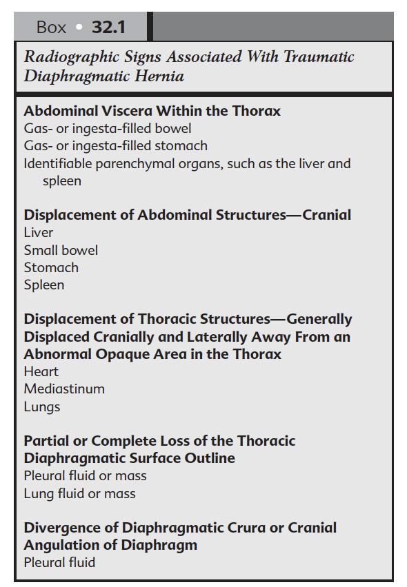
Table: Radiographic features of traumatic diaphragmatic hernia
Incidence of laterality of traumatic hernia?
- In one report equal….
BUT in dogs has been reported R>L
Which organs are most commonly herniated in traumatic diaphragmatic hernias? And when chronic?
Acute (IN ORDER)
Liver > small intestine > stomach > spleen > omentum
Chronic (IN ORDER)
Liver > small intestine > omentum > spleen > stomach > colon > pancreas
What are the most consistent radiographic features of traumatic diaphragmatic hernia?
- Abdo organs in thorax
- Displacement of abdo/thoracic organs
- loss of thoracic diaphgramatic surface
- assym / altered slope on lateral
- Pleural fluid
What specific life-threatening complication occurs secondary to tension gastrothorax?
- Potential / actual cardiovascular tamponade
What feature is a consistent finding with chronic diaphragamatic hernias?
- Pleural fluid
=> also consistent if strangulated organ is present
Approximately what % of diaphragmatic hernias are congenitally predisposed?
15%


-
1. Radiation Protection and Physics of Diagnostic Radiology36
-
2. Digital Radiographic Imaging23
-
3. Dental technique8
-
4. Physics of Ultrasound30
-
5. Principles of CT and MRI33
-
6. Contrast media34
-
7. Intro to radiographic interpretation2
-
8. Radiographic anatomy of axial skeleton10
-
9 - Principles of Interpretation of Axial skeleton16
-
10 - Canine and Feline dental disease65
-
11. Nasal cavity37
-
12. MRI Brain47
-
14. XR Vertebrae41
-
15. CT / MR Spine69
-
16. Radiographic anatomy of appendicular skeleton28
-
17. Principles Appendicular skeleton5
-
18. Orthopaedic Diseases of Young and Growing Dogs and Cats51
-
19. Fracture Healing and Complications in Dogs48
-
20. Bone tumours and infections21
-
21. Joint Disease68
-
28. Principles of interpretation - Thorax6
-
29. Larynx and trachea38
-
30. Oesophagus64
-
31. Thoracic wall12
-
33. Mediastinum35
-
32. Diaphragm38
-
34. Pleural Space17
-
35. Cardiovascular system (+ extra bits)72
-
36. Lung31
-
38. Principles of interpretation - Abdomen2
-
Chapter 39 - Peritoneal Space52
-
40 - Liver and Spleen43
-
41 - Kidneys and Ureters40
-
42 - Urinary Bladder31
-
43 / 44 - Urethra / Prostate30
-
45 - Uterus, Ovaries and Testes30
-
46 - Stomach35
-
47 - Small intestine49
-
48 - Large Bowel22
-
THE MOST IMPORTANT QUESTIONS!328
