Which radiographic technique should be used for TXR?
- Low mAs, high kVp
What 3 characteristics of an alveolar pattern are described?
1) Air bronchogram
2) Lobar sign
3) Area of intense opacity without sharp margins (e.g. not a mass)
What does a lobar sign describe?
Region of opaque lung bordering adjacent aerated lobe
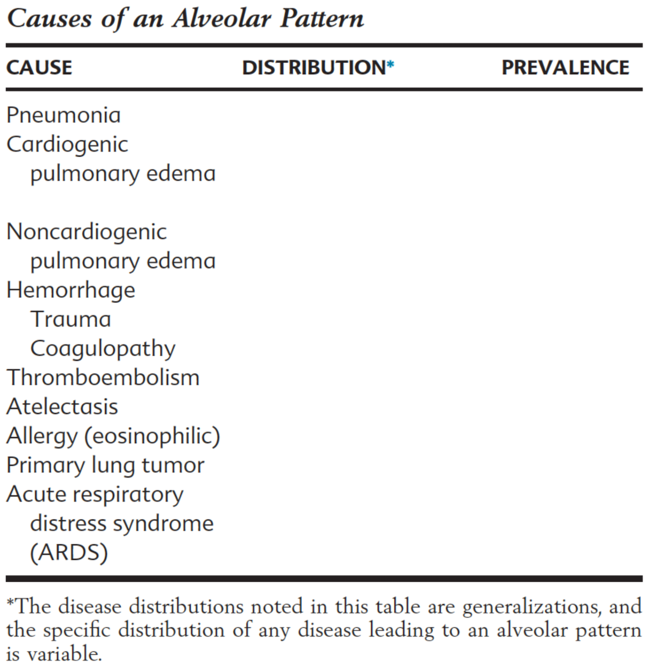
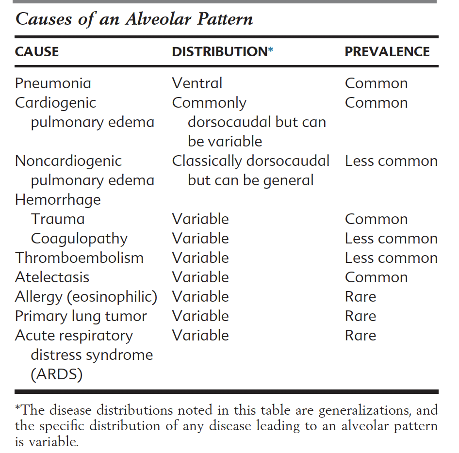
Detail 9 causes of an alveolar pattern (consider distribution and prevalence too..)
What radiographic feature should be identified with atelectasis? What must be considered when interpreting atelectatic lung?
- MEDIASTINAL SHIFT
- > Can mask alveolar pathology. Cannot distinguish radiographically -> SAMPLING may be required
In a bronchial pattern, where it the pathology?
- In the bronchial wall OR peribronchial space (actually a component of interstitium)
List 5 additional radiographic features that can be seen in association with bronchial pathology
1) Lobar collapse
2) Rib fractures
3) Hyperlucent lung
4) Bronchiectasis
5) Bronchial mineralisationa
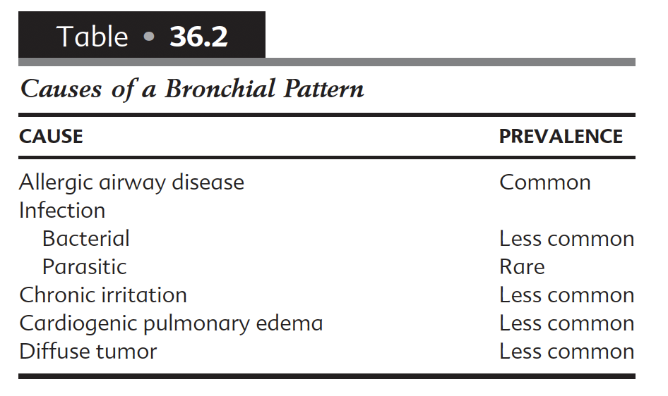
List 5 causes of a bronchial pattern
Which lung lobe most classically collapses in feline asthma?
Right middle
What diseases have been associated with bronchiectasis? List radiographic features of bronchiectasis
DDx: Pneumonia, Eosionophilic bronchopneumopathy, inflammatory airway disease
Rx: Increased diameter, failure to taper in periphery, non-linear nature of wall, thickened wall (if bronchitis also)
What 3 categories of disease have been associated with rib fractures in cats?
1) Respiratory (Most common)
2) Neoplasia (E.g. myeloma)
3) Metabolic (E.g. renal)
=> Osteopaenia associated with old age may play a role, as commonly older
Which ribs are classicallly affected by spontaneous fracture in cats with respiratory disease?
9-13, mid-portion.
What features of pulmonary hyperinflation may be recognised?
- Increased lucency
- Increased volume (increased cardiac / diaphragm distance)
- Tenting of diaphragm
What bronchial structures may become mineralised
- Bronchial wall
- Bronchial plug
- Bronchial mucous glands
In what instances is bronchial mineralisation typically seen?
- Cats with chronic bronchial disease -> bronchial pattern present
- Dogs with CUSHINGS -> No thickening of bronchi seen, and more diffuse
What threshold value is suggested for pulmonary nodules on XR?
7-9mm
=> likely can vary depending on patient and technical factors
What size can be used to distinguish mass from nodule?
20mm
What features may be used to distinguish a pulmonary nodule from an end-on vessel?
Vessels:
- More opaque (as more summated)
- “tail” of vessel
- Proximity to bronchus
- Smaller diameter than expected of pulmonary nodule (visible due to increased opacity)
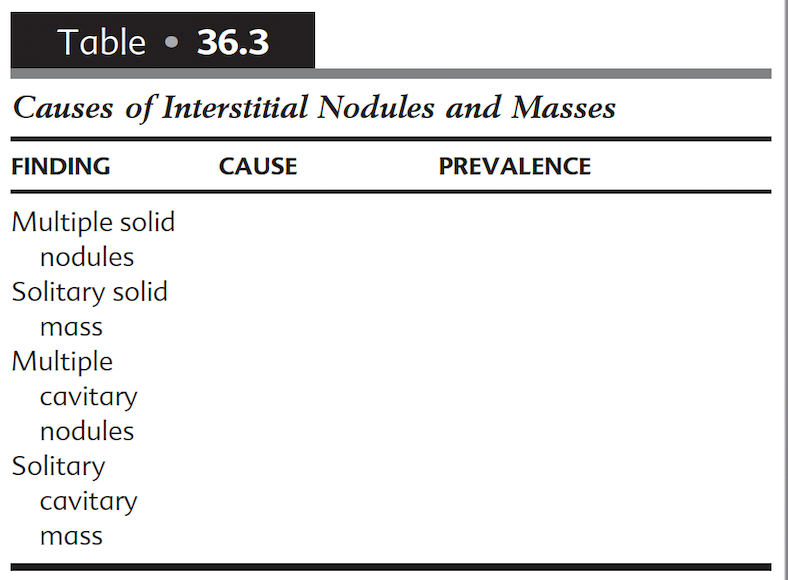
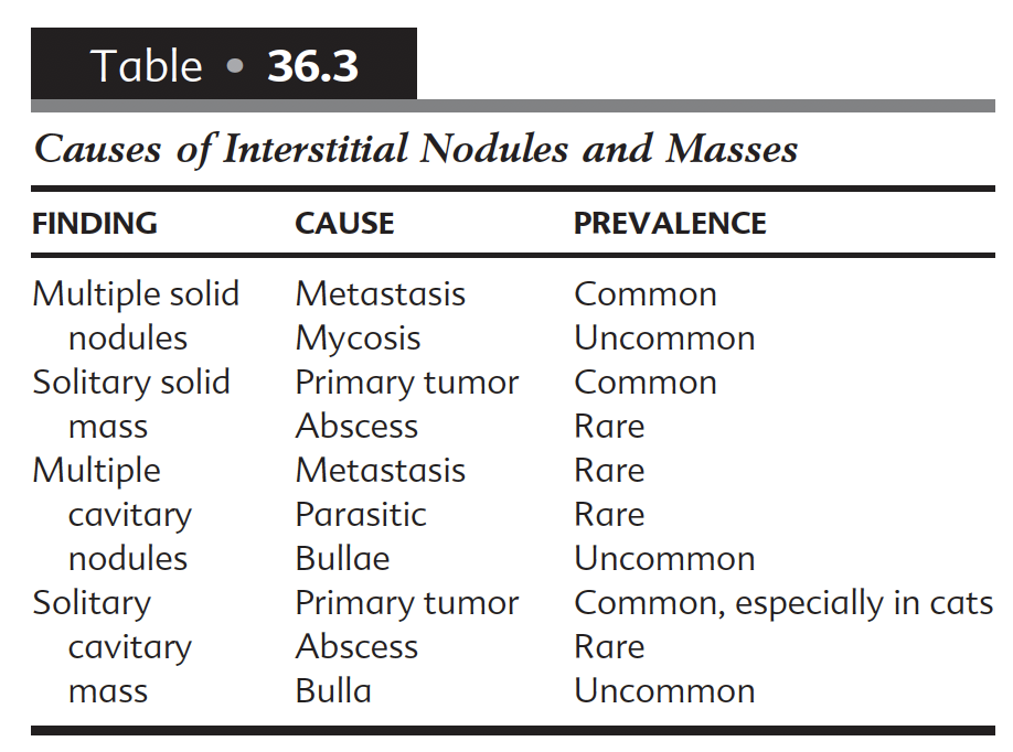
Causes of interstitial nodules and masses
What is the alternative name for heterotopic bone?
Pulmonary osseous metaplasia
What methods can be used to confirm pulmonary location of nodule on XR?
- Fluoro -> coincedent movement of lung
- CT
- Check for superficial structures!
- Repeat TXR
What radiographic features of adenocarcinoma and histiocytic sarcoma are described?
- Adenocarcinoma:
LEFT CAUDAL LOBE
- HS:
Tend to be larger
RIGHT MIDDLE, LEFT CRANIAL
Internal air bronchogram (>50%)
What features of an acute traumatic bulla may distinguish it from a chronic bulla / congenital bulla?
- Irregular wall -> can have haemorrhage in wall
- Other features of trauma: contusion, pleural effusion, fractures etc.
What is the difference between unstructured and structured interstitial patterns?
- Sturctured: Usually organised into discrete lesions
- Unstructured: NOT!


-
1. Radiation Protection and Physics of Diagnostic Radiology36
-
2. Digital Radiographic Imaging23
-
3. Dental technique8
-
4. Physics of Ultrasound30
-
5. Principles of CT and MRI33
-
6. Contrast media34
-
7. Intro to radiographic interpretation2
-
8. Radiographic anatomy of axial skeleton10
-
9 - Principles of Interpretation of Axial skeleton16
-
10 - Canine and Feline dental disease65
-
11. Nasal cavity37
-
12. MRI Brain47
-
14. XR Vertebrae41
-
15. CT / MR Spine69
-
16. Radiographic anatomy of appendicular skeleton28
-
17. Principles Appendicular skeleton5
-
18. Orthopaedic Diseases of Young and Growing Dogs and Cats51
-
19. Fracture Healing and Complications in Dogs48
-
20. Bone tumours and infections21
-
21. Joint Disease68
-
28. Principles of interpretation - Thorax6
-
29. Larynx and trachea38
-
30. Oesophagus64
-
31. Thoracic wall12
-
33. Mediastinum35
-
32. Diaphragm38
-
34. Pleural Space17
-
35. Cardiovascular system (+ extra bits)72
-
36. Lung31
-
38. Principles of interpretation - Abdomen2
-
Chapter 39 - Peritoneal Space52
-
40 - Liver and Spleen43
-
41 - Kidneys and Ureters40
-
42 - Urinary Bladder31
-
43 / 44 - Urethra / Prostate30
-
45 - Uterus, Ovaries and Testes30
-
46 - Stomach35
-
47 - Small intestine49
-
48 - Large Bowel22
-
THE MOST IMPORTANT QUESTIONS!328
