Chapter 50 The Shoulder Flashcards

(80 cards)

1
Q

What proportion of shoulder motion comes from the shoulder joint and what proportion from the synsarcosis?

A

2/3rds from joint

1/3rd from synsarcosis

2
Q

At what age does glenoid physis fuse?

A

6 months

3
Q

At what age does proximal humeral physis fuse?

A

1 year

4
Q

What is the ratio of glenoid to humeral head coverage?

What increases the ‘functional’ size of this ratio?

A

1: 2.5
i. e. moderately congruent.

Cartilaginous glenoid lip increases glenoid joint surface area by 25%

5
Q

What is approximate thickness of shoulder joint cartilage in 25kg dog?

A

1 mm

6
Q

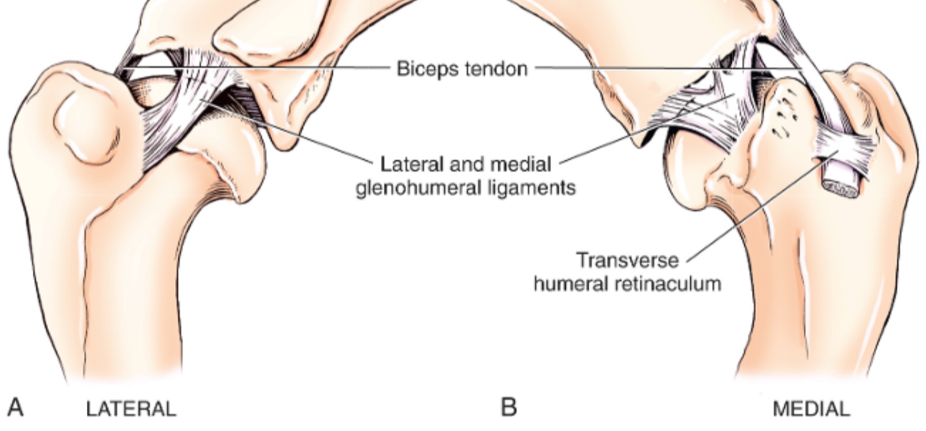
What is the name of the humeral groove through which the biceps runs?

A

Intertubercular groove

7
Q

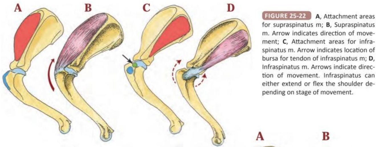
With which 5 muscles does the shoulder joint capsule blend?

A
  • Infraspinatus
  • Supraspinatus
  • Subscapularis
  • Teres minor
  • Coracobrachialis
8
Q

Label the diagram

A

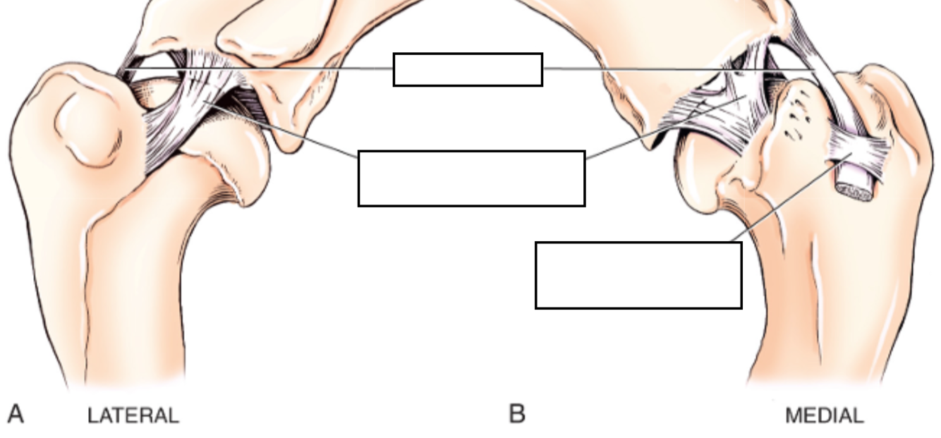
N.B. Cranial arm of Y of exists as separate intra-articular structure

9
Q

What are normal shoulder joint angles in dog?

And in cat?

What is recommended arthrodesis angle?

A

Dog:

Flexion 57º

Extension 165º

Cat:

Flexion 32º

Extension 164º

Recommended arthrodesis angle 105 - 110º

10
Q

List 3 passive stabilisers of the shoulder joint

List 8 active stabilisers of the shoulder joint

A

Passive shouder stabilisers:

  • Limited joint volume + adhesion/cohesion principles
  • Concavity compression
  • Capsuloligamentous constrainst
    • Glenohumeral ligaments
    • Joint capsule
    • Labrum
    • Tendon of origin of biceps brachii

Active shoulder stabilisers

_​_Major:

  • Infraspinatus
  • Supraspinatus
  • Subscapularis
  • Teres minor

Minor:

  • Biceps brachii
  • Long head of triceps
  • Deloideus
  • Teres major
11
Q

Type 1, 2 a dn 3 mechanoreceptors are present in collateral ligaments of shoulder - which type is most common

A

Type 1 (=Ruffini) receptors

12
Q

What contrast agent is used fro arthrography?

A

Iohexol (doent need dilution, unless also performing CT angio in which case dilute to 60 mg/mL

13
Q

What % of dogs identified by CT as having mineralization of peri-articular structures of shoulder were NOT lame?

A

40%

And 90% of those with lameness has other shoulder/elbow pathology

i.e. high % of incidental mineralization

14
Q

What % of shoulder pathology has been reported to be extra-articular (i..e would be missed with arthroscopy alone)?

A

15%

15
Q

Name 2 salvage procedures for shouder joint

A
  • Glenoid excision arthroplasty
  • Arthrodesis
16
Q

What structure has to be protected/avoided during glenoid excision arthroplasty?

A

Suprascapular nerve

(also note correct angle of cut: distolateral-proximomedial)

17
Q

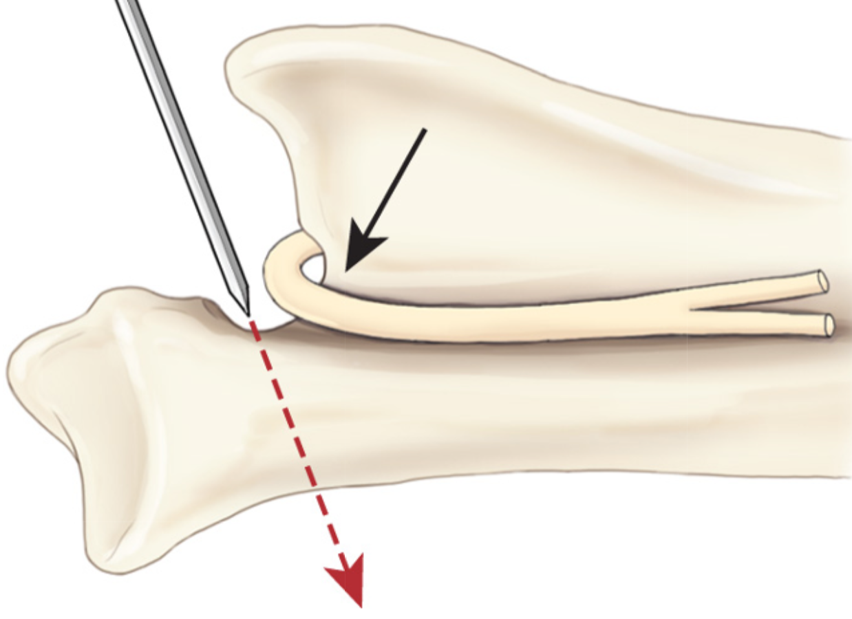
What approach is used for glenoid excision arthroplasty?

A

Craniolateral

18
Q

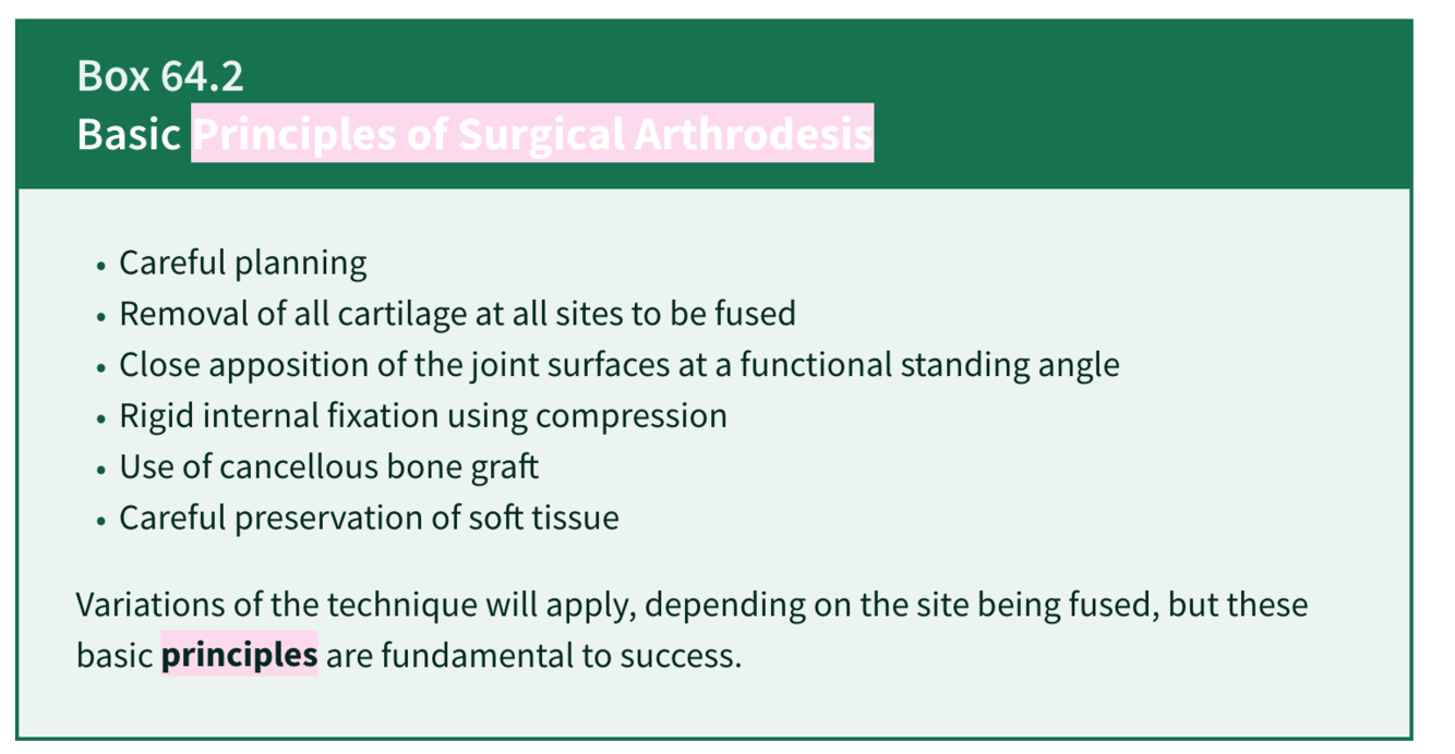
What are the 6 principles of arthrodesis?

A
19
Q

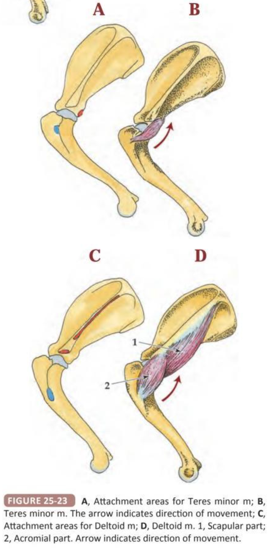
What muscle insterts on greater tubercle?

What muscle originates on acromion

A

Supraspinatus m inserts on greater tubercle

Acromial head of deltoid originates from acromion

i.e. may need to perform ostectomy or tenotomy to allow access to joint

20
Q

Describe the apprach to perform shoulder arthrodesis

A

Approach to body, spine and acromion combined with craniolateral approach to shoulder joint (inc tenotomy of acromial part of deltoid, supraspinatus, infraspinatus)

  1. Elevate insertion of trapezius muscle and origin of the omotransversarius
  2. The incision is continued distally along the cranial border of the acromial head of the deltoideus muscle.
  3. The omobrachial vein (and cephalic vein, if necessary) is divided, and the incision follows the lateral aspect of the brachiocephalicus muscle to its insertion.
  4. The insertion of the superficial pectoral muscle is incised and the muscle elevated and retracted cranially.
  5. Although osteotomies of the acromion and greater tubercle can be performed, tenotomy of the respective tendons of insertion of the deltoideus and supraspinatus muscles may result in fewer complications due to osteotomy nonunion, if proper hemostasis and reattachment of the muscles is achieved.
  6. Elevation of the supraspinatus muscle is continued proximally through the entire supraspinous fossa until the muscle can be retracted cranially. The suprascapular nerve is identified, carefully retracted, and protected at all times.
  7. The tendon of insertion of the infraspinatus muscle is transected and the muscle retracted caudodorsally. This approach exposes the entire craniolateral aspect of the scapula and the cranial aspect of the humerus.
  8. The lateral collateral ligament is transected and the joint capsule incised to allow luxation of the humeral head from the glenoid fossa.
21
Q

What are two options for creating a ‘congruent” surface for shoulder arthrodesis?

A
  • Burr cartilage (preferred if normal shape)
  • Ostectomy of glenoid + humeral head
22
Q

What 3 locking plate designs are available?

Name a disadvantage of each

A
  • SOP. Cant generate compression
  • LCP: Cant contour in plane
  • Reconstruction plate: Can compress but weaker plate
23
Q

Where is plate placed for shoulder arthrodesis?

How can stabilisation be augmented?

A

Apply plate along craniolateral scapula and cranial humerus

  • Can add ancillary fixation e.g. 2nd plate caudal to first one.
  • Or post-op spica splint (possibly innecessary if 2 plates used)
24
Q

What is recommended shoulder arthrodesis angle?

How else can an arthrodesis angle be derived?

A

105 - 110º

Compare to opposite shoulder joint angle during weightbearing

25
What are the most common sites for shoulder OCD?
Caudocentral and caudomedial humer head
26
What % of large/giant breed male dogs with OCD are bilaterally affected?
25 - 75%
27
List 2 earliest radiographic signs of OCD
* Loss of trabecular bone structure * Subchondral lysis
28
What is radiographic diagnosis?
Humeral OC(D) lesion
29
What step can be performed during radiography if OC lesion not immediately visible?
* Inernal or external rotation of humeral head * (Arthrography)
30
Name a (counterintuitive) step of medical management of shoulder OCD
Vigorous exercise (to break flap off) Surgical tx superior
31
What approached can be used for arthrotomy for OCD flap removal
* **Caudal** approach (avoids osteotomies/tenotomies, but axillary nerve right ver joint capsule). p. 138 * **Caudolateral inter-deltoid** approach with *crandiodorsal* retraction of infraspinatus and teres minor tendons (rather than separating and retracting the tendons which --\> less exporure) p. 130 * **Craniolateral** with infraspinatus tenotomy (or **modified craniolateral "Cheli's approach'** = vertical incision between supraspinatus and infraspinatus tendons) p.120
32
What does OATS stand for
Osteochondral autograft transfer surgery
33
What procedures can be performedin addition too OCD flap removal for shoulder OCD
* Osteochondral transfer e.g. OATS * Synthetic osteochondral resurfacing e.g. SynACart * Custom hemi-arthroplasty
34
In Murphy et al study (vetSurg, 2019), what approach was used for implantation of SynACart for treatment of shoulder OCD What were the 3 most common complications?
Caudal approach, without tenotomy of infraspinatus (internal rotation of antebrachium) SSI 4/24 Seroma 3/24 Septic arthritis 2/24
35
Comment on rad. What is recommended treatment?
Glenoid dysplasia (usually unilateral) Tx = excision arthroplasty or arthrodesis
36
What condition is this? What is usual management? Name 2 ddx
* **Multiple epiphyseal dysplasia** = defect in ossification of epiphyses of long bones, vertebrae, cuboidal bones, apophyses * Usually require PTS * Ddx: **Focal humeral head dysplasia** or **congenital hypothyroidism**
37
What condition is shown? How is it assessed further? What else should be checked?
Incomplete ossification of caudal glenoid * If lame --\> arthroscopic assessment to see if loose, if so remove. * Check rest of joint for other pathology!
38
What is another name for pseudogout? What breeds are usually affected in shoulder? And other breed/location
*Chondrocalcinosis* = HA deposition in articular cartilage. Greyhound shoulders GSD femoral head Often incidental, clinical relevance unknown. Necropsy photograph demonstrating chondrocalcinosis in a Greyhound. Observe the small, multifocal, raised white areas on the humeral head.
39
Hos is biceps tendinopathy classified?
Primary or secondary
40
List tests that can be performed to assess biceps tendon
* **Biceps tendon test** * Flex shoulder + palpate beceps tendon * **Drawer test** * Stabilise scapula + translate humerus cranially. Asseses for *pain* not instability * **Biceps retraction test** * Grasp tendon of insertion of biceps brachii near cranial aspect of elbow and pull caudally (if painful is consistent with tendinopathy) (German biceps tendon test = see if can fully flex shoudler while extending elbow. Consistent with complete rupture)
41
What additional rad image should be obtained to assess biceps tenon
Skyline view (= flexed, craniodistal - cranioproximal) Rad: a small mineralization within the tendon of the supraspinatus muscle (arrow). The tendon of origin of the biceps brachii muscle lies medial to the greater tubercle, within the intertubercular groove.
42
What imaging modalities can be used in work up of biceps tendinopathy?
* Rad (inc skyline view) * Contrast artrography (identified irregularities with tendon and surrounding synovium) * MRI * US
43
List 4 sonographic abnormalities seen with biceps tendinopathy
* Sonolucent line around tendon * Enlarged, hypoechoic tendon with fibre pattern disruption * Irregular/proliferative synovium * If chronic, irregular surface of bicipital groove
44
List anadvantage of MRI/US vs arthroscopic assessment of biceps tendon
US/MRI can assess changes *within* tendon
45
When is medical management for biceps tendinopathy recommended? List recommended medical tx for biceps tendinopathy?
If acture, try medical management _Tx:_ * Intra-articular steroids (10 - 40 mg methyl pred or 5 mg triamcinolone) * STRICT rest 4-6 weeks * IM injection of polyunsulfated GAG * (+- NSAIDs depening on whether had intra-articular steroids or not) * (Others reported: extracorporeal shockwave therapy, PRP/ste cells, therapeutic US, electrical nerve stim) --\> rupture reported in other species, but not reported in dogs
46
What are surgical options for biceps tendinopathy? When is one recommended over the other
* Tenotomy (arthrotomy or arthroscopic - N.B. different (cranial!) instrument portal for this) * Tenodesis Tenodesis if young/active/working dog (speculated faster return to function)
47
What is a theoretical concern re biceps tenotomy?
?contribution to shoulder stability.
48
Name 3 biceps tendon conditions in additon to biceps tendinopathy
* Calcifying tendinopathy * Medial displacement * Rupture (rupture of distal tendon sheath also reported)
49
What breeds are affected by medial displacement of tendon of origin of biceps brachii? How is it treated? Post-op support?
Greyhounds, Afghan, GSD, Border collie Tx: * Primary restoration of transverse ligament * Transverse ligament augmentation (Screws + PDS/Nylon/staple/mesh) Post-op * Velpeau sling
50
What breeds are most affected by calcifyign tendinopathy of tendon of origin of biceps brachii (and supraspinatus tendinopathy)
Lab and Rottweilers
51
What two conditions has supraspinatus contracture been described with
Trauma von Willebrands
52
How does PE of supraspinatus tendinopathy differ from biceps tendinopathy
More painful (N.B. mineralization medially i.e next to biceps) potentially more clinically relevant)
53
HOw does mRI apperance of normal supraspinatus tendon differ from adjacent infraspinatus and subscapularis
Can appear T2W hyperintense (suspected due to higher collagen content)
54
What are management options for supraspinatus tendinopathy?
* Medical (NSAIDS + rest (+ extracorporeal shockwave)) * Sx (excision of calcified tissue - use c-arm/hypodermic needles to locate)
55
What is aetiology of shoulder joit istability in small dogs vs larger?
Small dogs usually means congenital ST laxity Larger breed instability considered overuse injury
56
What % of shoulder instability cases are medial?
80% But cranial, caudal and lateral can happen
57
What was difference in shoulder abduction angle in dog affected by medial shoulder instability vs those not affected
Affected = 50º Unaffected = 30º Very variable thorugh so always compare to contralateral side
58
What was most common arthroscopic finding in dogs with MSI?
Superficial erosion of cartilage of caudal region of humeral head N.B. significance of ligament fraying not necessarilty proportional to clinical signs (i.e authors says has seen fraying with no lameness)
59
List 6 techniques to manage MSI. Which is preferred?
1. Biceps tendon transposition 2. Supraspinatus transposition 3. Subscapularis imbrication 4. Prosthetic ligament (preferred) 5. Radiofrequency induced thermal modification (RITM) 6. Arthrodesis/excision arthroplasty
60
What is recommended post-op management for MSI?
6 weeks velpeau sling for 6 weeks
61
What was reported rate of good-excellent outcome following biceps tendon transposition
85% Ligament augmentation "better' outcome Arthodesis also 85% good-excellent
62
What is conservative management of medial shoulder lux And lateral? When is conservative management attempted?
Medial: Velpeau, rest, analgesia Lateral: Spica or neutral sling (not velpeau and --\> shoulder abduction!) Attempt conservative if stable after reduction
63
List 5 surgical options for shoulder luxation
1. Biceps transposition 2. Supraspinatus transposition 3. Prostetic ligament (preferred) 4. Temporary transarticular bridging with bone plate also reported 5. Arthroplasty/arthrodesis
64
What muscles (broadly speaking, 4 points) are more prone to strains?
* Those that cross two (or more) joints * With high % fast twitch (type 2) fibres * Those that function in eccentric manner * Those that limit ROM across joint by their intrinsic tightness *
65
What 8 thoracic limb muscles are susceptible to strains?
1. Biceps 2. Triceps 3. Superficial pec 4. Deep pec 5. Serratus ventralis 6. Rhomboideus 7. extensor carpi radialis 8. Flexor carpi ulnaris
66
How are muscle strains graded?
Grade 1 - 4 Grade 1 = minimal tearing of indicidual fibres Grade 4 = complete rupture. recommend debride + repair in these cases
67
What other shoulder muscle can be affected by myopathy? How is it diagnosed? What is recommended treatment?
Teres minor - v painful. Dx: Us or MRI + histo Tx: excision of whole muscle!
68
What breeds get infraspinatus contracture?
Lab, Brittany spaniel, Pointer
69
What is typical infraspinatus hx?
Biphasic: initial lameness/swelling/pain --\> non-painful gait abnormality
70
What are US findings in infraspinatus contracture? MRI?
US: hyperechoic changesin muscle MRI: * Acute: Hyperintense * Chronic Uniformly hypointense centre of muscle, heterogenous hypointense periphery. Reduced muscle volume
71
How is infraspinatus contracture treated List an unusual ddx
Tenotomy Neoplasia - author seen fibrosarcoma
72
What is typical presentation of villonodular synovitis?
Large breed, severe lameness, joint distention
73
What are arthrocentesis findings in villonodular synovitis? Arthroscopy findings? Tx options:
Arthrocentesis: * Orange/red colour * Moderate numbers of neuts + macrophages Arthroscopy: * Yellow, hemorrhagic nodules/masses of synovium and capsular haemorrhage Treatment: Medical therapy is ineffective (although Dempsey (VCOT, 2018) report good outcome for 3 years w medical = firocoxib and controlled exercise) * Excisioon arthroplast/arthrodesis * (In humans, synovectomy, radiation, anti-TNA-a administration)
74
What condition is shown? How is it treated?
**Synovial chodrometaplasia** = proliferative disorder of undifferentiated stem cells. Described as primary or secondary in humans _Treatment:_ * Nodule debridement * Loose body removal * Synovial stripping * Arthroplasty/arthrodesis
75
What is histo in synovial chondrometaplasia (primary vs secondary)
Histo: * Primary: **Metaplastic cartilage**, containing crowded, **atypical chondrocytes** and **patchy calcification**, but devoid of fragments of articular cartilage or subchondral bone. * Secondary: Differs histologically from the primary variant; osteochondral fragments with **metaplastic cartilage** are present but contain **normal, evenly distributed chondrocytes** and **regular calcification**
76
List ddx to mineralization around shoulder (i.e. intra and extra articular)
* Calcifying tendinopathy of tendon of origin of biceps * Supraspinatus tendinopathy * Infraspinatus bursal ossification * Synovial chondrometaplasia * Joint mouse * Miscellaneous ST mineralization (trauma, neopasia, fibrodysplasia oddificant progressiva)
77
List broad ddx for intra-articular mass lesions
* Neoplasia * Synovial chondrometaplasia * Villonodular synovitis
78
In what joints (in dogs) has synovial chondrometaplasia been reported?
* Shoulder * Hip * Stifle * Tarsus
79
What is the difference between dystrophic and metaststic mineralization?
_Metastatic calcification_ can occur if the calcium–phosphate product is increased _Dystrophic calcification_ occurs in areas of damaged soft tissue (with normal levels of serum calcium and phosphate.)
80
What is tx for fibrodysplasia ossificans progressiva?
Corticosteroids and disodium etidronate (bone density conservation agent)