What is the radiographic appearance of a GDV?
- Usually stomach rotates 180-220 degrees clockwise
- Pylorus and duodenum move dorsal and leftward and are separated (compartmentalized) by Antrum wall folding back
- On a right lateral the pylorus is gas dilated and dorsally locates (as apposed to fluid filled and ventral)
- Because the spleen is attached to the greater curvature by the gastrosplenic ligament, the volvulus usually displaces the spleen to the right ventral side of the abdomen, and causes congestion and splenomegaly.
- There is often megaesophagus and has distended SI due decreased motility from sympathetic stimulation (pain, shock, etc.)
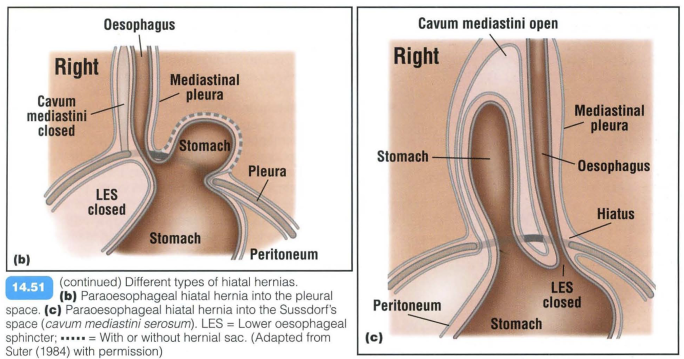
Paraesophageal hiatal hernia

Paraoesophageal hiatal hernia:
The lower oesophageal sphincter remains in place and is competent, but part of the stomach surrounded by a peritoneal sac herniates through the oesophageal hiatus parallel to an immobile oesophagus.
Two subtypes exist:
- Abdominal organs may herniate to the left of the oesophagus in which case they come to lie in the pleural cavity
- Abdominal organs may also slide to the right of the oesophagus and herniate into the potential space between the two mediastinal pleural leaves (Sussdorf’s space - cavum mediastini serosum or bursa infracardiaca). This space is normally closed by a thin membrane, which may be congenitally weak or ruptured following trauma. The prolapsed organs, usually part of the stomach, liver or small intestines, can move in and out as the space widens during inspiration and shrinks during expiration.
Clinical signs may range from none to recurrent gastrointestinal signs, including anorexia, retching, vomiting or bloating of the stomach without gastric torsion .
Radiographic findings include:
Left-sided paraoesophageal hernia:
- Changes in the diaphragmatic contour associated with a mass in the mid-caudal thoracic cavity
- Extrapleural sign with the base of the mass toward the diaphragm and very sharp margins on the lung side
- Presence of gas-filled tubular structures in the caudal thoracic cavity
- Presence of a hyperlucent cyst-like structure surrounded by a thin soft tissue rim filling the left hemithorax, may be seen if the stomach herniates and becomes incarcerated
- Mediastinal shift to the right and pulmonary atelectasis
Right-sided paraoesophageal hernia:
- Soft tissue mass in the mid-caudal thorax on the lateral view, caudal to the heart
- Widening of the caudal mediastinum on the DV view, associated with displacement of the cardiac silhouette to the left. As the pericardium is intact, the cardiac silhouette remains a normal size
- Gas in the stomach or small intestines may be identified in the caudal mediastinum.
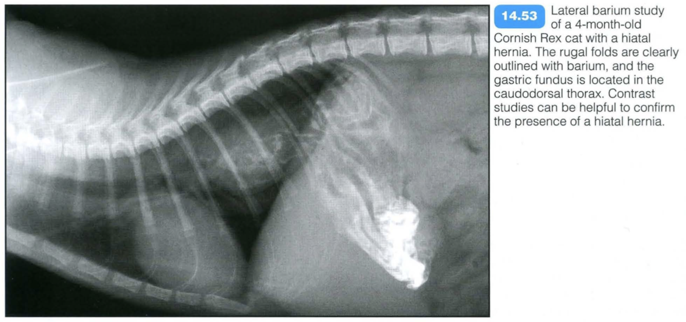
- Barium studies can confirm a suspicion of paraoesophageal hiatal hernia by outlining the abnormal position of the stomach or the small intestines in the caudodorsal thorax.
Sliding Hiatal Hernia
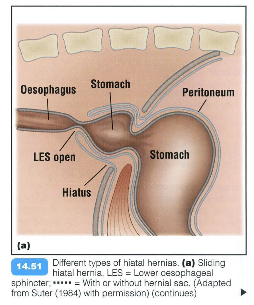
A hiatal hernia corresponds to a herniation of any abdominal organ through the oesophageal hiatus. It is most often congenital in origin but can be acquired:
- Following the repair of a chronic diaphragmatic rupture
- Secondary to a traumatic event
- Oesophageal/upper respiratory tract pathologies
- Neuromuscular disorders.
Three main types of hiatal hernia are recognized in animals:
- Short oesophagus hiatal hernia
- Sliding hiatal hernia
- Paraoesophageal hiatal hernia.
Sliding and paraoesophageal hiatal hernias can coexist. Chinese Shar Pei dogs are predisposed for hiatal hernias.
Short oesophagus hiatal hernia:
The lower oesophageal sphincter lies in the caudal mediastinum due to a short oesophagus. The cardia is pulled through the oesophageal hiatus. This type of hernia is very rare and has only been described in the dog.
Radiographic findings include:
- Ovoid soft tissue opacity in the mid-caudal thorax with a locally obliterated diaphragmatic silhouette on a lateral radiograph.
- Central, slightly left-sided location of opacity on DVNDviews.
- Permanent appearance.
Sliding intramediastinal hiatal hernia:
The lower oesophageal sphincter moves freely back and forth into the caudal mediastinum, followed by parts of the stomach, through a loose oesophageal hiatus. Clinical signs include variable degrees of vomiting or regurgitation, hypersalivation and chronic weight loss. If the stomach is incarcerated, severe respiratory distress can be present. Aspiration pneumonia is frequently associated with this
Radiographic findings include:
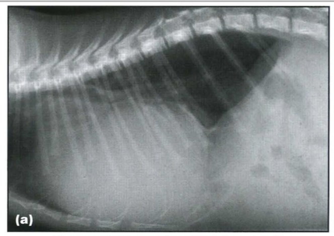
- Semicircular soft tissue mass, merging with the liver shadow localized between the CdVC and the aorta on the lateral view and in the caudal mediastinum on the DV view, superimposed over the hepatic shadow
- Gas present in the herniated stomach may allow recognition of rugal folds. If a large portion of the stomach is herniated and incarcerated, a large hyperlucent cyst-like structure surrounded by a thin soft tissue rim may be seen. In this case, lung collapse and mediastinal shift to the contralateral side is also visible
- Radiographic signs of aspiration pneumonia may be an associated finding.
- Radiographic signs might be absent, particularly during expiration, due to the sliding nature of the hernia.
- Positional radiography is usually unsuccessful in demonstrating a sliding hiatal hernia.
Contrast studies may aid diagnosis:
- A barium oesophagram may confirm suspicion of a sliding hiatal hernia. As the lower oesophageal sphincter moves back and forth, it can be in a normal position at the time of the examination
- Gastric rugae may be visible both in the caudal mediastinum and in the cranial abdomen, separated by a marked narrowing at the level of the oesophageal hiatus
- The lower oesophageal hiatus can be seen cranial to the cardia as a shallow indentation. It also allows assessment of concurrent oesophageal disorders, such as strictures or masses.
Fluoroscopy:
- This, together with an oesophagram, allows dynamic assessment of the position of the lower oesophageal sphincter and of the stomach. As the lower oesophageal sphincter function is altered due to its abnormal position, gastro-oesophageal reflux of contrast medium may be seen associated with swallowing.
Peritoneal-Pericardial Diaphragmatic Hernia
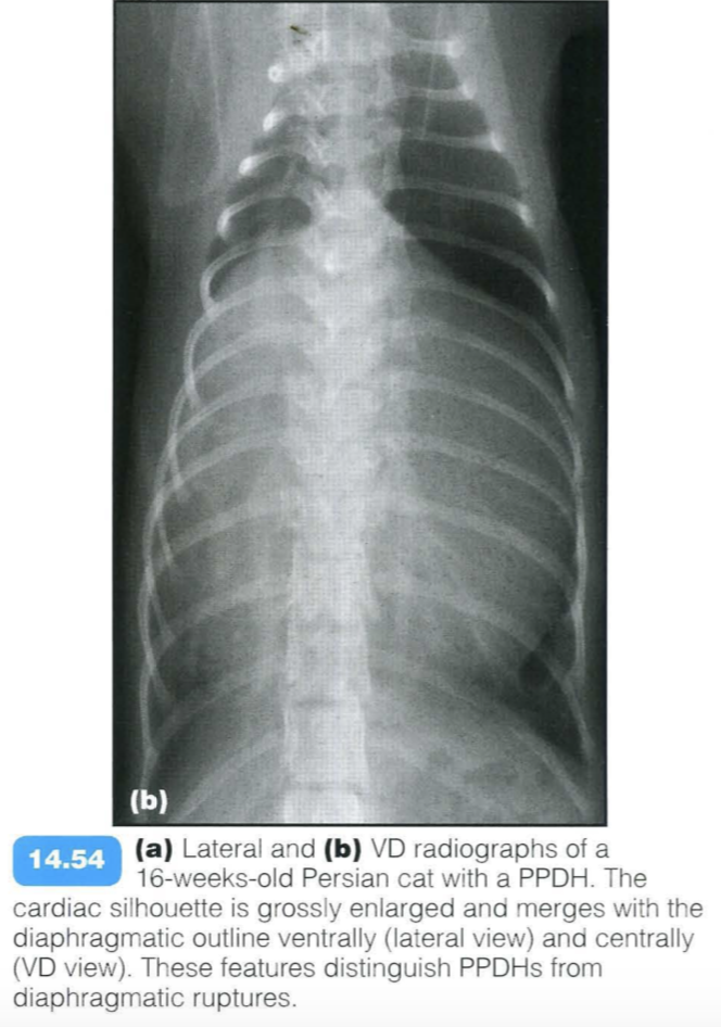
Peritoneopericardial diaphragmatic hernia (PPDH) is the most common diaphragmatic hernia in dogs and cats. It is due to an embryological fusion defect in the ventral diaphragm, leading to a communication between the pericardial and the peritoneal cavities. PPDHs are often associated with sternal or cardiac defects. The liver is the most frequently herniated organ, and may be associated with gallbladder, stomach, small intestine or omental herniation.
Clinical signs depend on the organs herniated and the size of the defect, and include respiratory, cardiovascular and gastrointestinal disorders. Auscultation may reveal muffled heart sounds or borborygmy over the cardiac area. Sudden onset of clinical signs may arise secondary to strangulation of a herniated organ. Most frequently no clinical signs are present and a PPDH is found incidentally.
Radiographic findings include:
- Moderate to gross enlargement of the cardiac silhouette, merging caudally with the diaphragmatic outline without associated radiographic signs of heart failure
- Dorsal displacement of the trachea
- Dorsal displacement of the CdVC
- Heterogenous opacity of the cardiac shadow due to the presence of falciform or omental fat, and/or gas or mineralized speckled structures contained in the herniated small or large intestines
- Persistence of a dorsal mesothelial remnant between the cardiac silhouette and the diaphragmatic outline, at the level or slightly dorsal to the CdVC, can be seen in cats
- Cranial displacement of the stomach and other abdominal organs
- Sternal abnormalities, including various deformities, reduction in the number of sternebrae and the absence or split of the xiphoid process
- Umbilical hernia or other abdominal wall defects
- Absence of radiographic signs of cardiac failure.
Contrast studies:
- Barium studies may outline the abnormal position of the stomach orthe small intestine in the pericardium if they are herniated
- Positive- or negative-contrast peritoneography may be useful when barium studies are inconclusive.
Fluoroscopy:
- The normal cardiac and diaphragmatic motion is not visible due to obliteration by herniated soft tissue structures.
Ultrasonography:
- This is an excellent modality to differentiate between cardiomegaly and a PPDH.
- An abdominal organ, usually part of the liver, is visible next to the heart, within the pericardial sac.
- Ultrasonography allows evaluation of the herniated organs as well as providing an assessment of cardiac function, if cardiovascular impairment was the reason for investigation.
Aortic Hilar Hernia
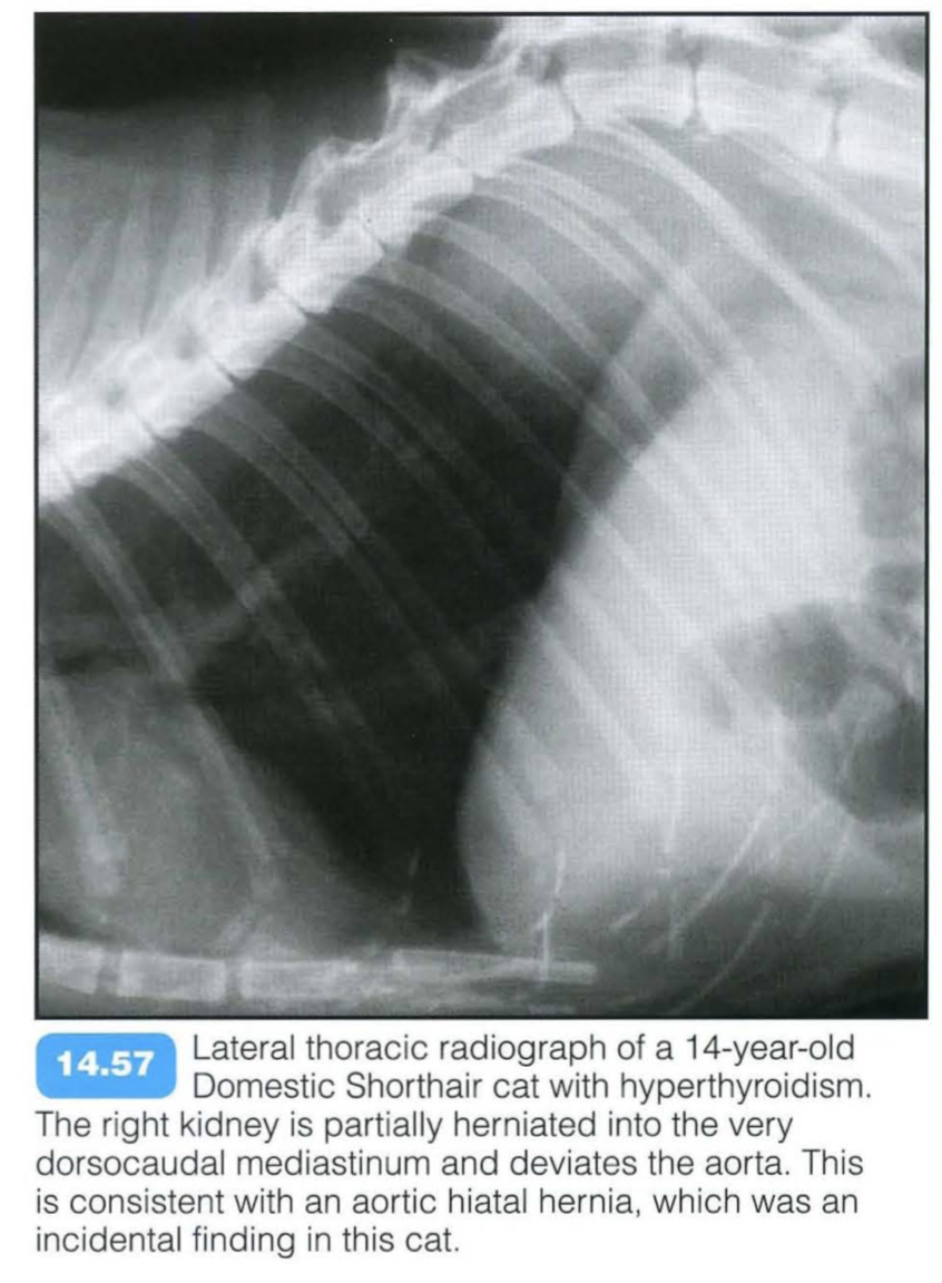
Aortic hilar hernia
The aortic hilus is the only diaphragmatic opening that connects the mediastinum with the retroperitoneal space. Free mediastinal gas can pass through the hilus and enter the retroperitoneal space relatively easily. If defects in this hilus exist, retroperitoneal organs such as the adrenal glands and kidneys can prolapse into the mediastinum. Intramediastinal kidneys are rare in dogs and cats and are usually detected incidentally.
Radiographic findings include:
- Caudodorsal mediastinal location of a kidney
- Should be differentiated from a dorsal circumferential diaphragmatic tear and renal prolapse.

