Radiographic findings with PTE:
- Pleural effusion
- Loss of the pulmonary artery
- Alveolar infiltrates (can be wedge shaped)
- Cardiomegaly
- Hyperlucent lung regions
- Enlargement of MPA
Acute Respiratory Distress Syndrome
(ARDS)
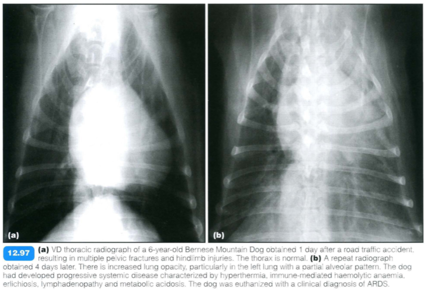
ARDS is a human term defined as acute fulminating respiratory failure, resulting from a variety of diseases , leading to diffuse lung injury. It may also be called adult respiratory distress syndrome or shock lung.
ARDS is considered to be a subgroup of non- cardiogenic oedema. It is part of the systemic inflammatory response, leading to increased vascular permeability, pulmonary hypertension and airway constriction and obstruction.
Inciting factors may be:
- Pulmonary (e.g. smoke inhalation , bacterial pneumonia)
- Non-pulmonary (e.g. endotoxaemia, pancreatitis, trauma, paraquat and other toxicities, fat embolism)
Histologically, ARDS is characterized by alveolar inflammation, oedema, haemorrhage, necrosis with formation of hyaline membranes or vascular congestion in conjunction with type 2 alveolar cell proliferation or interstitial fibrosis.
Clinical signs are acute onset of severe and progressive respiratory distress; in some cases signs are associated with an underlying cause (e.g. vomiting , evidence of trauma, etc .) . Progressive tachypnoea and dyspnoea are seen in most animals with altered lung sounds.
The most common reported underlying causes in dogs are:
- Bacterial pneumonia (which may be secondary to smoke inhalation or parasitic pneumonia)
- Sepsis and aspiration pneumonia
- Ventilator-acquired pneumonia
- Lung lobe torsion
- Non-pulmonary causes: gastric torsion, splenic torsion, trauma, laryngeal obstruction, pancreatitis, parvovirus infection, uraemia, disseminated intravascular coagulation (DIC), snake venom, drug toxicity
- Hyperoxia due to high inspired oxygen concentration (may result in lung injury similar to ARDS).
ARDS is possibly more common in younger dogs; no sex predisposition has been reported. A familial form is suspected in young Dalmatians (< 1 year old), some of which also have renal aplasia or hydrocephalus.
Radiographic findings include:
- May be normal in initial 24 hours
- Diffuse interstitial ± alveolar lung pattern, affecting all lobes is most common. Increasing severity with time, with air bronchograms visible by 36 hours after onset of lung injury
- Opacities relatively slow to change due to high protein content of fluid within the alveoli
- Bilateral distribution
- Pneumothorax and pneurnomediastinum may occur in later stages
- Radiographic signs associated with the underlying cause may be present
- In the familiar form, pneumomediastinum and gastro-oesophageal intussusceptions are seen in the late stages of the disease. Radiographic changes are mixed alveolar, interstitial and peribronchial patterns.

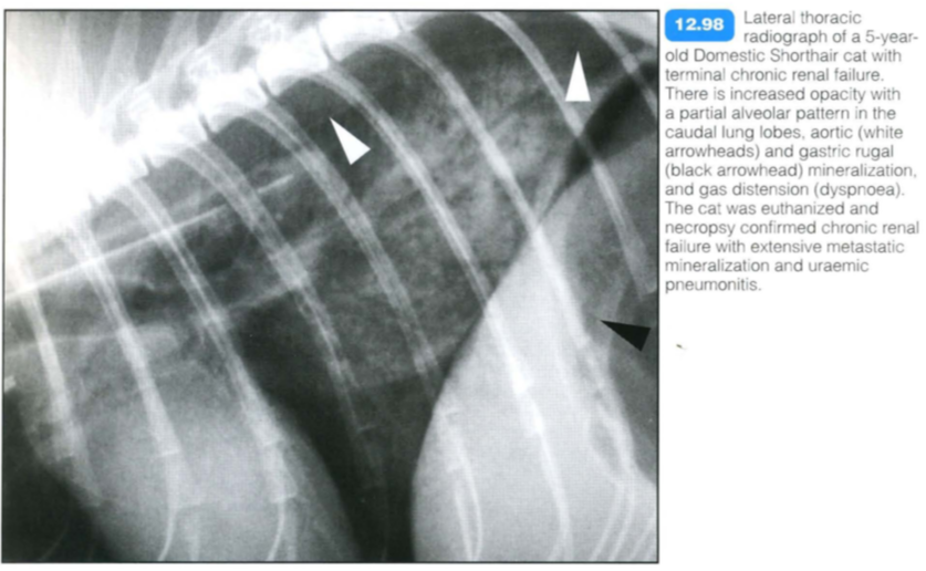
Uremic pneumonitis

Histopathological changes are similar to those of ARDS, and uraemic pneumonitis may be considered part of this syndrome. The high protein content of the oedema fluid suggests permeability oedema due to toxic lung damage. Other factors also are likely to play a role, e.g. reduced oncotic pressure and card io- vascular effects.
Clinical signs are related to severe renal disease (polyuria, polydipsia, anorexia, etc.) plus respiratory signs associated with oedema.
Uremic pneumonitis should be differentiated from other renal-induced respiratory diseases:
- Volume-overload pulmonary oedema
- Thromboembolic disease
Radiographic findings include:
- Pulmonary oedema within the caudodorsal lobes may be associated with ARDS or non-cardiogenic oedema due to fluid overload
- Abdominal radiographs may show evidence of underlying renal disease
- In cases of renal secondary hyperparathyroidism, mineralization of the blood vessels and myocardium may be present. There may also be demineralization of the skeleton
- Chronic uraemia may lead to degeneration and calcification of connective tissue.
Pulmonary hemorrhage
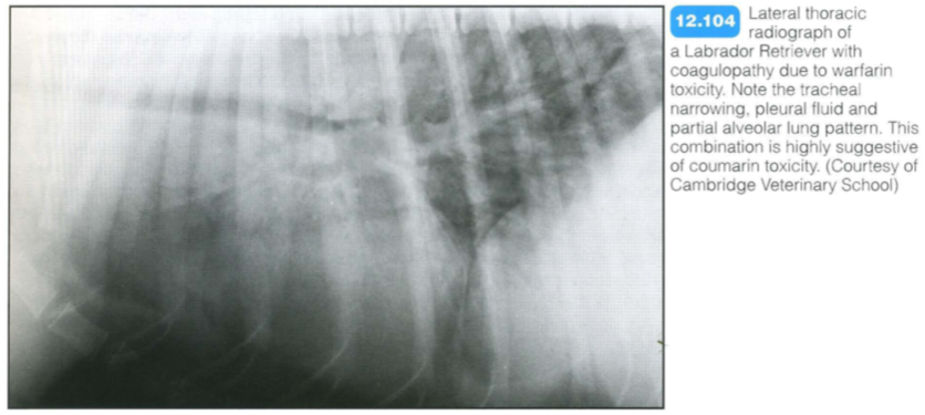
Most cases are associated with coagulopathies or trauma. Rodenticide poisoning (coumarin derivatives) is the most common cause of severe pulmonary haemorrhage; outdoor cats and dogs are at risk.
Other causes include:
- DIC
- Trauma
- Angiostrongylus infe-etion (dogs)
- Other coagulopathies
- Neoplasia (usually mass-related signs predominate)
Clinical signs include:
- Generalized blood loss (weakness, bruising, haematochezia, haematuria)
- Respiratory bleeding (cough, dyspnoea, haemoptysis)
- Upper airway obstruction
Thoracic changes may include haemorrhage of the
mediastinum, pleura and tracheobronchial airways.
Radiographic findings include:
- Generalized patchy interstitial/alveolar pattern with random distribution
- Possible pleural or mediastinal haemorrhage
- Tracheal narrowing due to submucosal/mucosal haemorrhage or extratracheal haematoma with narrowing of tracheal lumen - often generalized.
The combination of tracheal narrowing with pleural and mediastinal fluid and lung changes is suggestive of anticoagulant toxicity. Thoracic changes due to coumarin toxicity should resolve within 1-5 days of starting therapy. Haemorrhage due to Angiostrongylus vasorum infection usually also has radiographic changes of parasitic pneumonia. The pattern is classically peripheral in distribution.

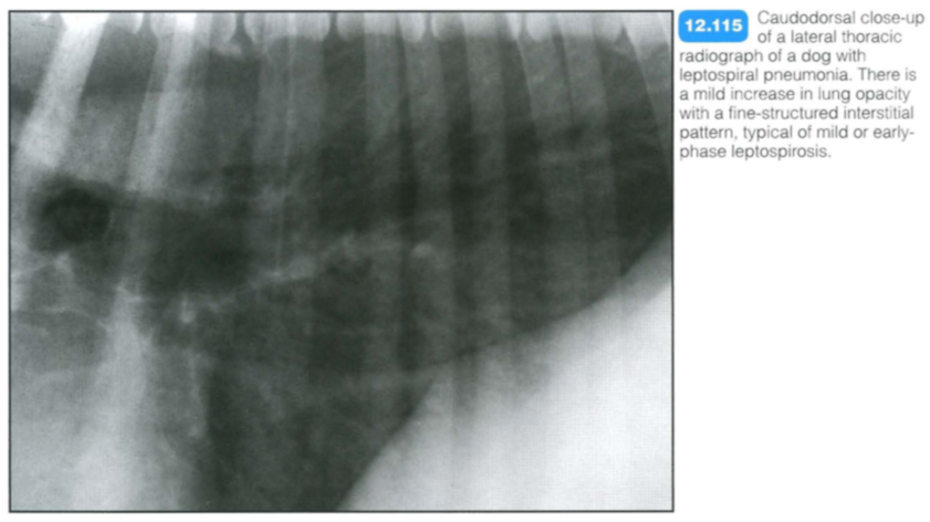
Canine leptospiral pneumonia
Clinical signs relate to acute renal failure rather than pulmonary involvement.
Radiography:
- A mild to marked un- or fine-structured interstitial pattern is seen, most noticeable in the caudodorsal lung fields or disseminated patchy opacities.
- In severe cases there may be a generalized increase in opacity in all lobes.
Other imaging techniques:
- Renal ultrasonography may be used to demonstrate increased renal cortical echogenicity, subcapsular fluid and an echogenic medullary band.

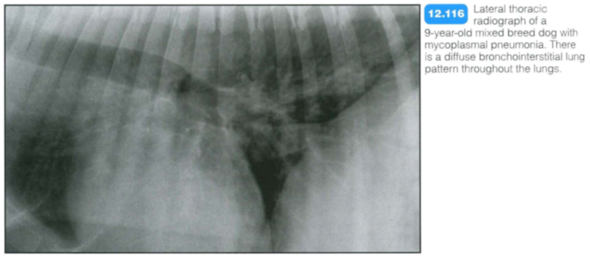
Mycoplasmal pnuemonia

Mycoplasmal pneumonia:
Mycoplasma spp . are small bacteria that are part of the normal oral flora in dogs and cats. In the lungs, mycoplasmal bacteria are involved in lower airway and lung disease, along with other bacteria. Immunodeficient or otherwise compromised animals are predisposed to mycoplasmal disease .
Radiography:
- A diffuse bronchointerstitial pattern is seen.
- An alveolar component is less common.
Viral pneumonia
Common pneumonia-inducing viruses include:
Dogs:
- canine distemper virus
- canine adenoviru s 2
- canine parainfluenza
Cats:
- feline calicivirus (FIP)
Young, unvaccinated or immunocompromised animals are most commonly affected. Clinical signs include pyrexia, coughing and oculonasal discharge.
Thoracic involvement with feline coronavirus infection (which causes feline infectious peritonitis) usually presents as pleural effusion rather than lung disease.
Radiographic findings include:
- Radiographs may be normal
- Mild diffuse interstitial lung pattern, often in the caudodorsal lung fields
- In severe cases, an alveolar lung pattern
- Peribronchial cuffing, if there is secondary bacterial infection; the radiographic signs resemble those seen with bacterial and aspiration pneumonia.
Fungal Pneumonia
Dimorphic fungi have a relatively well defined geographical distribution due to their specific growth requirement. Species include:
- Histoplasma capsulatum
- Blastomyces dermatitidis
- Coccidioides immitis
Histoplasmosis:
This is caused by Histoplasma capsulatum. It affects dogs and cats, most commonly less than 4 years old:
- In dogs there is usually gastrointestinal involvement
- In cats there is usually pulmonary involvement
The fungus requires a humid environment and has a wide distribution throughout temperate and tropical regions of the world; in the USA it is found in the drainage system of the Ohio, Missouri and Mississippi rivers (midwestern states).
Clinical signs are often absent; they may include coughing, dyspnoea, weight loss, lethargy and fluctuating fever. Thoracic radiographic findings include:
- Active phase: diffuse interstitial lung pattern and/or areas of coalescing opacities
- Chronic/healed phase: numerous 2-4 mm soft tissue or mineralized nodules; tracheobronchial lymphadenopathy and mineralization
- Disseminated form: polyostotic aggressive bone lesions are the most obvious radiographic feature
Blastomycosis:
This is caused by Blastomyces dermatitidis. It is common in dogs and uncommon in cats; most often seen in animals less than 4 years old. It is endemic in southeastern and eastern parts of the USA and southeastern Canada.
Routes of infection and primary focus include:
- Direct skin inoculation
- Inhalation and primary pulmonary form
- Dissemination to abdomen, skeleton and central nervous system possible.
Clinical signs of primary pulmonary form include dyspnoea, tachypnoea, coughing, fever, anorexia and weight loss. Thoracic radiographic findings include:
- Active phase:
- Miliary or mixed patterns
- Focal lung nodule/mass/consolidation
- Pleural effusion
- Tracheobronchial lymphadenopathy common
- Chronic/healed phase:
- Tracheobronchial lymphadenopathy possible
- Numerous non- mineralized nodules
- Disseminated form:
- Polyostoti c aggressive bone lesions are the most obvious radiographic feature
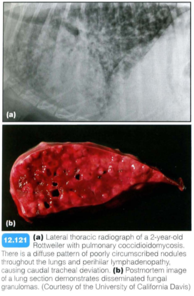
Coccidioidomycosis:
This is caused by Coccidioides immitis. It is common in dogs and rare in cats. The fungus requires semi-arid conditions and is endemic in southwest and western regions of the USA, and widespread in parts of Mexico, Central and South America.
Routes of infection and primary focus include:
- Direct skin inoculation (very rare)
- Inhalation and primary pulmonary form
- Dissemination to abdomen, skeleton and central nervous system possible.
The primary pulmonary form often has no clinical signs (subclinical and self-limiting). There may be a mild cough and fever, partial anorexia and weight loss. Thoracic radiographic findings include:
- Active phase: interstitial lung pattern; tracheobronchial lymphadenopathy possible
- Chronic/healed phase: disseminated, ill defined soft tissue nodules; tracheobronchial lymphadenopathy is uncommon
- Disseminated form: polyostotic aggressive bone lesions are the most obvious radiographic feature.
- Inhalation and primary nasal form in cats
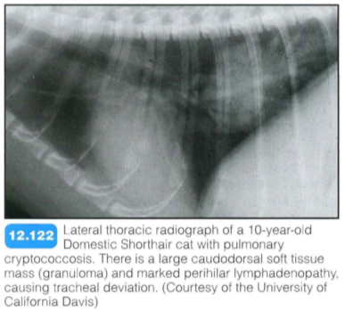
Cryptococcus:
This is caused by the yeast Cryptococcus neoformans. It is common in cats and uncommon in dogs. It has a worldwide distribution.
Routes of infection and primary focus include:
- Inhalation and primary nasal forms in cats
- May extend to central nervous system and cause neurological disease
- Primary or secondary lung infection is possible.
Thoracic radiographic findings include:
- Multiple nodules or small masses throughout the lungs
- Lung consolidation and pleural effusion possible
Aspergillosis:
This is caused by Aspergillus spp. It is common in dogs and rare in cats; the disseminated form is rare in both. German Shepherd Dog bitches are predisposed to the disseminated form. Aspergillosis has a worldwide distribution.
Routes of infection and primary focus include:
- Inhalation and primary nasal form with Aspergillus fumigatus; usually restricted to nose
- Inhalation and usually primary pulmonary form with A. terreus and A. deflectus; haematogenous spread to other organs possible.
There is a variety of non-specific clinical signs with disseminated aspergillosis, often including signs of spinal disease (discospondylitis). Thoracic radiographic findings include:
- Non-specific interstitial lung pattern
- Soft tissue nodules throughout the lungs
- Usually less important than other organ changes,
- such as discospondylitis.
Parasitic Pneumonia
Toxoplasmosis:
Toxoplasma gondii is a protozoa with a worldwide distribution. Cats and other felidae are the only definitive host; they may also serve as intermediate hosts. Clinical toxoplasmosis occurs during the intermediate phase. Cats and dogs may be affected; cats are most commonly infected and have non-specific multiorgan signs. Respiratory involvement is common in acute disease.
Thoracic radiographic findings include:
- Diffuse interstitial and patchy alveolar infiltrate
- Random distribution of changes.
Heartworm disease:
This is caused by Dirofilaria immitis, a filarial nematode that resides primarily in the pulmonary arteries. It has a worldwide distribution in temperate and tropical climates, including most of the USA, Central and South America, Japan, Australia and southern Europe.
Clinical signs include exercise intolerance, weight loss, coughing, right-sided heart failure and dyspnoea in severe cases. It is a predisposing factor for pulmonary thromboembolis. Heartworm disease is common in dogs and less common in cats.
Radiographic findings include:
- Patchy to extensive alveolar pattern, most marked in the caudal lobes in severe cases
- Changes have a periarterial distribution
- Enlarged and tortuous arteries (caudal in most cases but also often in the cranial lobes) may be seen
- Right-sided heart enlargement
- Pulmonary granulomas present in some cases.
Angiostrongylosis: (French heartworm disease): Angiostrongylus vasorum is a metastrongylid parasite of dogs and foxes. The adult worm lives in the main pulmonary artery, the right side of the heart or the pulmonary arterioles. It is most commonly reported in the southern UK and southern Europe; it is rarely diagnosed in the USA.
There are two main clinical syndromes:
- Respiratory disease due to an inflammatory response to the eggs and migrating larvae
- Haemorrhagic diathesis, possibly due to antigenic factors secreted by the parasite.
The disease is more common in young dogs, with Cavalier King Charles Spaniels and Staffordshire Bull Terriers possibly over-represented. Clinical signs include coughing, dyspnoea, ecchymotic haemorrhage, haemoptysis, haematomas, gastrointestinal bleeding, vomiting, diarrhoea and neurological disease.
Radiographic findings include:
- Diffuse bronchial and interstitial pattern initially (5 weeks after infection)
- Patchy alveolar pattern with peripheral distribution, preferentially affecting the caudodorsal lung lobes. Maximal changes 7-9 weeks after infection. May have lobar opacification. Bronchial component present in most cases
- Small volume of pleural fluid in some cases
- Mild hazy interstitial pattern remains following treatment
- Cardiovascular changes are uncommon.
Allergic Pneumonia
(Eosinophilic Bronchopneumopathy)
Pulmonary infiltrate with eosinophilia:
Pulmonary infiltrate with eosinophilia (PIE; eosinophilic broncho- pneumonopathy) is a manifestation of immunological hypersensitivity. The underlying cause cannot be found in most cases. It usually occurs in young adult or middle-aged dogs. Siberian Huskies and Alaskan Malamutes are possibly predisposed. The main clinical sign is coughing, but signs may include dyspnoea, exercise intolerance and nasal discharge.
Multicentric lymphoma
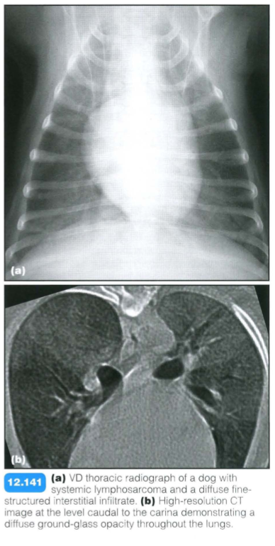
Radiographic findings:
- Diffuse un- or fine-structured interstitial pattern most common. This is often described as reticular, honeycomb or micronodular
- Most apparent in the caudodorsal and perihilar lung fields
- Bronchial wall thickening (neoplastic lymphatic infiltrate)
- The pulmonary vessels are generally well seen despite the interstitial pattern
- May develop within days and regress similarly quickly with chemotherapy
- Very rarely, a nodular pattern or large masses can occur
- Additional common findings are pleural effusion, lymphadenopathy of the tracheobronchial, cranial mediastinal and sternal lymph nodes, and mediastinal widening
An interstitial pattern with lymphadenopathy is very suggestive of lymphoma, with the other main differential diagnosis being fungal pneumonia.
Radiographic diagnoisis is usually sufficient. CT may be performed for other reasons and typical features should be recognized:
- Ground-glass opacity, which may be more prominent in the caudal lung lobes
- Enlarged sternal, cranial mediastinal and tracheobronchial lymph nodes

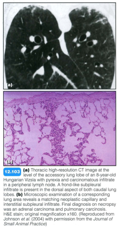
Alveolar Septal Metastasis

Alveolar septal metastases:
These are common with anaplastic mammary carcinoma and less common with salivary or pulmonary carcinoma or transitional cell carcinoma of the urinary bladder. Metastatic spread to the interstitium of the lung occurs via the lymphatic system and microvasculature. Neoplastic cells infiltrate the alveolar septal walls causing thickening, and may also cause thrombosis of small vessels.
Alveolar septal metastases and bronchoalveolar carcinoma: Radiographic findings are not specific but may include:
- Diffuse interstitial disease with occasional alveolar infiltrates or ill defined nodules
- Hyperlucent lung areas (regional embolic oligaemia)
- Peribronchial cuffing (filling of lymphatic system with tumour cells)
- Pleural thickening
- Mild pleural effusion
- Sternal lymphadenopathy
CT provides exceptional detail of the lung tissue and vasculature, and is very useful for diagnosis and staging of radiographically occult neoplastic disease. Imaging findings in dogs may include:
- Normal pleura
- Subpleural zone:
- Subpleural lines due to an interstitial infiltrate and fibrosis
- Subpleural wedge-shaped interstitia thickening (base parallel to the pleura) caused by neoplastic infiltration and fibrosis
- Patches of ground-glass opacity caused by tumour cells, necrosis and haemorrhage
- Subpleural emphysema due to small airway obstruction caused by fibrosis
- Mosaic perfusion: patchy hyperattenuating and hypoattenuating areas with small pulmonary arteries due to altered perfusion patterns, secondary to neoplastic embolization
- Distortion of architecture
- Small nodules
- Areas of consolidation.
- Peribronchial zone:
- Peribronchovascular interstitial thickening due to tumour nodules or atelectasis
- Small nodules
- Areas of consolidation
Interstitial Mineralization
Mineralization of the pulmonary parenchyma may be caused by:
- Dystrophic mineralization
- Metastatic mineralization
- Idiopathic mineralization
Dystrophic mineralization:
- Damaged or necrotic lung tissue in dogs with normal serum calcium
- Calcium embolism secondary fungal granulomas (histoplasmosis), abscessation, neoplasia or meta- bolic disease (hyperadrenocorticism).
- Diffuse inflammatory lesions that result in fibrosis, such as chronic uraemia, can also have a component of mineralization.
- Heterotopic bone (pulmonary osteoma) is considered a form of dystrophic mineralization
- Osteosarcomas occasionally form osteoid pulmonary metastases
- Hyperadrenocorticism (Cushing ‘s disease): This causes dystrophic mineralization of the soft tissues, including the skin, stomach, arteries, skeletal muscles and kidneys.
Metastatic mineralization:
- This is caused by altered serum calcium and phosphorus levels with deposition of minerals in the normal tissues.
- Causes of hypercalcaemia include: “GOSHDARNIT”
- primary and secondary hyperparathyroidism
- hypervitaminosi s D
- lymphosarcoma
- cholecalciferol rodenticide toxicity
- disseminated bone cancer - multiple myeloma.
- Tumours (e.g. anal gland carcinomas) can also produce a parathormone-like hormone, which causes hypercalcaemia as a paraneoplastic syndrome.
Idiopathic and iatrogenic mineralization:
- This includes pulmonary alveolar and bronchiolar microlithiasis (idiopathic) and mineralization secondary to barium aspiration (iatrogenic)
