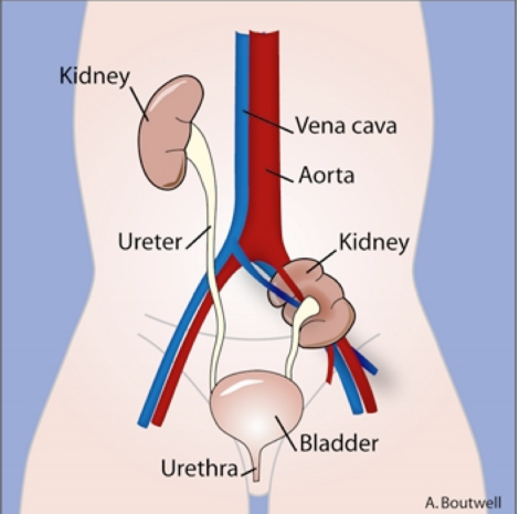
The urogenital system is composed of:
- The kidneys (3 of them, bilateral, from the intermediate mesoderm)
- The ureters (ureteric bud, also from IM)
- The bladder (from the cloaca/urogenital sinus and allantois)
- The urethra (from the urogenital sinus)
What are the three separate pairs of nephric structures that arise successively in a cranio-caudal fashion with the previous structure degenerating and being replaced?
- The first is the pronephros (before kidney)- never functional in humans.
- The second is the mesonephros (middle kidney). All but a small portion of this structure degenerates- the portion that remains will become ducts of the male reproductive system, the Wolffian or mesonephric ducts, which become the ductus deferens.
- The final and definitive kidney and ureter are formed from the metanephros (final kidney).
- These connect to the cloaca (end of the gut tube, thus endoderm coated with mesoderm), which divides into the allantois, rectum and urogenital sinus, which eventually forms the bladder and urethra.
The Urogenital Ridge
- The urogenital system is derived from intermediate mesoderm, which lies between the somites and the lateral plate mesoderm.
- During the fourth week of development, lateral folding of the embryo results in the location of the intermediate mesoderm to two dorsolateral ridges. These ridges run the length of the embryo and are called the urogenital ridge (because they form the kidneys (uro), gonads (genital) and their tubules).
Development of the Pronephros
In humans, the pronephros is a rudimentary kidney structure that develops bilaterally in the intermediate mesoderm in neck and head region.
- They begin to form and regress during 4th week (4 heart chambers, 4 branchial arches, 4 limb buds all form during this week – rule of 4s)
- Based upon morphological observations it is almost certain that the pronephros (pleural=pronephroi) are not functional. However, they start the developmental interactions followed by the next structures, the mesonephros.
Development of the Mesonephros
The mesonephros appears in a cranial to caudal manner, just caudal to the pronephros. It is quite large, and over time extends from the 1st thoracic to 3rd lumbar vertebrae.
A. Early in the fourth week, mesonephric tubules begin to form in the intermediate mesoderm. These paired structures are produced in a cranial to caudal fashion.
B. The mesonephric tubules do differentiate into excretory units. They form a Bowman’s capsule at the end of each tubule and branches of the dorsal aorta extend into each and form a glomerulus.
C. As the mesonephric tubules are forming, a mesonephric duct begins to form as a solid rod of cells in the urogenital ridge from the thoracic region down to the cloaca (embryonic bladder/rectum combo). After fusion with the cloaca, the rod begins to form a lumen, resulting in a duct. The mesonephric tubules fuse with the mesonephric duct, opening the passage from the mesonephric excretory unit to the cloaca.
D. Even as caudal mesonephric tubules are forming, the cranial portion of the mesonephros has begun to degenerate- this all occurs between 5-8 weeks of age.
E. In the male, mesonephric tubules will persist as the efferent ductules of the testis and the mesonephric (Wolffian) duct will persist as the ductus deferens. In the female (without substantial testosterone), they will all regress and a parallel duct system will form the oviduct and uterus.
Development of the Metanephros
A. The metanephric kidney is the last/third and definitive kidney. It begins to form early in the fifth week (thus overlapping with the mesonephros). It arises from two structures: the ureteric bud (ducts) and the metanephric blastema (kidney).
B. The ureteric bud buds off of the mesonephric duct. It grows dorsally and induces the intermediate mesoderm to form the metanephric blastema. A blastema is a blob of undifferentiated tissue.
C. The ureteric bud gives rise to the urine collecting elements- collecting tubules, major and minor calyces, and the ureters.
D. The ureteric bud undergoes repeated branching to form many generations of collecting tubules. The first generations become confluent to form the major calyces, the next generations form the minor calyces, and the remainder forms the true collecting tubules.
E. The metanephric blastema gives rise to urine producing and processing portions of the kidney between the glomerular capillary and the collecting duct. These are the Bowman’s capsule, proximal convoluted tubule, Henle’s loop, and the distal convoluted tubule.
F. The metanephric blastema is induced by the ureteric bud to form vesicles and, eventually, S-shaped tubules. i. S-shaped tubules become nephrons. ii. Vascular endothelial cells grow into this primordium to form the glomerulus iii. Older nephrons mature to form elongated loops of Henle, newer nephrons are formed in more cortical areas.
G. At birth, each kidney contains 800,000 to 1,000,000 nephrons. Nephron formation is thought to be complete at birth. Growth of the kidneys after birth is the result of elongation of the proximal convoluted tubules and loops of Henle, as well as increased interstitial tissue.
Metanephric blastema
- Bowman’s capsule
- Tubular system
- Proximal convoluted tubules
- Loops of Henle
- Distal convoluted tubules
Ureteric bud forms…
- Collecting tubules
- Major and minor calyces
- Renal pelvis
- Ureters
Regulation of Kidney Development and Defects
- The events that regulate induction of the different structures in the kidney are highly coordinated.
- Reciprocal induction of the ureteric bud and the metanephric blastema are required for normal branching morphogenesis and tube formation. Proteins secreted by one tissue (e.g., the ureteric bud) induce the adjacent tissue (e.g., the metanephric blastema) to change morphologically (e.g., tubule formation) and to secrete new proteins that reciprocally induce changes in the first tissue (e.g., budding).
- Mutations of the genes anywhere in this pathway may cause a disruption leading to renal agenesis, renal hypoplasia (reduced number of nephrons but otherwise normal architecture), or renal dysplasia (with malformed elements).
- Renal hypodysplasia (malformed and small) is more common than hypoplasia without dysplasia.
- When these renal proteins are used in other regions of the body, syndromes of defects in several organ systems may be seen.
Ascent of the Kidneys
A. The metanephric kidneys originate deep in the pelvic region, adjacent to the bladder. During the late embryonic (week 6-8) and early fetal period (week 9), they shift into the abdominal region. This is largely due to the growth of the caudal region of the embryo.
B. Blood supply to kidney begins at sacral level from dorsal aorta branches. As the kidneys ascend, they develop new connections with the aorta at each level. Sometimes these vessels will persist in the adult. Kidney Development 6
C. During ascent, the kidneys rotate 90° so that the hilum faces medially.
D. If the inferior poles of the metanephric blastema grow into one another while they are still in the pelvis, they can become fused into a horseshoe kidney. When ascending into the abdomen, this horseshoe kidney will get caught under the Inferior Mesenteric Artery (IMA) and the kidney will remain in the pelvis (see below).
Formation of the Urinary Bladder
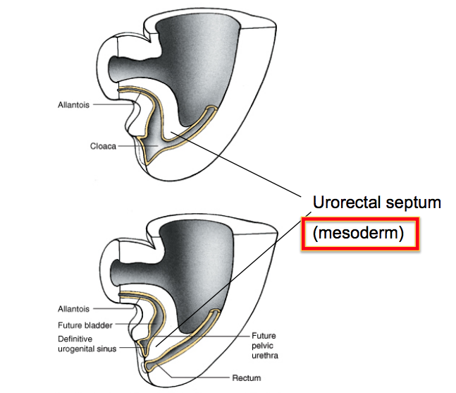
A. The urorectal septum, a mesodermal cone, divides the cloaca into the urogenital sinus and the rectum. Failure of this cone to create a good division between the rectum and bladder can lead to imperforate anus and other malformations of the external genitalia, and recto-vesicle fistulas (internal connections allowing leakage between the rectum and bladder). Although the lining of the rectum and bladder are associated with the endodermal germ layer, the segregation requires the mesodermal cone, and thus associates with mesodermal germ layer disorders (VACTERL), similar to the tracheo-esophageal separation you will learn aout in DMH.
B. The base of the allantois at its attachment to the urogenital sinus expands to form the urinary bladder, with an embryonic connection (urachus) to the extra embryonic allantois (for early waste removal). The connecting urachus closes and becomes the median umbilical ligament in the adult (STEP1). Failure of the urachus to close leads to leaking of urine out the umbilicus in the newborn (patent urachus).
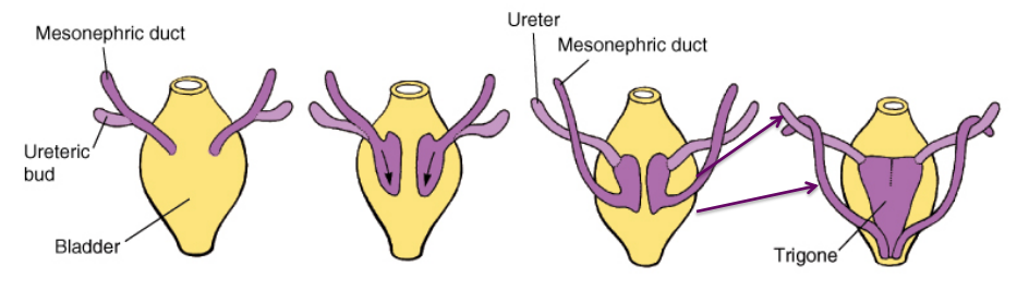
C. Growth of the dorsal bladder incorporates the mesonephric ducts and ureteric buds (of the metanephros) into the bladder wall. The remaining ureteric bud forms the ureter. The smooth area formed and bounded by the entry of the mesonephric ducts (future ductus deferens that empty into the prostate along with the seminal vesicles) and ureters is known as the trigone of the bladder. Stretch receptors here signal bladder fullness (flashback to Neuro).
E. The area of the urogenital sinus where the mesonephric ducts enter becomes narrowed and forms the beginning of the urethra.
Patent Urachus
Failure of the urachus to close leads to leaking of urine out the umbilicus in the newborn
Horseshoe Kidney
Horseshoe Kidney (1/600) is usually asymptomatic and forms when the bilateral metanephric blastema get stuck together in the midline. Because the inferior mesenteric artery comes off of the ventral side of the aorta in the lower abdomen, the now horseshoe-shaped kidney gets caught under this vessel and cannot rise up to its normal location along the dorsal, retroperitoneal wall of the abdomen.
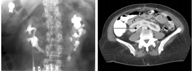
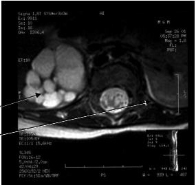
Unilateral Multicystic Dysplasia
- Unilateral multicystic dysplasia (1/2400) forms when the kidney epithelia overexpress PAX2 and are often associated with an atretic ureter. PAX2 expression may be due to increased hydrostatic pressure.
- Multicystic dysplastic kidney is a form of renal dysplasia characterized by the presence of multiple, noncommunicating cysts of varying size separated by dysplastic parenchyma and the absence of a normal pelvocaliceal system. The condition is associated with ureteral or ureteropelvic atresia, and the affected kidney is nonfunctional. Other terms used to describe this condition include multicystic kidney and multicystic renal dysplasia. Multicystic dysplastic kidney is the most common cause of an abdominal mass in the newborn period and is the most common cystic malformation of the kidney in infancy
Bilateral Multicystic Kidney or Bilateral Renal Agenesis
- When bilateral (more common in females, 1:3000) multicystic kidney or bilateral renal agenesis is incompatible with life. Two babies have now survived c.2013.
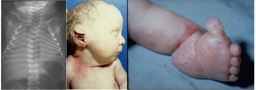
- In cystic kidneys with low but some urine production (oligohydramnios), little amniotic fluid is generated (from urine), causing abnormal face and limb development and poor lung formation due to decreased lung distension (Potter’s sequence, for Edith Potter (1901-1993)
POTTER
P – Pulmonary hypoplasia (urine/amniotic fluid is needed for lung development)
O – Oligohydramnios (insufficient amniotic fluid = trigger)
T – Twisted face (compressed features due to oligohydramnios)
T – Twisted skin (ditto)
E – Extremity defects (ditto)
R – Renal failure in utero (primary cause)
Pelvic Kidney
•One-third of all children born with a pelvic kidney have other complications either with their cardiovascular system, the central nervous system or their urinary system
Trigone
•The trigone starts out mesodermal in origin whereas the rest of the bladder lining is endodermal in origin. Eventually the endoderm overgrows it.
Imperforate Anus
- Imperforate anus typically groups with mesodermal anomalies (VACTERL)
- VACTERL stands for
Vertebral defects
Anal atresia
Cardiac defects
Tracheo-esophageal fistula
Renal anomalies
Limb abnormalities.
-
Kidney Structure27
-
Urinary Tract Structure20
-
Renal Blood Flow and Glomerular Filtration Rate50
-
Tubular Function I: Sodium Transport21
-
Osmoregulation and Vasopressin System34
-
Volume Regulation and RAS9
-
Tubular Transport II: Potassium8
-
Tumors of Kidney and Urinary Tract46
-
Acid Base52
-
Kidney Development19
-
Hyponatremia and Hypernatremia39
-
Cystitis, Pyelonephritis and Interstitial Nephritis57
-
Hypokalemia and Hyperkalemia40
-
Bladder Function and Dysfunction64
-
Mineral Metabolism13
-
Hematuria and Renal Stones14
-
Acute Renal Injury I and II39
-
Diuretics10
-
Urinalysis Introduction and Vascular Disease in the Kidney62
-
Nephrotic Syndrome I and II53
-
Nephritic Syndrome I and II70
-
Drugs Acting on the Angiotensin System9
-
Drugs Mentioned in Lecture (aka Misc)14
-
Chronic Kidney Disease I and II43
-
Secondary Hypertension36
-
Genetic Diseases of the Distal Nephron15
