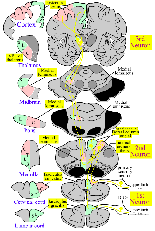
Dorsal Columns System
(DC)
Carries afferent epicritic sensation.
Fine touch, vibration sense, proprioception.
Type A𝛼 and A𝛽 (or Ia, Ib, and II)
[Lumbar Spinal Cord]
Primary neuron enters via medial division of dorsal root ganglion (DRG).
Carries lower limb information.
Axons form the gracile fasciculus.
[Cervical Spinal Cord]
Primary neuron enters via medial division of dorsal root ganglion (DRG).
Carries upper limb information.
Axons form the lateral cuneate fasciculus at or above T5.
Somatotopy: cervical fibers located laterally next to gray matter
[Medulla]
Gracile and cuneate fasciculi run ipsolaterally up to medulla.
Axons terminate on secondary neurons in gracile and cuneate nuclei.
Second neurons cross midline as internal arcuate fibers.
Somatotopy: lower limb info ventral; upper limb info dorsal
[Pons and Midbrain]
Axons form the medial lemniscus (ML).
Somatotopy: lower limb info lateral; upper limb info medial
[Thalamus]
Medial lemniscus terminates in the ventral posterior lateral (VPL) nucleus.
Third neurons in VPL thalamus.
Somatotopy: lower limb info lateral; upper limb info medial
[Cortex]
VPL axons terminate in the postcentral gyrus (primary somatosensory cortex)
Somatotopy: lower limb info medial; upper limb info lateral
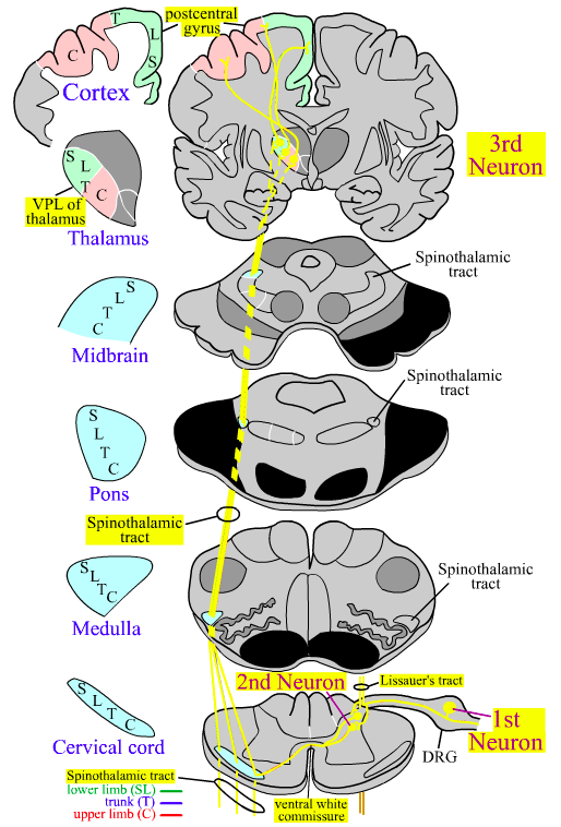
Anterolateral System
(ALS)
Carries protopathic sensation.
Includes 3 pathways:
- Spinothalamic tract (STT)
- Spinoreticular tract
- Spinomesecephalic tract
Spinothalamic Tract
(STT)
Part of the anterolateral system along with spinoreticular and spinotectal fibers.
Carries protopathic sensation from contralateral side of the body.
Pain, temperature, and crude touch.
Type A-𝛿 and C fibers.
[Spinal cord]
Primary neurons in dorsal root ganglion (DRG)
Axons enter via lateral division of DRG
Sends collaterals into Lissauer’s tract
(~ 1 level rostrally and caudally)
Axons terminate in dorsal horn (lamina I, II, and V)
Second neurons in Lamina I and V
Axons decussate immediately in ventral white commissure
Axons form the spinothalamic tract (STT)
Stomatotopy: cervical fibers located ventrally next to gray matter
[Pons, Medulla, Midbrain]
Axons ascend as the STT carrying information from contralateral side of the body.
Somatotopy: cervical ⇒ sacral goes ventral ⇒ dorsal
Medulla: STT is lateral/dorsal to inferior olive
Pons/Midbrain: STT lateral to medial lemniscus
[Thalamus]
STT axons terminate at the ventral posterior lateral nucleus (VPL)
Third neurons in the VPL thalamus.
Somatotopy: lower limb lateral, upper limb medial
[Cortex]
VPL axons terminate in the postcentral gyrus
Somatotopy: lower limb medial, upper limb lateral
Facial
Epicritic Pathway
Fine touch and proprioception transmitted via the trigeminal nerve (CN V).
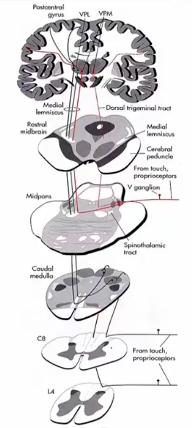
[Pons]
Enters at the mid-pons.
Majority of fibers crosses midline.
Travels medially within longitudinal pontine fibers.
Some fibers travel via Dorsal trigeminal tract.
[Midbrain]
Anterior position within medial leminiscus.
Some fibers travel via Dorsal trigeminal tract.
[Thalamus]
Project into the ventral posterior medial (VPM) thalamus.
[Cortex]
Project to lateral somatotopy of post-central gyrus.
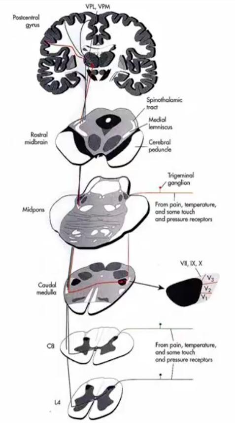
Facial
Protopathic Pathway
Pain and temperature information carried via
Trigeminal nerve (CN V)
[Pons]
Enters spinal cord at the mid-pons.
Descends down to the caudal medulla and crosses midline.
Ascends via the Spinothalamic tract (STT).
[Midbrain]
Anterior position within the STT
[Thalamus]
Fibers terminate within the ventral posterior medial (VPM) thalamus.
Third neuron originates in the VPM.
[Cortex]
VPM fibers terminate laterally within the postcentral gyrus.
Horner’s Syndrome
Preganglionic sympathetic neurons in the T1 intermediolateral nucleus (aka ciliospinal center of Budge) ⇒ postganglionic neurons in cervical ganglion.
Postganglionic sympathetic neurons ⇒ ipsilateral dilator muscle of pupillae, superior tarsal muscle, and sweat glands of face.
Lesion of this pathway results in Horner’s syndrome.
Characterized by ipsilateral:
miosis (constricted pupil)
ptosis (drooping eyelid)
anhidrosis (lack of sweating) on the face
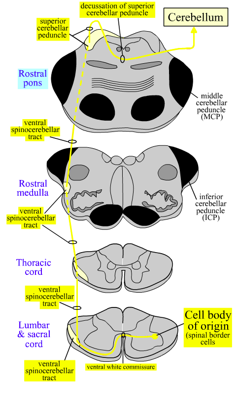
Ventral Spinocerebellar Tract
(VSCT)
VSCT sends to cerebellum an efferent copy of spinal cord motor neuron output.
Used for motor control of lower limbs.
Fibers cross twice ending on ipsilateral side to origin.
Lesion of VSCT fibers produces contralateral deficits.
- Spinal Cord
- neurons in lamina VII & ventral horn of lumbar spine (spinal border cells)
- 1st crossing:
⟾ axons cross in ventral white commissure
⟾ forms contralateral VSCT
- Rostral Pons:
- VSCT enters superior cerebellar peduncle (SCP)
- 2nd crossing:
⟾ axons cross again in decussation of superior cerebellar peduncle to reach ipsilateral cerebellum to origin
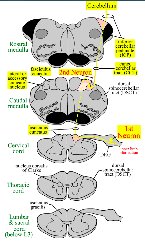
Cuneocerebellar Tract
(CCT)
Carries unconscious proprioception from upper body to cerebellum.
Used for motor control.
- Spinal cord
- 1st neuron in DRG
- enter via medial division of dorsal root
- travel in cuneate fasciculus
- send collaterals to lateral (accessory) cuneate nucleus
- 1st neuron in DRG
- Caudal medulla
- 2nd neuron in lateral cuneate nucleus
- axons form cuneocerebellar tract
- 2nd neuron in lateral cuneate nucleus
- Rostral medulla
- CCT fibers travel via inferior cerebellar peduncle (ICP)
- Cerebellum
- Fibers enter ipsilateral cerebellum
- tract lesions produces ipsilateral deficits
- Fibers enter ipsilateral cerebellum
Pyramidal System
Major motor pathway from the cortex.
Corticospinal, corticobulbar, corticoreticular tracts.
- Originates
- primary motor cortex (main)
- premotor cortex
- primary somatosensory cortex
- posterior parietal area
- Terminates
- alpha & gamma motor neurons in spinal cord
- cranial nerve motor neurons in brainstem
- Susceptible to major insult including vascular damage.
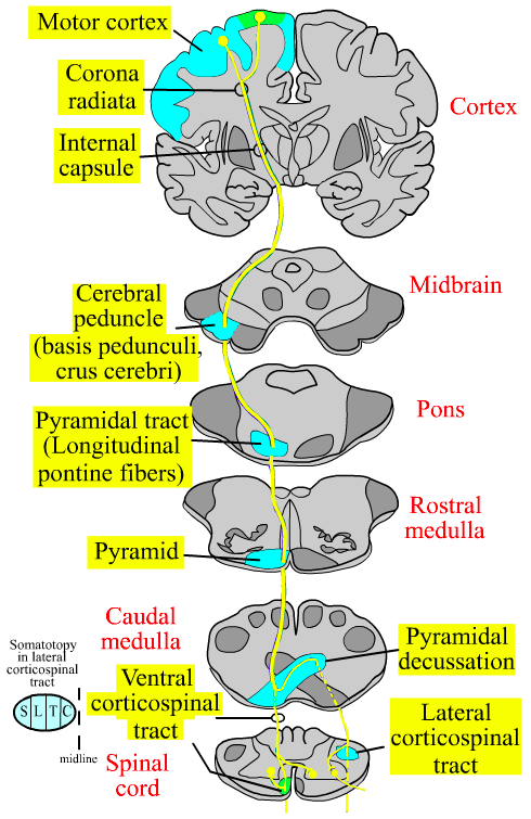
Cortical Spinal Tracts
(CST)
Control of voluntary movements.
Suppression of innate reflexes.
[Cortex]
Pyramidal neurons (lamina V of cerebral cortex) mostly in precentral gyrus but also other cortical areas
⇒ corona radiata
⇒ posterior limb of internal capsule
[Midbrain]
Middle 1/3 portion of cerebral peduncle
(Crus cerebri and Basis pedunculi)
[Pons]
Pyramidal tract in basal pons
(Longitudinal pontine fibers)
[Open Medulla]
Pyramid
[Closed Medulla]
85% of fibers cross at the pyramidal decussation
15% of fibers remain on ipsilateral side
[Spinal Cord - starting at cervical]
Decussated fibers form Lateral Cortical Spinal Tract (LCST)
Cervical fibers closer to gray matter
Controls distal motor neurons for fine movements
Runs within lateral funiculus w/ RuST & MRST
Remainder of fibers form the Ventral Cortical Spinal Tract (VCST)
Cross at segmental level
Influences bilateral neurons controlling axial muscles
Lateral Corticospinal Tract (LCST)
Characteristics
- Terminates on LMN in ventral horn for distal muscles
- Synapses on spinal interneurons
- Ia & Ib interneurons
- Renshaw cells
- Lesions:
- spasticity → d/t mix with corticoreticular and reticulospinal tracts
- Babinski sign ± Grasp reflex
- Isolated LVST lesion
- rare
- see w/ lesion in primary motor cortex or pyramids
- get flaccid paralysis & substantial atrophy
- no UMNs
Ventral Corticospinal Tract (VCST)
Characteristics
- Originates in trunk & limb extensors region of contra. motor cortex
- Runs in ventral funiculus
- Bilaterally innervates:
- axial motor neurons
- interneurons in ventral horn
- Functions to maintain posture
- Lesion → little deficit if unilateral
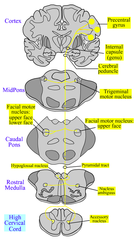
Corticobulbar Tract
(CBT)
Controls cranial nerve motor functions.
- Cortex
- originates from pyramidal neurons in lamina V of contralateral motor cortex head region
⟾ corona radiata
⟾ genu of internal capsule
- originates from pyramidal neurons in lamina V of contralateral motor cortex head region
- Midbrain
- axons in medial 1/3 of cerebral peduncle
- Pons
- controls bilateral trigeminal motor nucleus
- controls bilateral upper face facial motor neurons
- controls only contralateral lower face facial motor neurons
- unilateral CBT lesion = contralateral drooping of corner of mouth
- Medulla and Spinal Cord
- controls bilateral hypoglossal nucleus
- controls bilateral nucleus ambiguus
- controls bilateral spinal accessory nucleus
⟾ nuclei often receive mostly crossed CBT fibers
⟾ CBT lesion may cause contralateral weakness
Extrapyramidal Tracts
Involves multiple UMNs.
Originates primarily in premotor cortex and cerebellum.
Synapses in brainstem before reaching LMN.
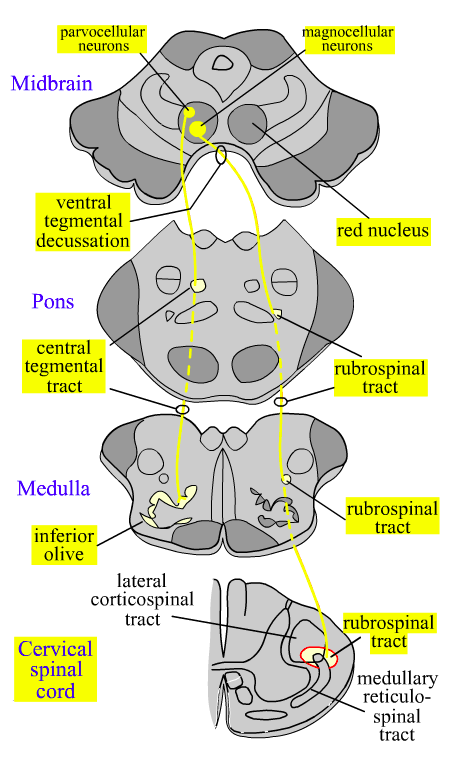
Rubrospinal Tract
(RuST)
Functions to control upper arm flexion.
Specialized for brachiation in primates.
- Path:
- Cortex
- magnocellular neurons of contralateral red nucleus
- Midbrain
- crosses in ventral tegmental decussation
- Travels with LCST in lateral funiculus
- Terminates in cervical spinal cord
- synapses with ventral horn LMN & interneurons
- Cortex
- Controls upper arm musculature
- flexors > extensors
- Lesions → upper arm spasticity
- Contributes to decorticate posture

Rubro-olivary Fibers
- Originates from parvocellular neurons in red nucleus
- Axons project via central tegmental tract → ipsilateral inferior olive
- Functions in motor learning
- regulates cerebellar function
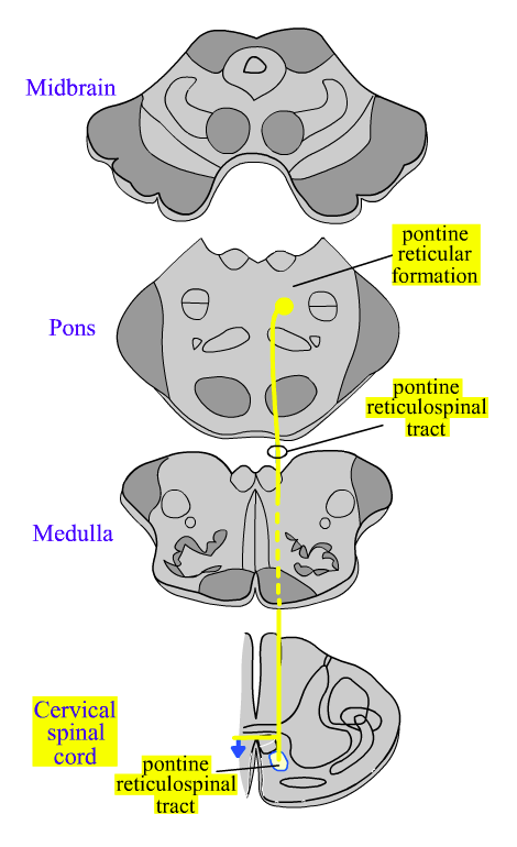
Pontine Reticulospinal Tract
(PRST)
- Function:
- Facilitates extensor and inhibits flexors.
- Involved in control of posture and gait-related movements.
- Maintains muslce tone.
- Opposes MRST.
- Controlled by contralateral motor cortex.
- Lesions = spasticity.
- Tract:
- Originates from 2 pairs of neurons within pontine reticular formation
- nucleus reticularis pontis oralis (rostral pons)
- nucleus reticularis pontis caudalis (caudal pons)
- Originates from 2 pairs of neurons within pontine reticular formation
- Axons descend ipsilaterally
- travel in ventral funiculus with VCST
- Terminate bilaterally in all spinal cord levels
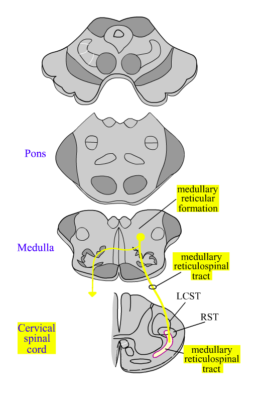
Medullary Reticulospinal Tract
(MRST)
- Characteristics:
- Inhibits extensors and facilitates flexors.
- Involved in control of posture and gait-related movements.
- Terminates mostly on gamma MN and interneurons that control proximal extensors.
- Opposes PRST.
- Primarily controlled by contralateral motor cortex.
- Lesion = spasticity
- Originates from gigantocellular neurons of ipsilateral medullary reticular formation
- Axons descend bilaterally, mostly uncrossed
- Terminates in all spinal cord levels
- Travels within lateral funiculus with LCST
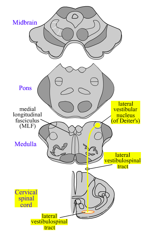
Lateral Vestibulospinal Tract
(LVST)
- Characteristics:
- Controls ipsi LMN & interneurons innervating extensor antigravitic muscles
- Controls contralateral limb via interneurons
- Maintain muscle tone by exciting extensors
- Balance at rest and during movement
- Equilibrium
- Maintain/adjust posture
- Labyrinthine Reflex
- Lesion = balance problems
- Path:
- Medulla
- Originates from ipsilateral lateral vestibular nucleus (Deiter’s nucleus)
- Fibers pass through inferior vestibular nucleus
- Axons descend ipsilaterally to all spinal cord levels
- Medulla
- Spinal cord
- Travels in ventral funiculus with VCST
Controlled by inhibitory input from Purkinje neurons in cerebellum:
- Vermis and flocculonodular lobes.
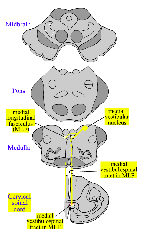
Medial Vestibulospinal Tract
(MVST)
- Characteristics:
- Primarily controls LMNs innervating muscles of the head and neck.
- Role in positioning of head/neck in relation to balance and eye movement.
- Right reflex
- Allows suppression of vestibular ocular reflex (VOR)
- Synchronize head and eye movements
- Path:
- Originates mostly from medial vestibular nuclei
- Axons descend bilaterally to cervical and high thoracic spinal cord
- Forms the descending branch of MLF
- Travels in ventral funiculus with VCST
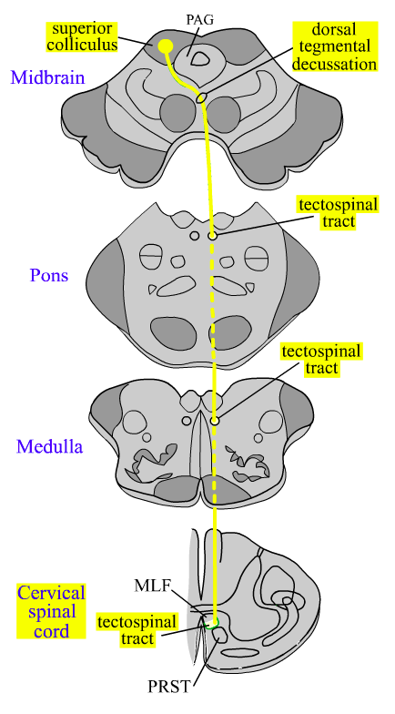
Tectospinal Tract
(TST)
Important in directing head movement towards novel visual, auditory, or somatic stimuli.
- Midbrain
- Originates in contralateral superior colliculus
- Axons curve around PAG
- Crosses in dorsal tegmental decussation
- Continues caudally as tectospinal tract
- Travels in midbrain, pons, and medulla near midline, ventral to MLF
- Travels in ventral funiculus with MLF and PRST
- Terminates in high cervical spinal cord
Spasticity
- Reticulospinal tracts from PRF & MRF → spinal cord
- both ⊕ and ⊖ interneuronal connections
- both terminate directly & indirectly to MNs in IML
- Act on somatic LMNs to ∆ muscle tone & segmental reflexes
- Lesion of reticulospinal tract or corticoreticular fibers ⟾ imbalance of MN ⊕ and ⊖
- Spasticity due to net overexcitation of reflex pathways
- clonus
- clasp-knife
- hyperreflexia
- Treatment:
- activate reciprocal ⊖ systems via
- Ia muscle stretch to opposing muscle
- Ib GTO reflexes with message/vibration
- Deafferentation of DRG to remove afferent limb of stretch reflex
- Intrathecal Baclofen → GABA-B agonist
- activate reciprocal ⊖ systems via
Loss of Consciousness
- Consciousness depends on intact RF
- Major insult to brainstem RF → LOC
- Often due to central tegmental tract damage
- Part of Ascending Reticular Activating System (ARAS)
- Often due to central tegmental tract damage
- Severity varies
- Extensive damage to lower medulla RF incompatible with life
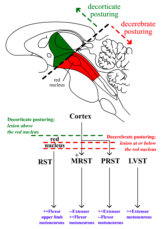
Decorticate Posturing
Damage in the brainstem above the level of the red nucleus.
Decortication interrupts cortical fibers providing tonic activation of MRST.
Upper limbs flexed ⟾ d/t red nucleus via rubrospinal tract (RST)
Lower limbs extended ⟾ due to ⊕ influence by PRST and LVST without ⊖ influence from MRST.


-
Craniofacial Complex37
-
Nervous System Development33
-
Blood Brain Barrier21
-
Neurotransmitters27
-
Intro to Nervous System29
-
Action Potentials and Channelopathies18
-
Neuronal Degeneration and Regeneration19
-
Neuronal Interactions24
-
Nerves, Spinal cord, and Pathways124
-
Spinal Cord Lesions16
-
Pain35
-
Hypothalamus63
-
CNS Pathways & ANS35
-
Limbic System70
-
Cerebral Cortex107
-
Cerebellum50
-
Basal Ganglia30
-
Motor Systems18
-
Brainstem13
-
Cranial Nerves15
-
Thalamus23
-
Aging and Alzheimers15
-
CVA, TBI, and Coma17
