Functional Matrix
The functional relationship between soft tissue and skeletal units.
Size, shape, structure integrity, and location of each components results from secondary and compensatory responses to the operational demands of the functional matrix.
Consider biology, environment, and behavior.
Chondocranium
Base of the skull.
- Develops first during embryology
- Derived from endochondrial ossification
- Forms supportive platform on which remainder of skull develops
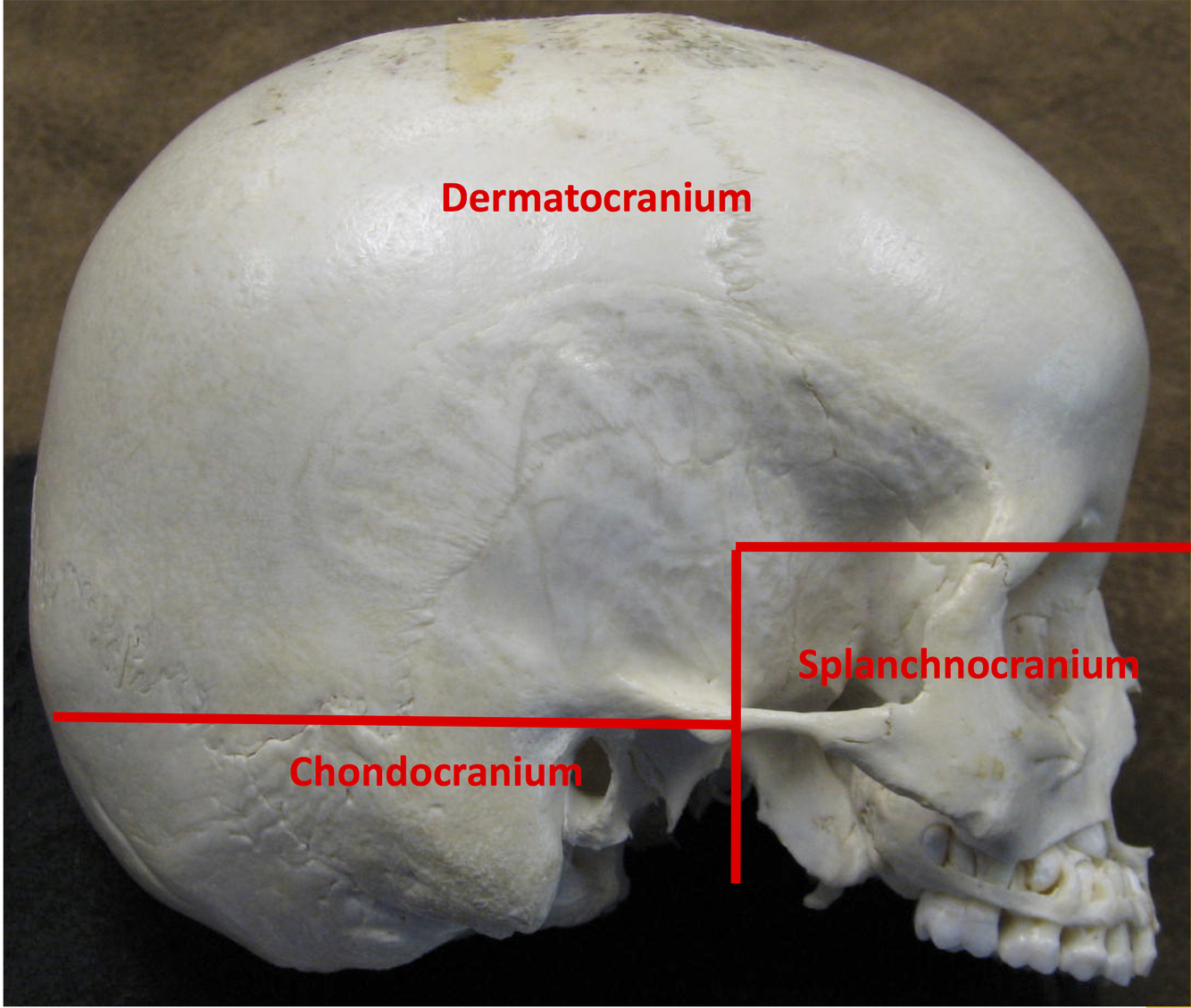
Dermatocranium
Creates the brain case.
- Develops second during embryology
- Derived from intramembranous ossification
- Fontanelle are reminants of the membrane at birth

Splanchnocranium
Contributes to facial structure.
- Formed last during embryology.
- Derived from intramembranous ossification.
- Does not complete formation until the end of puberty.

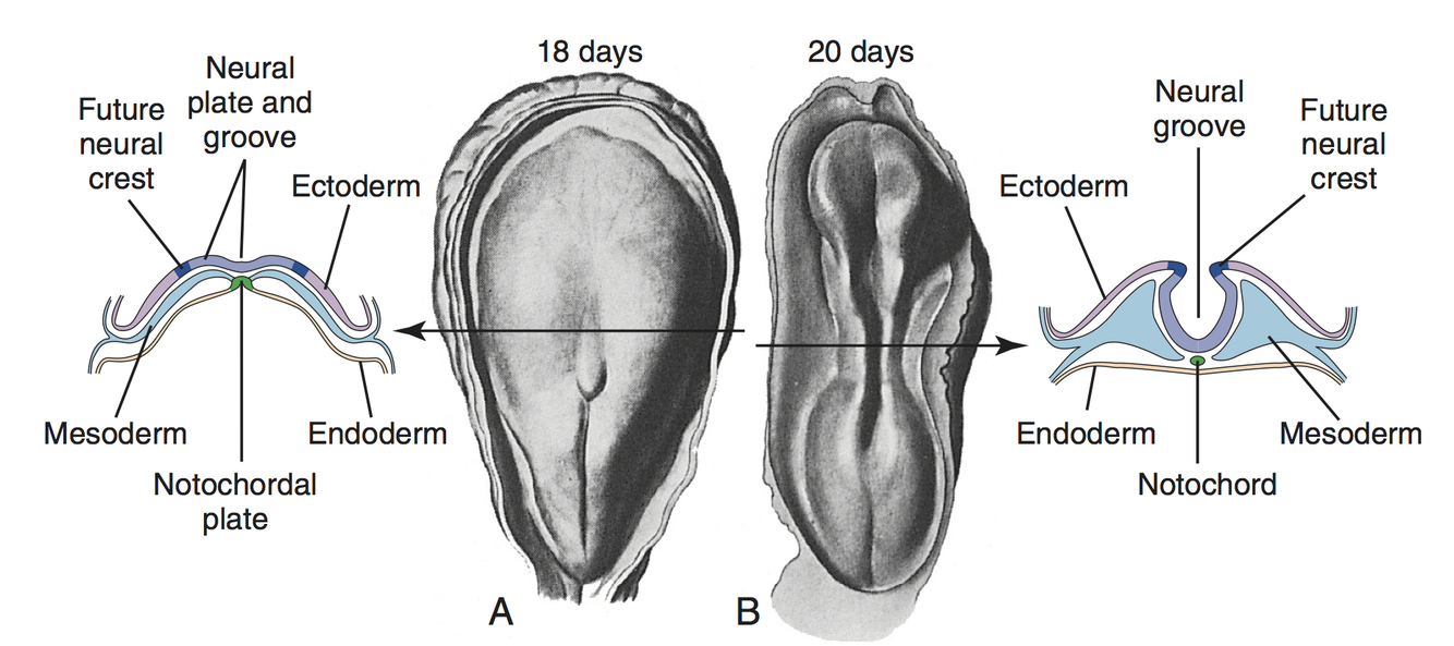
Primary Neurulation
- During 3rd week of development:
- Transition from bi-laminar disc to tri-laminar disc.
- Longitudinal band of ectodermal cells expands to form the neural plate.
- Neural plate continues to expand and folds in the middle forming the neural groove.
- Neural groove deepens as neural folds on either side approach each other at the dorsal midline.
- During 4th week of development:
- Neural folds begin to fuse centrally.
- Fusion proceeds both caudally and rostrally forming the neural tube.
- As neural tube forms, it seperates from overlying ectoderm and sinks into underlying mesoderm.
- Leaves behind a group of cells from the crest of each neural fold ⇒ future neural crest cells.
- Future neural crest cells migrate out of the neural folds, seperating the ectoderm from neural tube.
- Controlled by Hox Genes
- Neural tube expands forming the three primary vesicles:
- Prosencephalon ⇒ forebrain
- Mesencephalon ⇒ midbrain
- Rhombencephalon ⇒ hindbrain
Rhombomeres
The part of the neural tube that develops behind the rhombencephalon (hindbrain) develops a series of bulges ⇒ rhombomeres.
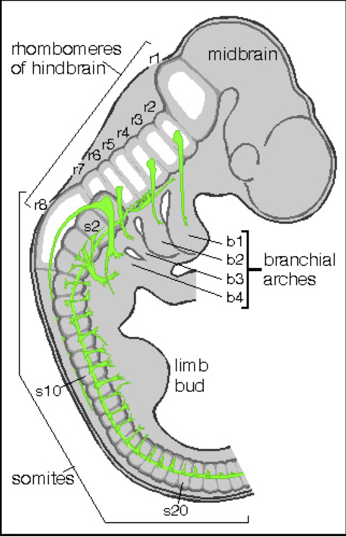
Neural Crest Cell
Migration
During 4th to 5th week of development:
Neural crest cells associated with specific rhombomeres migrate into the ventrolateral aspect forming the pharyngeal arches.
Six paired outpocketings:
1st pharyngeal arch: rhombomeres 1, 2, and 3
2nd pharyngal arch: rhombomeres 3 and 4
3rd pharyngal arch: rhombomeres 6 and 7
4th pharyngal arch: rhombomere 5
5th pharyngal arch: often never develops or degenerates quickly
6th pharyngal arch: rhombomere 5 (cannot be seen externally)
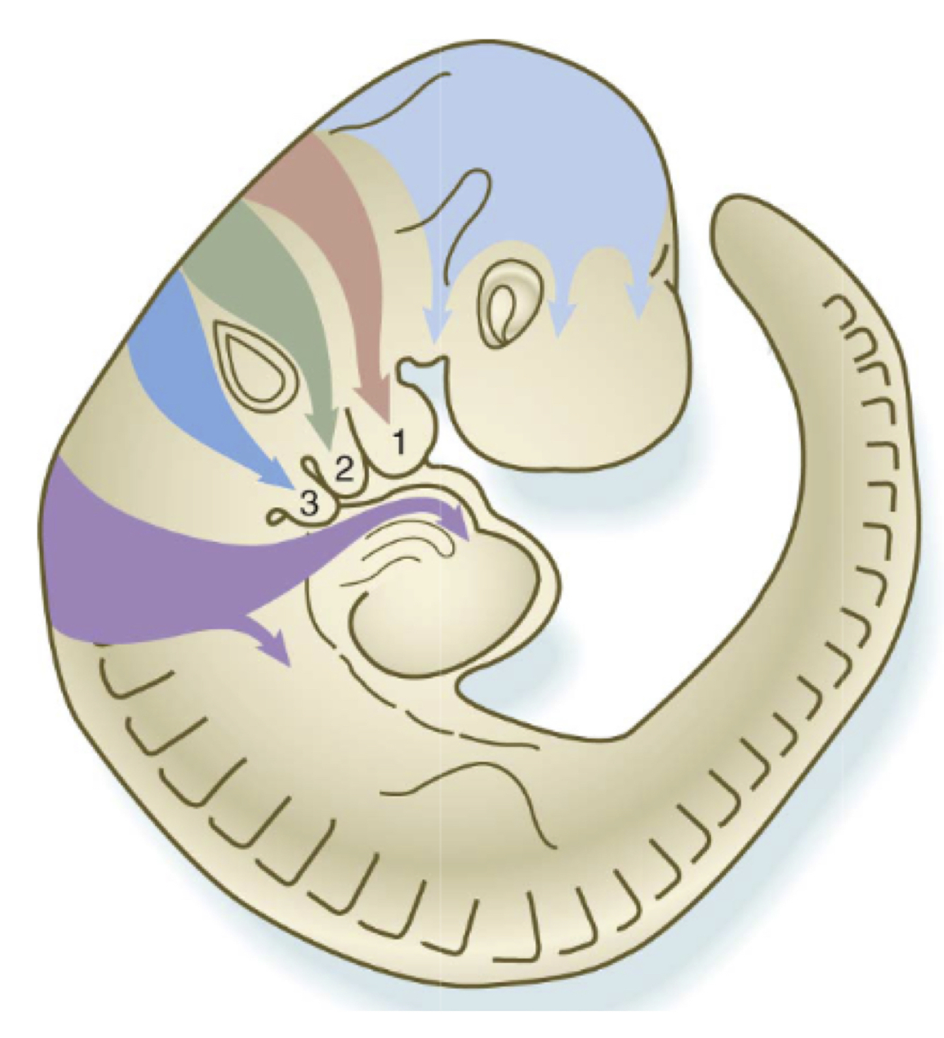
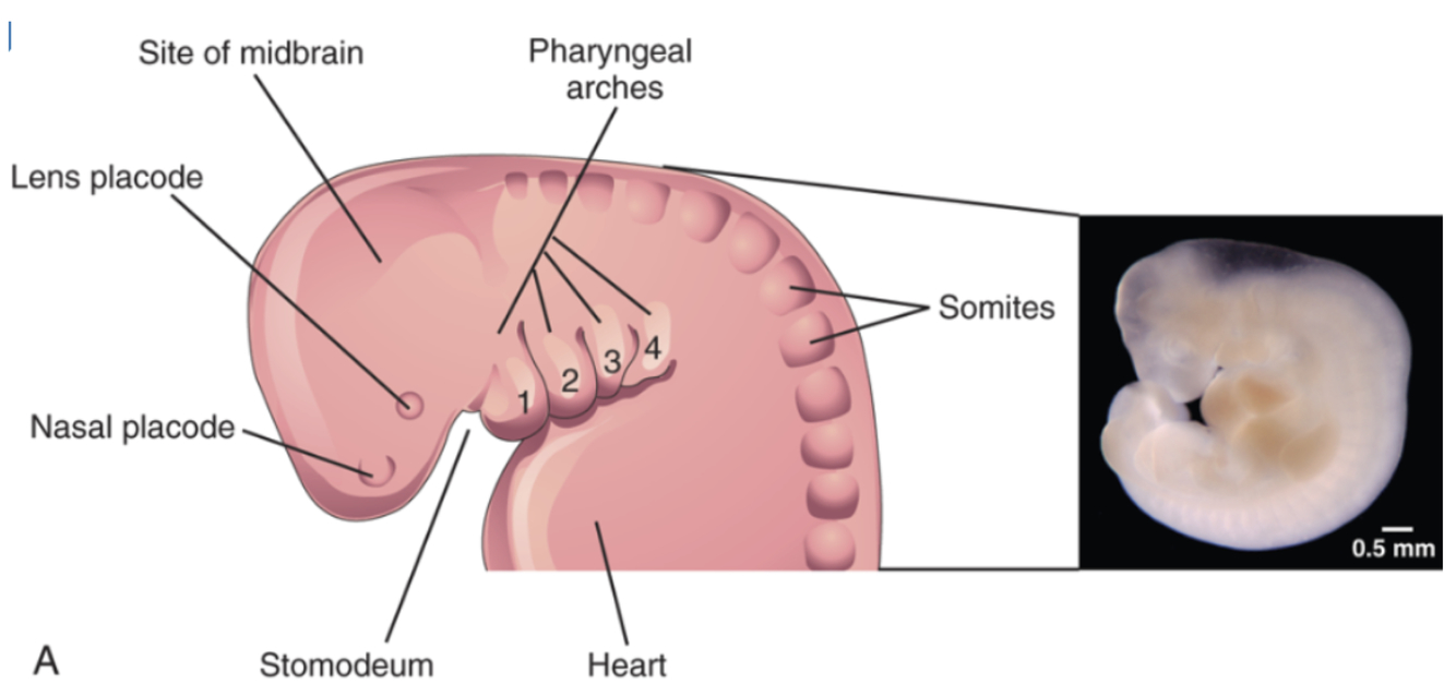
Pharyngeal Arches
Pharyngeal arches
- Composed of mesoderm
- Each arch is associated with a specific:
- cranial nerve
- muscle group
- artery
- skeletal or cartilaginous derivative
First Pharyngeal Arch
Derivatives
- Cranial nerves:
- V2: maxillary division of trigeminal nerve
- V3: mandibular division of trigeminal nerve
- Muscle:
- Muscles of mastication
- Tensor tympani
- Tensor veli palatini
- Cartilage & Skeletal Elements:
- Part of sphenoid
- Incus
- Malleus
- Maxilla
- Mandible
- Zygomatic
- Squamous portion of temporal bone
- Vessels:
- Maxillary artery
- Other structures:
- Mucous membranes of anterior 2/3 of tongue

Second Pharyngeal Arch
Derivatives
- Cranial nerves:
- VII: facial
- Muscle:
- Muscles of facial expression
- Posterior belly of digastric
- Stylohyoid
- Stapedius
- Cartilage & Skeletal Elements:
- Stapes
- Styloid process
- Stylohyoid ligament
- Lesser horns of hyoid
- Vessels:
- Stapedial artery

Third Pharyngeal Arch
Derivatives
- Cranial nerves:
- IX: glossopharyngeal
- Muscle:
- Stylopharyngeous
- Cartilage & Skeletal Elements:
- Greater horn of hyoid
- Vessels:
- Common carotid
- Proximal internal carotid

Fourth Pharyngeal Arch
Derivatives
- Nerves:
- CN X: vagus
- Superior laryngeal nerve
- Muscle:
- Pharyngeal constrictors
- Cricothyroid
- Levator veli palatini
- Cartilage & Skeletal Elements:
- Laryngeal cartilages
- Vessels:
- Proximal right subclavian
- Arch of aorta
- Other structures:
- Mucous membrane of posterior 1/3 of tongue
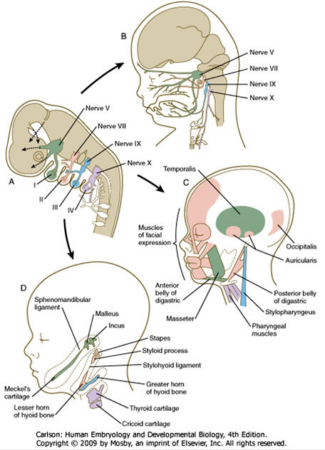
Sixth Pharyngeal Arch
Derivatives
- Nerves:
- CN X: vagus
- Recurrent laryngeal nerve
- Muscle:
- Intrinsic muscles of the larynx
- Cartilage & Skeletal Elements:
- Laryngeal cartilages
- Other structures:
- Mucous membranes of posterior 1/3 of tongue

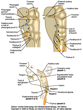
Branchial Clefts & Pouches
Connective tissue between the pharyngeal arches form:
Branchial clefts externally ⇒ ectoderm
Branchial pouches internally ⇒ endoderm

First Branchial/Pharyngeal Pouch
Derivatives
- Mucosa of eardrum
- Tympanic cavity
- Auditory tube
- Mastoid antrum
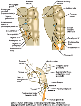
Second Branchial/Pharyngeal Pouch
Derivatives
- Tonsilar fossa
- Surface epithelium of palatine tonsils

Third Branchial/Pharyngeal Pouch
Derivatives
- Thymus
- Inferior parathyroid glands

Fourth Branchial/Pharyngeal Pouch
Derivatives
Superior parathyroid glands.
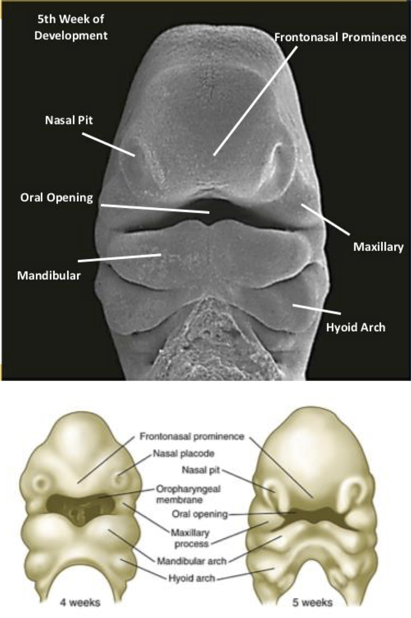
Development of the Face
Brain, eyes, and stomodeum recognizable by week 4.
Complete by 7th week of development.
Involves interaction of numerous embryonic tissues:
- Neural tube
- Paraxial Mesoderm
- Endoderm of pharynx
- Cranial ectoderm
Five facial prominences responsible for development of facial features:
- One frontonasal
- elevation of the margins of the nostrils
- sides develop into the medial and lateral nasal prominences
- Two maxillary
- Migrate medially and merge with frontonasal prominence.
- nose
- upper lip
- palate
- Migrate medially and merge with frontonasal prominence.
- Two madibular
- Merging of madibular prominences
- lower jaw
- lower lip
- lower cheek
- chin
- Merging of madibular prominences
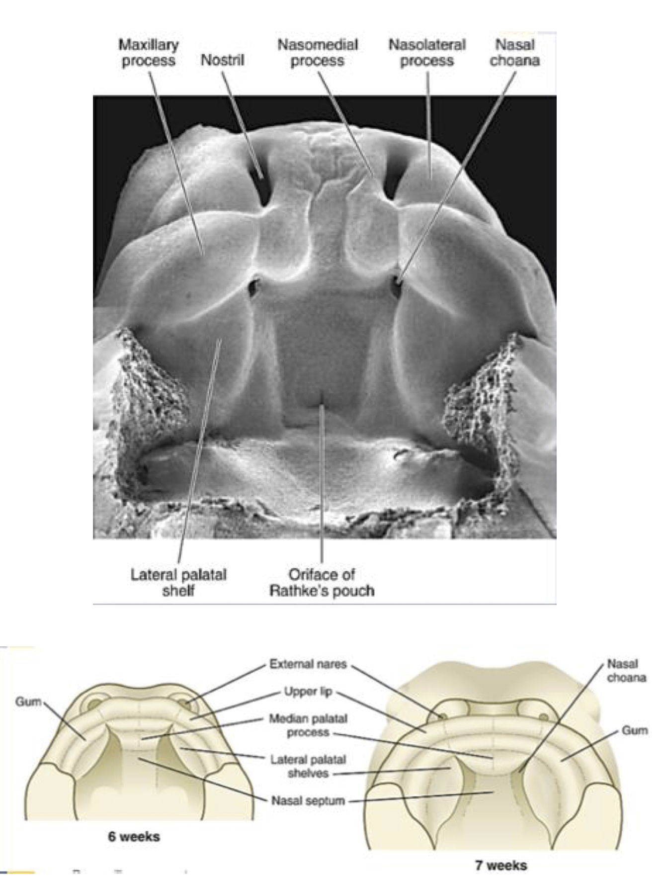
Development of the Palate
During 7th week of development.
Initially widely seperated.
Merging of frontonasal and maxillary prominences.
- Median palatine process drived from frontonasal prominence.
- Lateral palatine process drived from maxillary prominences.
By the time of birth, a tiny gap or complete union may exist.
Interruption of migration or failure to fuse results in cleft lip/palate.
Postnatal Growth
- Sexual dimorphisms account for 90% of development/maturation.
- Females complete development earlier than males.
- Behavior and environment influences ~ 10% of development/maturation.
- Majority of focus resides with brain development.
- Facilitates early learning
- Observation
- Cultural knowledge
- Facilitates early learning
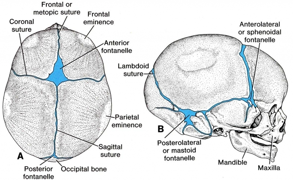
Fontanelle Closure
Skull’s basic morphology established at birth.
Fontanelle faciliate continued growth and expansion of skull and passage through birth canal.
Fontanelles are 38% closed by end of 1st year and 96% closed by end of second.
- Posterior fontanelle closes at 2-3 months.
- Sphenoidal fontanelle closes at ~ 6 months.
- Mastoid fontanelle closes after 6-18 months.
- Anterior fontanelle closes between 1-3 years of age.
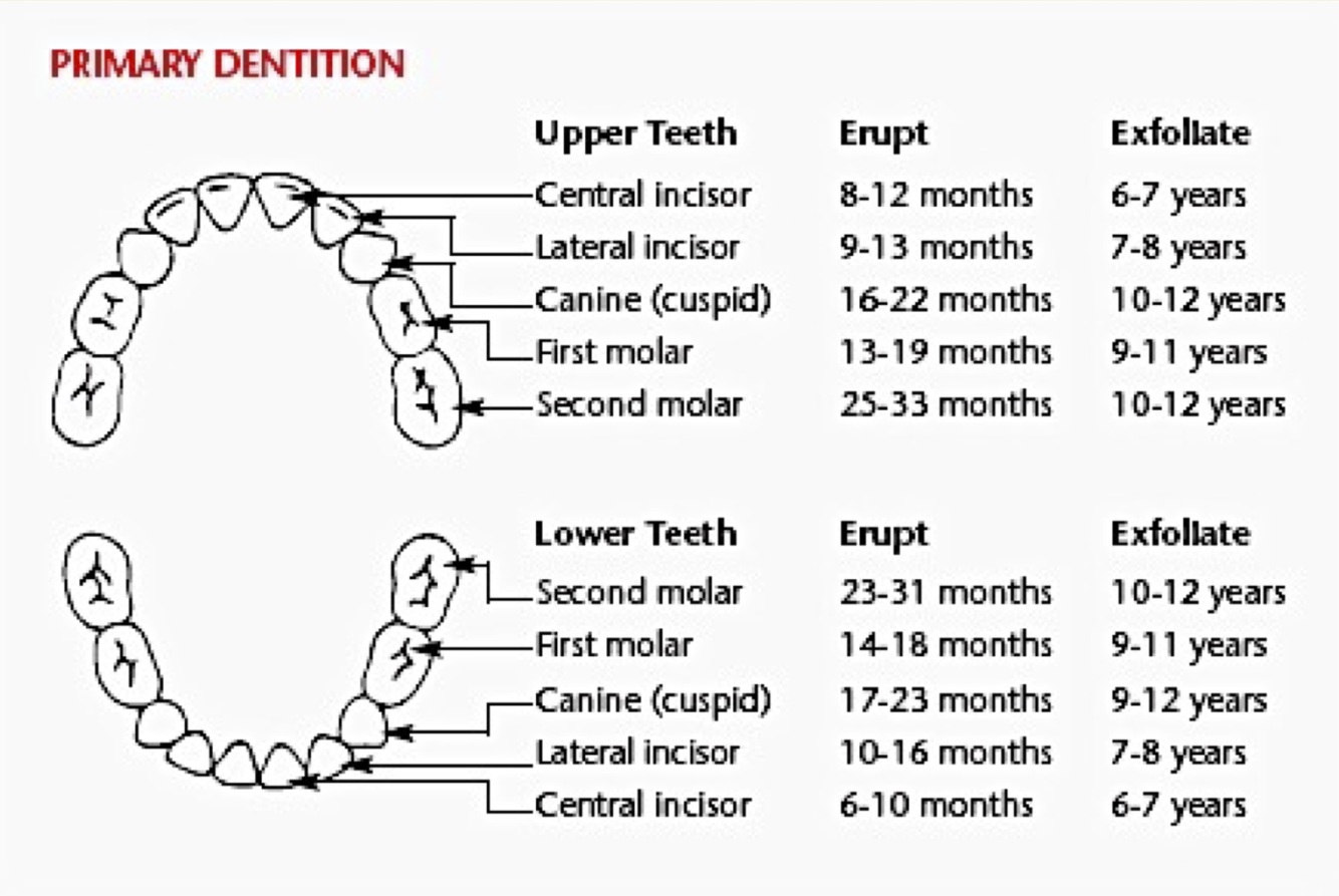
Development of Dentition
- Teeth develop independently during embryology but functions together morphologically
- Eruption & exfoliation timeline patches development/puberty.
Skull
Characteristics and Functions
- Formed by the articulation between the cranium and mandible.
- 22 bones
- 6 unpaired
- 8 paired
- 6 ear bones
- 22 bones
- Most complex region of skeleton
- Functions:
- Foundation for sight, smell, taste, and hearing.
- Framework for masticatory apparatus


-
Craniofacial Complex37
-
Nervous System Development33
-
Blood Brain Barrier21
-
Neurotransmitters27
-
Intro to Nervous System29
-
Action Potentials and Channelopathies18
-
Neuronal Degeneration and Regeneration19
-
Neuronal Interactions24
-
Nerves, Spinal cord, and Pathways124
-
Spinal Cord Lesions16
-
Pain35
-
Hypothalamus63
-
CNS Pathways & ANS35
-
Limbic System70
-
Cerebral Cortex107
-
Cerebellum50
-
Basal Ganglia30
-
Motor Systems18
-
Brainstem13
-
Cranial Nerves15
-
Thalamus23
-
Aging and Alzheimers15
-
CVA, TBI, and Coma17
