Muscle Anatomy
Review
actin-myosin filaments ⇒ sarcomere ⇒ myofibril ⇒ muscle fiber ⇒ fascicle ⇒ compartment
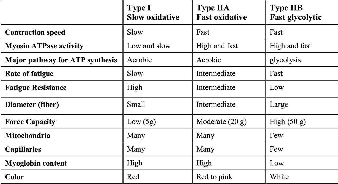
3 main muscle fiber types.
Fibers intermingled within individual muscles.
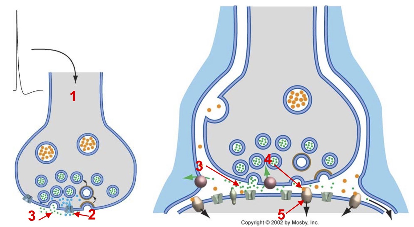
Neuromuscular Junction
(NMJ)
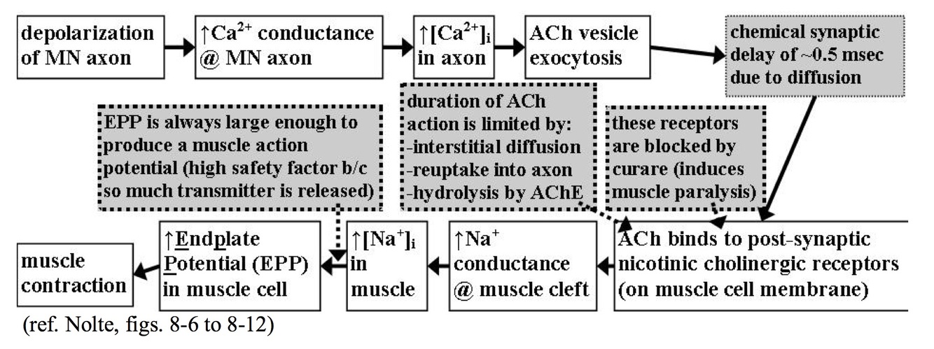
Chemical synapse between motor neuron and muscle.
Duration of ACh action limited by diffusion and hydrolysis by acetylcholinesterase.
EPP generated large and always sufficient to produce a muscle action potential.
- Motor neuron: electrical signal ⇒ chemical signal
- AP invades axon terminal causing opening of voltage dependent Ca2+ channels
- Ca2+ influx ⇒ release of ACh into synapse
- Skeletal muscle fiber: chemical signal ⇒ electrical signal
- ACh binds nicotinic ACh receptors opening voltage-gated cation channels
- Na+ influx results in depolarization ⇒ end plate potential (EPP)
- EPP generates AP ⇒ Ca2+ release from SR ⇒ contraction
Spinal Motor Neurons
- Final common pathway of the motor system
- Located in ventral horn (Rexed lamina IX)
- Axons leave ventral root to project to skeletal muscle fibers
- Three types of motor neurons
- Alpha motor neurons
- most numerous
- innervate extrafusal muscle fibers
- Gamma motor neurons
- innervate intrafusal fibers containing muscle stretch receptors
- compensate for silencing of stretch receptors during muscle contraction
- Beta motor neurons
- innervate both extrafusal and intrafusal muscle fibers
- similar function to gamma motor neurons
- Alpha motor neurons
Motor Unit
A single 𝛼-motor neuron and all the muscle fibers that it innervates.
- MN innervates many fibers of the same histochemical type
- Fibers of a unit fire synchronously
- MN action potential ⇒ EPP in all innervated fibers ⇒ contraction
Motor Nuclei
- Motor neurons organized into pools or motor nuclei
- Arranged in rostral-caudal columns spanning 1-4 segments of the spinal cord
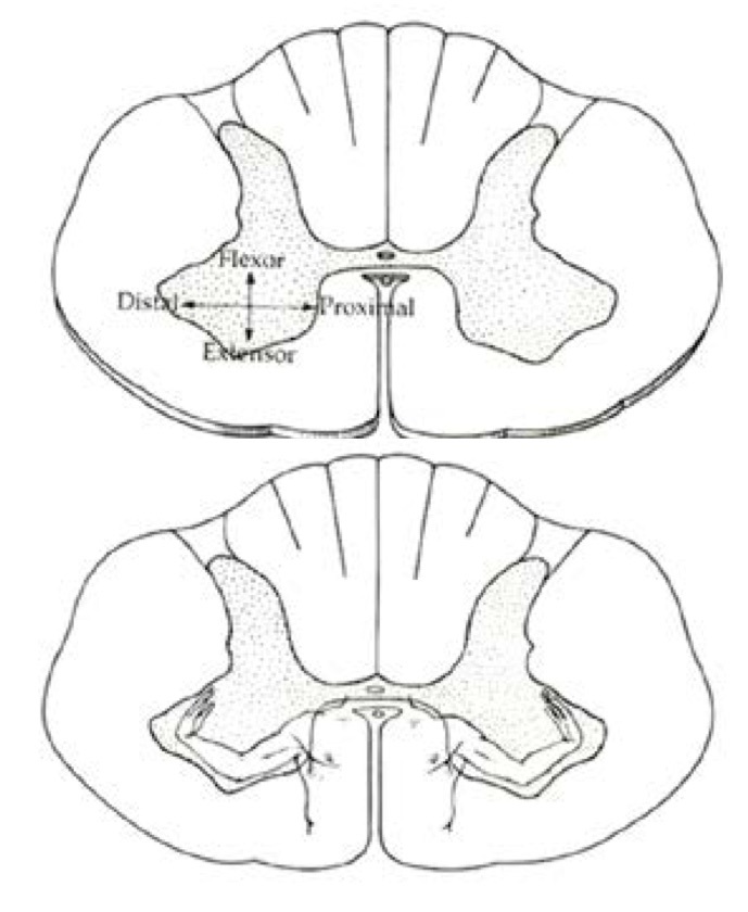
- Pools show somatotopy within the spinal grey matter
- flexor muscle MNs located more dorsally than extensor MN
- proximal (axial) MNs located more medial than distal (peripheral) MNs
Muscle Recruitment
Muscle force is graded in two ways:
- Increasing firing rate of individual MNs
- Results in summation of post-synaptic responses in muscle
- Tetanus = maintained contraction without relaxation
- Force greater than from single twitch
- Increasing the number of MNs recruited
- Follows Henneman’s size principle
- Motor units recruited small to large
- AP will depolarize smaller MN more strongly due to higher input resistance
- MN size related to type of fiber it innervates
- Slow muscle fibers innervated by small MN ⇒ recruited first
- Fast fatigue-resistant fibers (FFR) next
- Fast fatigable fibers (FF) recruited last
- Minimizes fatigue and ensures incremental force
Motor Unit Errors
- Action potential fails to propagate to all branch of an axon ⇒ neuropathy
- NMJ function compromised
- Myastenia gravis
- Botulinum toxin
- Mixed motor units
- Death of MN cause surriving axon sprouts to reinnervate nearby fibers regardless of type
- Results in uncoordinated contraction
- Susceptible to fatigue and failure even years later
- Aging, poliomyelitis, ALS
Golgi Tendon Organ
(GTO)
- Arranged in series at the muscle-tendon junction
- Sensitive to tension
- Group 1b sensory afferent fibers
- interweaved in collagen fibers
- Contraction of extrafusion fibers results in tension at tendon
- Distortion of GTO ⇒ opening of ion channels ⇒ depolarization
- GTO firing rate proportional to muscle force
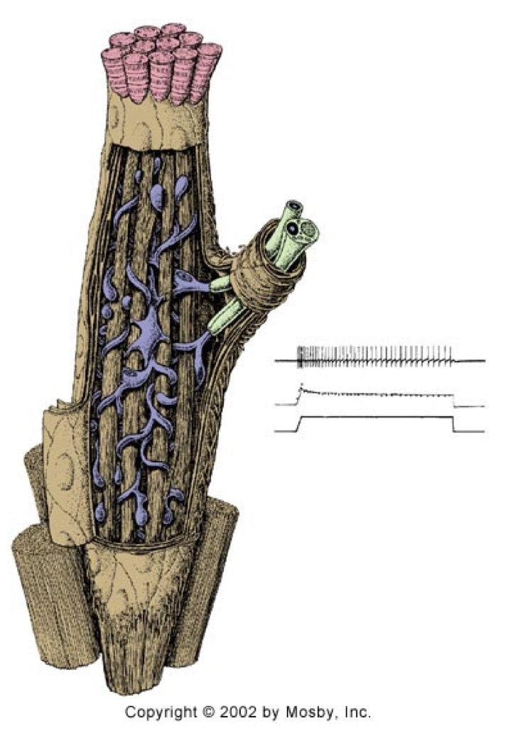
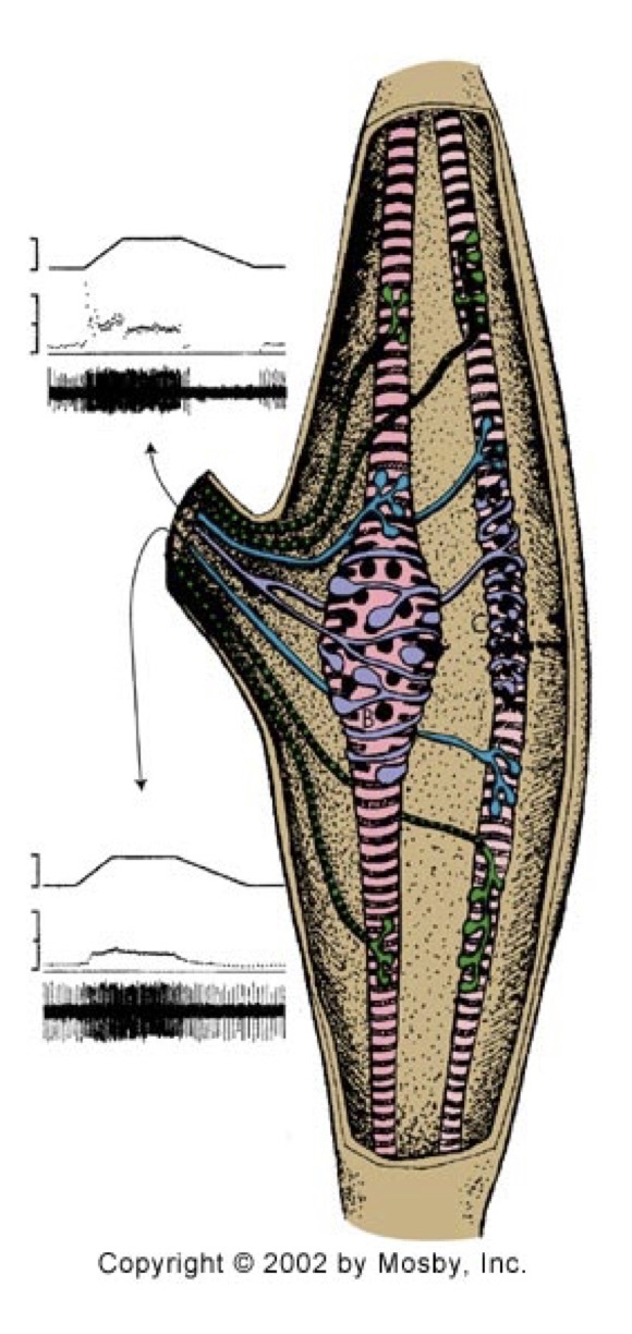
Muscle Spindle
- Encodes muscle length and velocity
- Encapsulated sensory endings arranged in parallel to muscle fibers
- Center of intrafusal fibers contain sensory fiber endings group Ia and II
- Ends of intrafusal fibers innervated by 𝛾-MN
- Group Ia sensory fibers (primary endings)
- sensitive to initial muscle stretch (velocity) and static length
- attached to dynamic nuclear bag fibers, static nuclear bag fibers, and nuclear chain fibers
- Group II sensory fibers (secondary endings)
- senstitive to static muscle length
- attached to static and nuclear chain fibers
- Spindles increase firing rate with muscle stretch and decrease with muscle contraction
- Spindle sensitivity can be adjusted ⇒ alpha-gamma co-activation
- contract ends of intrafusal fibers via 𝛾-MN to maintain tension
- allows continued spindle function during muscle contraction
Peripheral Nerve Fibers
Classification
- Type A𝛼 ⇔ Group Ia and Ib
- Largest, heavily myelinated axons
- Proprioception from spindles and GTO’s
- Type A𝛽 ⇔ Group II
- Large myelinated axons
- Muscle spindles and cutaneous corpuscles
- muscle length, fine touch, pressure
- Type A𝛿 ⇔ Group III
- Small lightly myelinated axons
- Free nerve endings
- Sharp fast pain, temperature
- Type B
- Small, slightly myelinated axons
- Preganglionic ANS
- Type C ⇔ Group IV
- Small unmyelinated axons
- Postganglionic ANS
- Slow pain
CNS
Synapses
- Chemical or electrical (mostly chemical in mammals)
- Uses excitatory or inhibitory neurotransmitters
- Net synaptic input controls neuronal firing
- Inputs can be on soma, dendrite, or axon
- Location impacts role of synapse
CNS
Postsynaptic Potentials
- Single postsynaptic potentials subthreshold for AP generation
- ~ 0.1 mV in CNS compared to 35-40 mV at EPP of NMJ
- Excitatory postsynaptic potentials (EPSPs)
- depolarizing
- brings closer to threshold
- Ex:
- Glutamate ⇒ AMPA (and/or NMDA) receptors
- Acetylcholine ⇒ nicotinic acetylcholine receptors
- Inhibitory postsynaptic potentials (IPSPs)
- hyperpolarizing
- brings further away from threshold
- Ex:
- Glycine ⇒ glycine receptors
- GABA ⇒ GABAA receptors
- Neuronal response is based on summation of simultaneously received postsynaptic potentials
Synaptic Summation
- Spacial summation
- Sum of simultaneous inputs from different synapses along the membrane surface
- Multiple excitatory inputs more likely to bring neuron to threshold
- Inhibitory inputs make simultaneous excitatory inputs less effective
- Temporal summation
- Single input fires repetitively in small enough time window so that PSPs add in postsynaptic cell
Neurons receive continuous barrage of EPSPs and IPSPs which summate over both space and time.
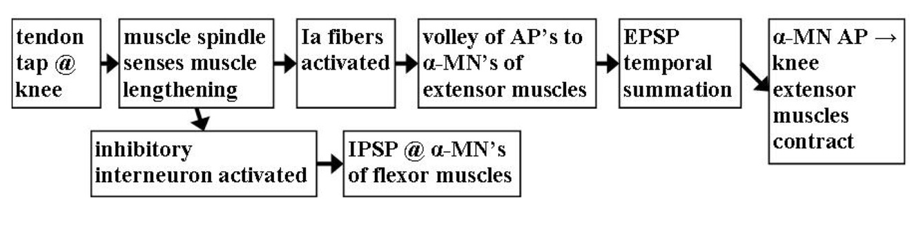
Stretch (Myotatic) Reflex
Negative feedback mechanism to stabilize muscle length.
Alows for automatic length adjustment without supraspinal involvement.
Reflex has both monosynaptic and disynaptic components.
- Monosynaptic excitation of 𝛼-MNs innervating the homonymous (same) muscle and its synergists.
- Disynaptic inhibition of antagonist 𝛼-MNs.
Steps:
- Muscle stretch activates muscle spindles.
- Ia afferents:
- Excite homonymous MNs and synergist MNs ⇒ muscle contraction.
- Excite Ia inhibitory interneurons
- Located in lamina VII
- Ia inhibitory interneurons inhibit antagonist motor neurons ⇒ relaxation of opposite muscle group
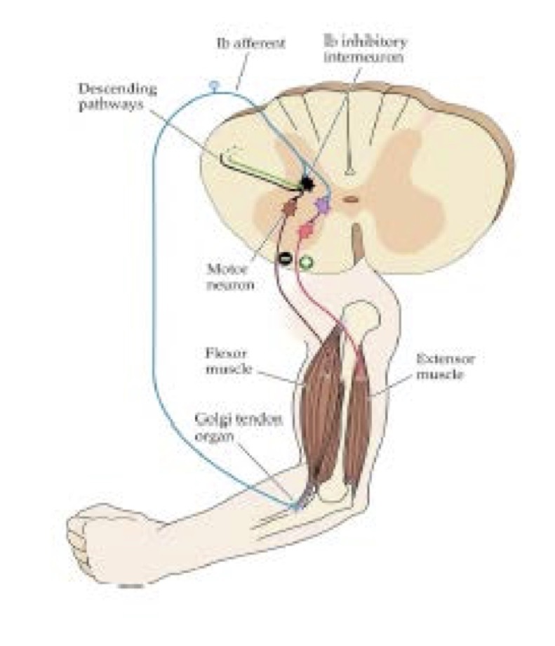
Inverse Stretch Reflex
Prevents muscle and tendon damage by limiting extreme force.
Along with stretch reflex, maintains posture and balance.
Involves disynaptic central components.
Steps:
- High force in tendon excites GTO
- Ib afferent:
- Excites Ib inhibitory interneurons
- Located in lamina V, VI, and VII
- Activates excitatory interneurons
- Excites Ib inhibitory interneurons
- Ib inhibitory interneurons inhibit homonymous and synergist MNs
- Excitatory interneurons activate antagonist motor neurons.
- Antagonist muscles contract.
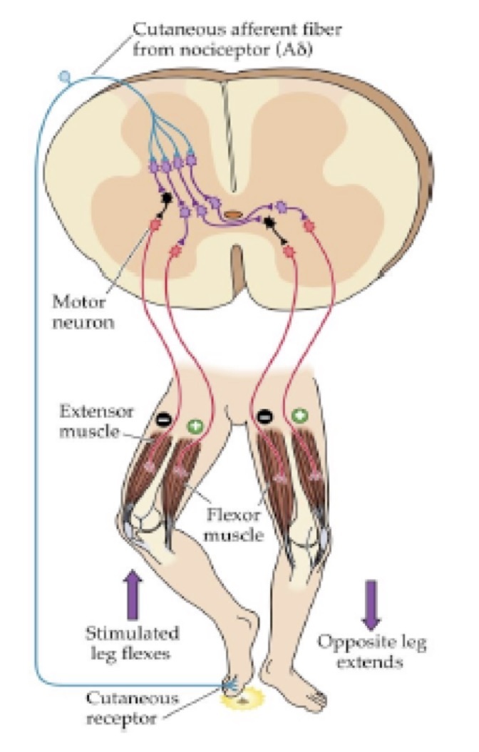
Flexion Withdrawal and Crossed Extension
Reflex
Exact pattern of muscles that are excited and inhibited depends on the location of noxious stimulus.
Spinally mediated.
Polysynaptic reflex.
- Painful stimulus activates high threshold afferent fibers in cutaneous nocireceptors.
- Nociceptive afferents activate several different excitatory interneurons in the dorsal horn.
- Flexor MNs activated via polysynaptic excitatory pathway ⇒ flexion of ipsilateral limb moves body away from noxious stimulus.
- Extensor MNs inhibited via polysynaptic excitatory pathway with last interneuron inhibitory.
- Same stimulus leads to activation of commissural (midline crossing) neurons.
- Via polysynaptic excitatory pathway activates contralateral extensor MNs ⇒ extension of contralateral limb to support body.
- Activate an inhibitory interneuron ⇒ inhibits contralateral flexor MN.
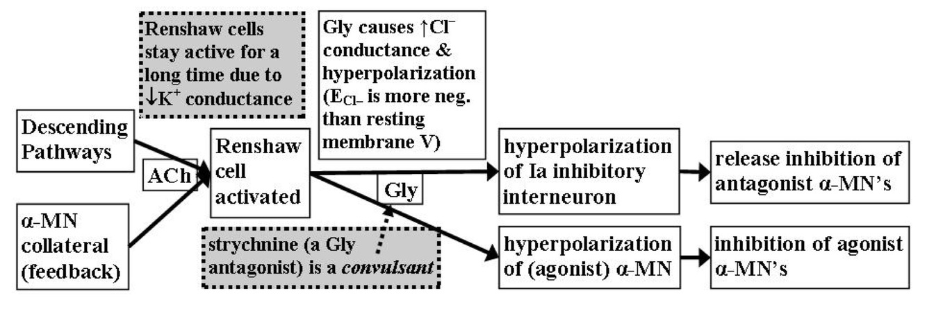
Renshaw Cell Circuitry
- Renshaw cells mediate recurrent inhibition
- Located near motor neurons in lamina IX
- Receive inputs from central collaterals of motor neurons
- Inhibit homonymous and synergistic motor neurons
- Inhibit Ia inhibitory interneurons
- Functions include:
- Regulation of fraction of MN pool activated
- Limit duration of MN firing
- Stabilize firing rate: desynchronize EPSPs, allowing smoother contraction
- Steps:
- Excited by motor neurons (Ach)
- Projects back to inhibit via glycine the same motor neuron, homonymous, and synergist MNs
- Inhibits Ia inhibitory interneurons
- Results in disinhibition of antagonist MNs
Inhibitory Synapse
Effectors
- Strychnine
- glycine receptor antagonist
- loss of inhibition results in tonic contractions
- Clostridium tetani
- blocks neurotrasmitter release at inhibitory interneurons
- results in tonic contractions
- Loss of normal excitatory/inhibitory balance could lead to spasmodic muscle contraction
Lower Motor Neuron
Signs
Clinical manifestations which present in diseases that directly affect spinal MNs or MN axons.
- Hyporeflexia
- Hypotonia ⇒ reduced contraction at rest, also decreased resistance to stretch
- Fasciculations ⇒ spontaneous contractions of a motor unit
- may be visible
- Fibrillations ⇒ spontaneous contractions of a single muscle fiber
- not perceptible, only detected by EMG
- Atrophy
- Paresis or Flaccid paralysis ⇒ weakness or paralysis with low muscle tone
Synaptic Plasticity
Definition
Use-dependent changes in synaptic strength.
- Synaptic strength dependent on history.
- PSP produced depends on the previous activity at that synapse.
- Can be short-term (ms to min) or long-term (hours to years).
- Plasticity that increases synaptic strength ⇒ potentiation or facilitation
- Plasticity that decreases synaptic strength ⇒ depression
Short-term Plasticity
- Frequency-dependent facilitation:
- PCP increased when presynaptic impulses arrive in close proximinity
- Effect decreases as interval period increases
- Frequency-dependent depression:
- PCP decreases when presynaptic impuses arrive close in time
- Effect decreases as interval period increases
- Mechanism:
- Bursts of activity cause transient calcium accumulation in presynaptic terminals
- [Ca2+] cuases change in the probability of neurotransmitter release.
- Mainly attributed to presynaptic effects.
- Can occur with postsynaptic receptor availability.
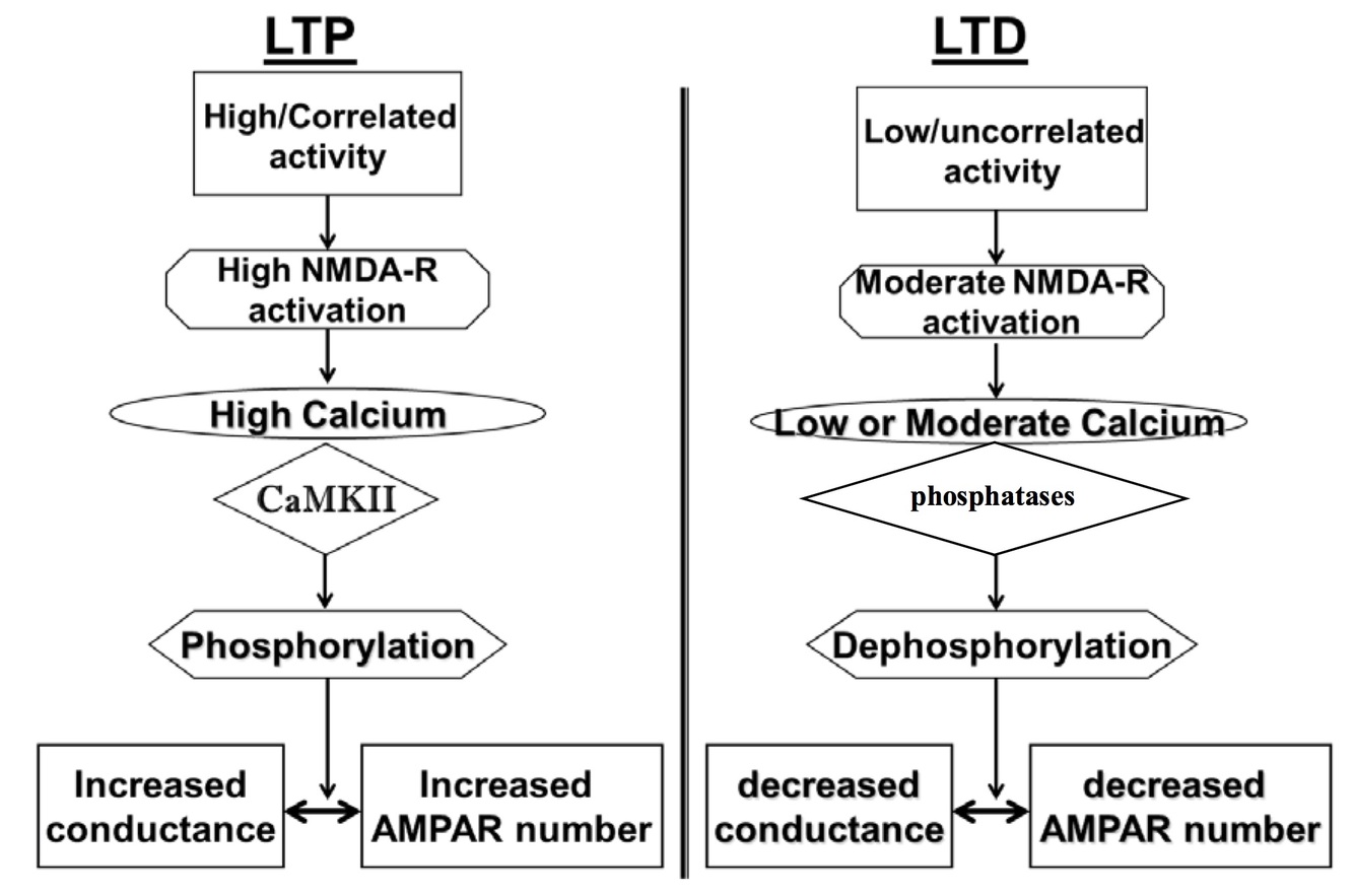
Long-Term Potentiation
(LTP)
- Persistent increase in synaptic strength after a brief presynaptic tetanus
- Best studied in hippocampus ⇒ memory formation
- Input specific ⇒ potentiation restricted to pool of excited synapses
- Cooperativity ⇒ two different presynaptic input pathways activated simulatenously can boost each other ⇒ Hebbian rule
- Associativity ⇒ presynaptic activity must coincide with strong postsynaptic depolarization
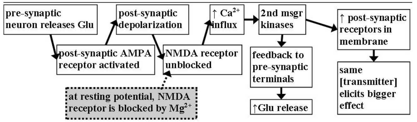
LTP Induction
- Requirements
- Postsynaptic depolarization
- NMDA receptor activation
- Postsynaptic calcium influx
- Calcium-dependent activation of second messanger pathways
- Increase AMPA receptors in postsynaptic membrane
- stablize current receptors through phosphorylation
- insertion of new receptors
- Gene transcription ⇒ formation of new synapses
- Increase AMPA receptors in postsynaptic membrane
- Experimental induction:
- Apply high frequency burst of stimuli to presynaptic neuron
- LTP only at that synapse (and other active at the same time) but not silent inputs
- Stimulate/depolarize postsynaptic neuron just after stimulating the presynaptic neuron
- Incoming EPSP paied with postsynaptic depolarization
- Apply high frequency burst of stimuli to presynaptic neuron
Long-Term Depression
(LTD)
Persistent decrease in synaptic strength after a brief low frequency stimulation.
- Mechanism:
- Lower levels of activity results in lower level of NMDA receptor activation
- Low to moderate calcium influx activates protein phosphatases
- Deposphorylation of substrates leads to AMPA receptor internalization
- Fewer AMPA receptors available at the synapse.
- Experimentally:
- Low frequency stimulation to presynaptic neuron.
- Stimulate/depolarize postsynaptic neuron just before stimulating presynaptic neuron.
- Incoming EPSP arrives 10-100 ms before postsynaptic depoarlization.
-
Craniofacial Complex37
-
Nervous System Development33
-
Blood Brain Barrier21
-
Neurotransmitters27
-
Intro to Nervous System29
-
Action Potentials and Channelopathies18
-
Neuronal Degeneration and Regeneration19
-
Neuronal Interactions24
-
Nerves, Spinal cord, and Pathways124
-
Spinal Cord Lesions16
-
Pain35
-
Hypothalamus63
-
CNS Pathways & ANS35
-
Limbic System70
-
Cerebral Cortex107
-
Cerebellum50
-
Basal Ganglia30
-
Motor Systems18
-
Brainstem13
-
Cranial Nerves15
-
Thalamus23
-
Aging and Alzheimers15
-
CVA, TBI, and Coma17
