which amino acids are glucogenic? [2]
which a.a are ketogenic? [2]
glutamine
Alanine
lysine and leucine are ketogenic
which cells regulate water contents in the gut?
what is the mechanism of this?
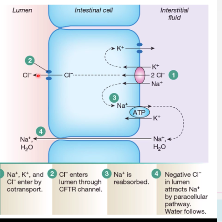
secretory cells of the intestinal crpyts:
- CFTR channel within these cells controls this:
a) Cl- moves from ECF via Na/K/CL2 cotransporter (as does Na & K)
b) Cl- enters lumen through CFTR channel
c) Na+ is reabsorbed via Na/K ATPase
d) negative Cl- in lumen attracts Na by paracellular pathway (through cell gaps)
e) water follows the Na into the lumen
gastric epithelial cells
- parietal cells produce HCl. But HCl is actually quite toxic. how does the body cell overcome this issue of not causing self harm via the HCl? (2)
1. HCl is only produced when food is in the stomach = get unstimualted and stimulated parietal cells:
a) unstimulated parietal cells have H+ ATPase Pumps in the cytosol
b) stimulated parietal cells have H+ ATPase Pumps on apical surface
- *2. surface mucus cells secrete mucus**
- without mucus = would directly interact with cells
- mucus works as:
a) physical barrier; gel layer
b) chemical barrier; bicarbonate
explain how diabetes disrupts gluconeogensis pathway ox
insulin doesnt work:
SO
- pyruvate dehydrogenase remains phosphorylated & therefore inactive
- = less acetyl co-A to go into krebs cycle from pyruvate
- instead fats are broken down to produce fatty acids & acetyl co-A & goes into krebs cycle instead
- means that pyruvate is available for gluconeogenesis
explain what happens to the stomach when food enters the intestines?
a) hormonal control
b) nervous control
Primarily inhibits gastric acid secretion when FOOD AND ACID ENTERS THE INTESTINES
NERVOUS CONTROL:
- *It signals the sympathetic system to stop gastric secretions**
- Inhibition of parietal and chief cells
HORMONAL CONTROL:
- *- Cholecystokinin, secretin and GIP (gastric inhibitory protein) produced by duodenum –> inhibit gastric secretions**
- Cholecystokinin and GIP released by presence of lipids and carbohydrates
- Secretin released when pH decreases (due to entrance of acidic chyme into the duodenum)
Q
PDH is regulated in two ways:
- PDH is de/-phosphorylated by which enzymes? what do they add / remove? what is their effect?
which substances control 1.?
:)
- PDH Kinases inhibit PDH by adding PO4
- PDH Phosphatases activate PDH by removing PO4
//
- *control of PDH kinases**
- PDH kinases are activated: by ATP, acetyl Co-A and NADH (last two are products of PDH) = switch off PDH.
- Pyruvate & insulin inhibits PDH Kinasese (as pyruvate wants PDH to be active to break pyruvate down) = switch on PDH.
control of PDH phosphatases
- Ca2+ ions activate PDH phosphatises - increases PDH. occurs in muscle -> eventually get more ATP production = switch on PDH
- insulin activates PDH phosphatases - actives PDH
what does Akt do in muscle?
Akt: phosphorylates and inactivates glycogen synthase kianse = activates glyocgen synthase
how is the TCA cycle controlled in response to exercise?
(3)
1.
- Ca2+ is an allosteric keeps the pyruvate deyhyrogenase complex activated.
- therefore causes **quicker conversion of pyruvate -> acetly co-A
- increases TCA to occur more**
(PDH controls the entry to the TCA, it’s activity is regulated. The enzyme is phosphorylated and dephosphorylated depending on whether it is active or inactive. The calcium is promoting essentially the active state of PDH, by influencing PDH phosphatase (this PDH phosphatase will remove phosphates from the PDC and activate it)
2:
calcium and ADP drive activity of two dehydrogenase enzymes in the TCA to maintain high ATP production
3.
Low levels of ATP/NAD pushes PDH into its active state.
what are the step changes in metabolism that undergo during starvation? (5)
- glucose starts falling; insulin goes down, glucagon up
- glycogen can keep brain alive for 2/3 days / 30 hrs
- swith to FA break down = energy for 2-3 days due to gluconeogenesis
- FA then switches to ketone bodies. (ketone bodies can go into brain - important !)
- final resource = break down proteins to release amino acids for gluconeogenesis
to put simply - insulin has what effect on PDH?
what effect does insulin have of kinases & phosphatases?
what do adrenaline and glucagon do to PDH? - why?
insulin caueses the activation of PDH & eventual production of acetyl co-A
insulin = -ve effect on kinases (which inhibit PDH)
+ve effect on phosphatases (which activate PDH)
adrenaline and glucagon: want pyruvate untouched, so it can be used to make glucose via gluconeogenesis = inhibit PDH
NADPH is a co-factor created by immune cells.
Name 2 functions of NADPH
- as a reducing agent (like NADH)
- to generate ROS: immune cells use NADPH oxidase to reduce O2 to free radical and the H202. H202 then used to kill engulfed pathogens = respiratory burst
what covers the rectus abodminis?
rectus sheath
what does tissue transglutaminase (tTG) do to gliadin in CD patients? [1]
removes amide group / deamination
Name the cells bordering the lumen [1]
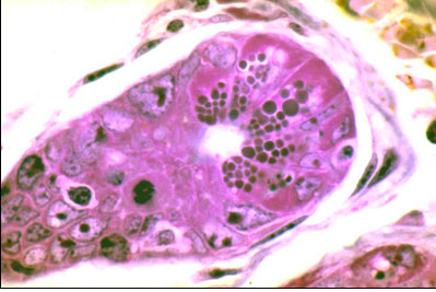
paneth cell
what region of the GI tract is this? [1]
how can you tell? [1]
duodenum [1]
brunners glands [1]
label A-E

A = **enterocyte brush border**
B = **lacteal**
C = **goblet cell**
D = **immune cells (lymphocytes)**
E = **lamina propria**

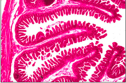
name this region of the intestine [1]
how can you tell [1]

jejunum
plicae circularis
Plicae circulares are out foldings of both the mucosa and submucosa. Projecting from these folds are numerous villi that are outfoldings of the mucosa.

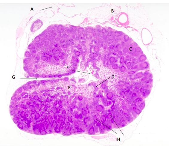
label A-H of the lymph node

A = afferent lymphatic, B =subcapsular sinus, C = cortex, D = medullary cords, E = medulla, F = efferent lymphatic, G = hilus, H = secondary follicles

label A-H of the lymph node

A = afferent lymphatic, B =subcapsular sinus, C = cortex, D = medullary cords, E = medulla, F = efferent lymphatic, G = hilus, H = secondary follicles
name this region of the intestine [1]
how can you tell [1]

jejunum
plicae circularis
Plicae circulares are out foldings of both the mucosa and submucosa. Projecting from these folds are numerous villi that are outfoldings of the mucosa.
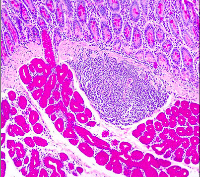
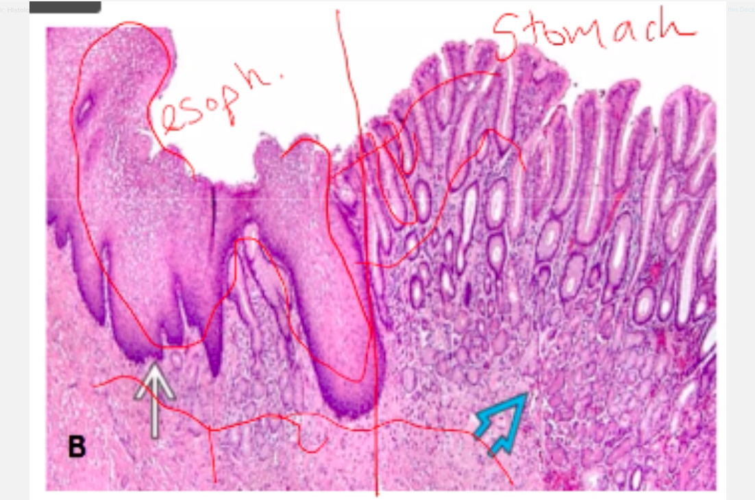
what is the squamocolumnar junction? - chang
squamocolumnar junction
- abrupt change in the mucosa from stratified squamous to columnar cells (and glands)
- Oesophagus joins at an acute angle
- only the mucosa changes, the underlying layers stay the same !!
(oespahus -> stomach?)
what is the squamocolumnar junction? - chang
squamocolumnar junction
- abrupt change in the mucosa from stratified squamous to columnar cells (and glands)
- Oesophagus joins at an acute angle
- only the mucosa changes, the underlying layers stay the same !!
(oespahus -> stomach?)

label A-E

A = **enterocyte brush border**
B = **lacteal**
C = **goblet cell**
D = **immune cells (lymphocytes)**
E = **lamina propria**
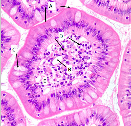
what region of the GI tract is this? [1]
how can you tell? [1]

duodenum [1]
brunners glands [1]


-
FunMed EOYS251
-
FunMedEOYS347
-
FunMedEOYS459
-
FunMedEOYS153
-
FunMedEOYS427
-
FunMed EOYS Long 16
-
big boy things51
-
CR EOYS142
-
CR EOYS248
-
CR EOYS351
-
Calculations6
-
CR EOYS421
-
MET EOYS154
-
big boys29
-
MET EOYS253
-
MET EOYS343
-
MET EOYS445
-
MET EOYS538
-
BB EOYS150
-
BBB2
-
BB EOYS255
-
Short king spring65
-
BB EOYS350
-
BB EOYS446
-
:O29
-
BB EOYS447
-
Loco EOYS43
-
LOCO SSS59
-
LOCO EOYS 441
-
Loco EOYS344
-
LOCO EOYS 553
-
LOCO SSS246
-
LOCO EOYS542
-
LOCO EOYS629
-
SSS FunMed52
-
Spotter Qs130
-
SSS FunMed252
-
METSSS44
-
METSS249
