how do you clinically test the superior and inferior rectus? [1]
how do you clinically test the superior and inferior olbique? [1]
how do you clinically test the medial / lateral rectus? [1]
how do you clinically test the superior and inferior rectus? [1]
abduct the eye
(do this to isolate the muscle so that the eye is aligned with the angle of the muscle pull)
how do you clinically test the superior and inferior olbique? [1]
adduct the eye
how do you clinically test the medial / lateral rectus? [1]
abduct / adduct the eye
how does the hypothalamus regulate water balance?
- where do you find osmoreceptors? [1]
- which hypothalamic nuclei are stimulated to increase water in ur body ? how do they work?
- where do you find osmoreceptors? [1]
- *subfornical organ (wall of third ventricle): detects osmolarity**
- subforrnical organ activates cells in the:
- *i) medial preoptic nucleus**
- this nucleus connects to the limbic system: regulates concious sense of thirst
- *ii) paraventricular nucleus & supraoptic nucleus**
- secrete ADH (makes more aquaporins in CD)
- oxytocin
which of the following is innervated by the superior laryngeal nerve?
thyroartyenoid
cricoartyenoid
cricothyroid
transverse arytenoid
olbique arytenoid
which of the following is innervated by the superior laryngeal nerve?
thyroartyenoid
cricoartyenoid
cricothyroid
transverse arytenoid
olbique arytenoid
others = recurrent laryngeal nerve
spinal accessory nerve comes from which spinal levels? [1]
C1-5
which part of the brain gives you the ability to track an object? [1]
what does ^ connect with?
which pathway mediates neck flexes triggered by seen objects? [1]
which part of the brain gives you the ability to track an object? [1]
superior colliculi
some optic nerve fibres go to the superior colliculi -> connects to the medial longitudinal fasciculi (MLF): links together and synchronises the oculomotor nuclei
which pathway mediates neck flexes triggered by seen objects? [1]
tectospinal tract
= together give synchronised eyes and neck movement
which 3 cells cause seeing stuff x
photorceptors (rods & cones - recive light signal & lose inhibitions) that connect to
bipolar cells that connect to
ganglion cels that send axons to optic nerve
where is aq humour made in the eye? [1]
ciliary body
what happens to photoreceptors in the dark? [2]
what happens to photoreceptors in the light? [1]
what happens to photoreceptors in the dark? [1]
constant inward leak of sodium in outerpart of the receptor: keeps the cell depolarised. causes the release of glutamate from its synaptic ending
what happens to photoreceptors in the light? [1]
light hyperpolarises the tonic glutamate release
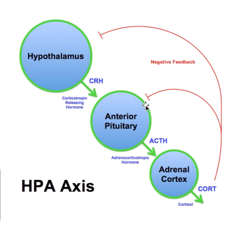
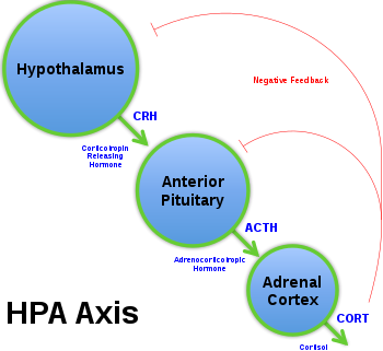
describe the mechanism of HPA axis (hypothalamus-pit-adrenal axis)
describe the mechanism of HPA axis (hypothalamus-pit-adrenal axis):
- Cells in hypothalamus release CRH (Corticotropin releasing hormone)
- CRH acts on anterior pit to releease ACTH (adrenocorticotropic hormone)
- ACTH acts on adrenal cortex to release cortisol
- *BUT: negative feedback system:**
- cortisol inhbits release of above
what are three mechanisms of ADH reducing water loss? [3]
- Increase aquaporins in CD
- increases perm. of CD to urea (water follows)
- stimulates sodium reab in thick loop of henle: Na/K/2Cl
describe the mechanism of HPA axis (hypothalamus-pit-adrenal axis)
which important structures run through the posterior triangle? [3]
- spinal accessory nerve
- external jugular vein
- part of subclavian artery
- part of brachial plexus
which of the following supplies the corpus callosum?
middle cerebral artery
anterior cerebral artery
posterior communicating artery
menigeal branch
opthalmic artery
which of the following supplies the corpus callosum?
middle cerebral artery
anterior cerebral artery
posterior communicating artery
menigeal branch
opthalmic artery
what is role of middle ear? [3]
- impedence matching (the middle ear transfers the incoming vibration from the comparatively large, low impedance tympanic membrane to the much smaller, high impedance oval window)
- pressure equalisation
- inner ear stimulation
which nuclei in the brain determins the source of sound? [1]
superior olivary nuclei
what does Rinne’s test test?
how do u do this?
what is normal [1]
conduction deafness [1]
sensorineural deafness [1]
Rinne test: Place the base of a struck tuning fork on the mastoid bone behind the ear. Have the patient indicate when sound is no longer heard. Move fork (held at base) beside ear and ask if now audible. In a normal test, AC > BC; patient can hear fork at ear. With conductive loss, BC > AC; patient will not hear fork at ear.
- normal response: sound is heard louder and longer by air conduction. sound from tuning fork stops, but if move the fork closer - sound is still heard (bc easier to hear air conducted sound)
- conduction deafness: take tuning fork off mastoid proces, tuning fork wont be heard (bc bone conduction is better than air conduction of affected side)
sensorineural deafness: air conduction is better than bone conduction in affected ear. sound is loudest in unaffected ear
how do u conduct webers test? [1]
what is normal? [1]
what is conduction deafness response [1]
what is sensorineural deafness response [1]
vibrating tuning fork on middle of forehead & ask patient which ear it is heard.
normal patient = equally heard
conduction deafness = sound is louder in affected ear
sensorineural deafness = sound is louder in unaffected ear
which 3 functionally distinct motor pathways does UMN use? [3]
corticospinal tracts: precise movements
- *rubrospinal** tracts: gross movements, flexor movement
- *vestibulospinal** and reticulospinal tracts: posture and balance, muscle tone and position of head and limbs
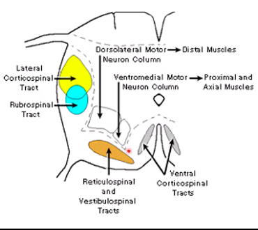
label the pathways of UMN x
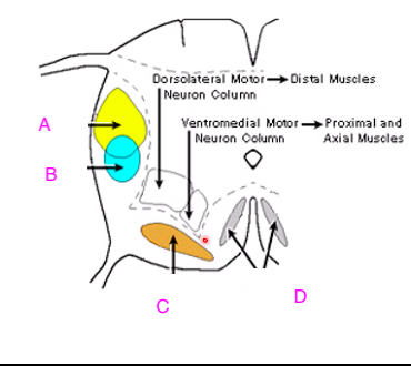
A: lateral corticospinal tract. key !!
B; rubrospinal tract
C: reticulospinal and vestibulospinal tracts
D: ventral and corticospinal tracts
what are functions of the:
vestibulo-cerebellum
spino-cerebellum
cerebro-cerebellum
what does damage of each of the above cause?
what are functions of the:
vestibulo-cerebellum:
- **balance & posture
- co-ordinates eye and head movements**
- damage = ability to stand and maintain posture impaired
spino-cerebellum:
- *- locomotion
- voluntary movements of arms and legs
- damage = overshoot and intention tremor, impaired gait**
cerebro-cerebellum
- **skilled motor tasks
- ataxia failure**
what are functions of the:
vestibulo-cerebellum
spino-cerebellum
cerebro-cerebellum
what does damage of each of the above cause?
what are functions of the:
vestibulo-cerebellum:
- **balance & posture
- co-ordinates eye and head movements**
- damage = ability to stand and maintain posture impaired
spino-cerebellum:
- *- locomotion
- voluntary movements of arms and legs
- damage = overshoot and intention tremor, impaired gait**
cerebro-cerebellum
- **skilled motor tasks
- ataxia failure**
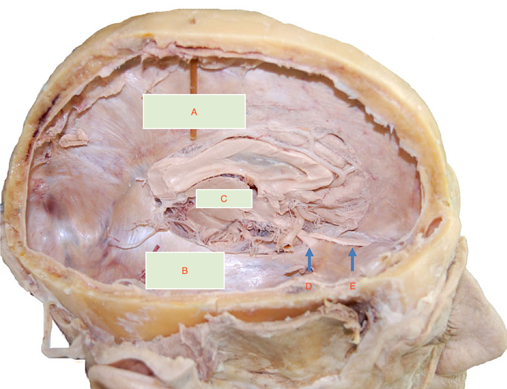
what is D?
carotid canal
optic tract
olfactory tract
optic chiasm
superior sagital sinus
what is D?
carotid canal
optic tract
olfactory tract
optic chiasm
superior sagital sinus
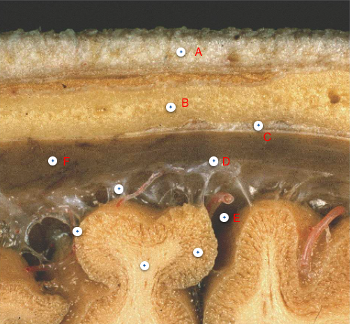
which of the following is arachnoid mater?
A
B
C
D
E
F
G
which of the following is arachnoid mater?
A
B
C
D
E
F
G
what is muscle tone due to? [3]
which structure detects tension in the tendon? [1]
muscle tone: due to a partial state of contraction in some fibres [1]. maintained reflexievly and adjuststed to the needs and posture of movement [1]
requires integity of monosynpatic reflex to occur [1]
= resting tension !!


-
FunMed EOYS251
-
FunMedEOYS347
-
FunMedEOYS459
-
FunMedEOYS153
-
FunMedEOYS427
-
FunMed EOYS Long 16
-
big boy things51
-
CR EOYS142
-
CR EOYS248
-
CR EOYS351
-
Calculations6
-
CR EOYS421
-
MET EOYS154
-
big boys29
-
MET EOYS253
-
MET EOYS343
-
MET EOYS445
-
MET EOYS538
-
BB EOYS150
-
BBB2
-
BB EOYS255
-
Short king spring65
-
BB EOYS350
-
BB EOYS446
-
:O29
-
BB EOYS447
-
Loco EOYS43
-
LOCO SSS59
-
LOCO EOYS 441
-
Loco EOYS344
-
LOCO EOYS 553
-
LOCO SSS246
-
LOCO EOYS542
-
LOCO EOYS629
-
SSS FunMed52
-
Spotter Qs130
-
SSS FunMed252
-
METSSS44
-
METSS249
