Anomalous origin of both coronary arteries from left sinus of Valsalva
RCA is ectopic
Anomalies of course:
- Retroaortic course
- Prepulmonic course
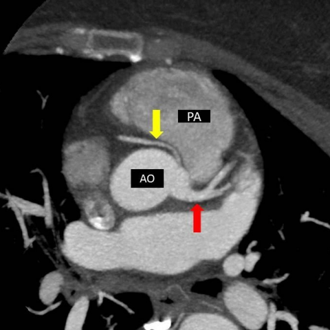
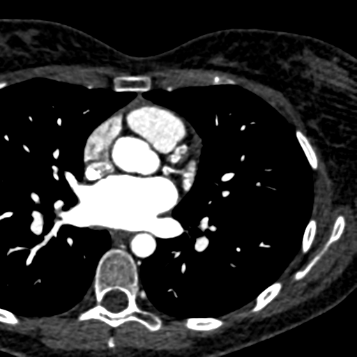
Axial MIP image shows common origin of RCA and left main coronary artery from left aortic sinus. Anomalous inter-arterial course of RCA between aorta (AO) and pulmonary artery (PA).
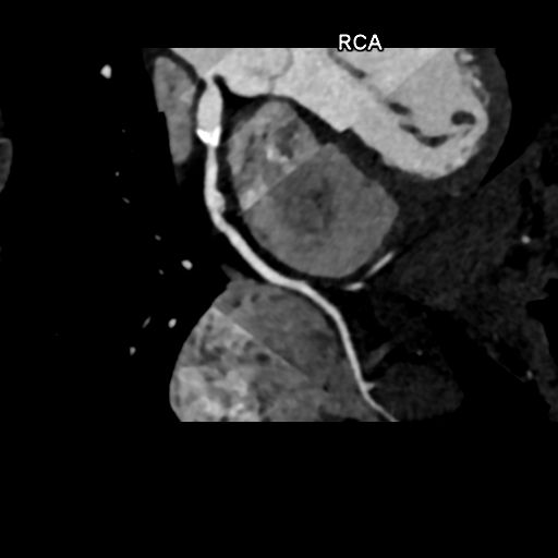
Anomalous origin from opposite coronary sinus
- Both coronary arteries from right sinus of Valsalva
- Ectopic LCA takes an acute angle behind PA
- 30% sudden death (infarction)
https: //www.jbsr.be/articles/872/print/
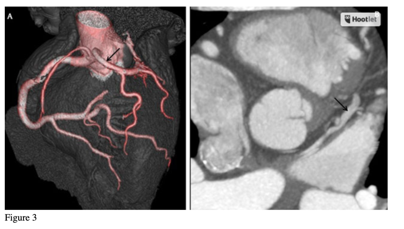
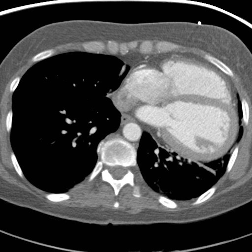
3a: Coronary CT angiography showing the origin of left main coronary artery (black arrow) from the right coronary sinus with prepulmonic course; 3b:Coronary CT angiography showing the anterior interventricular vein (black arrow) draining directly to the left atrium.
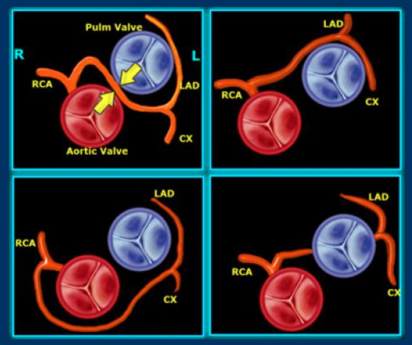
The illustration in the left upper corner is the most common and clinically significant anomaly.
There is an anomalous origin of the LCA from the right sinus of Valsalva and the LCA courses between the aorta and pulmonary artery.
This interarterial course can lead to compression of the LCA (yellow arrows) resulting in myocardial ischemia.
The other anomalies in the figure on the left are not hemodynamically significant.
https://radiologyassistant.nl/cardiovascular/anatomy/coronary-anatomy-and-anomalies
What are the most common forms of Congenital heart disease?
The most common structural defects are:
- bicuspid aortic valve
- mitral valve prolapse (MVP)
most of which are asymptomatic.
2 Anomalies of coronary artery termination
- Congenital coronary AV fistula
- Both arteries are orthotopic.
- Venous side of fistula originates in RA, coronary sinus, or RV
- Coronary cameral fistula
- Coronaries terminating in a cardiac chamber
- Case Discussion
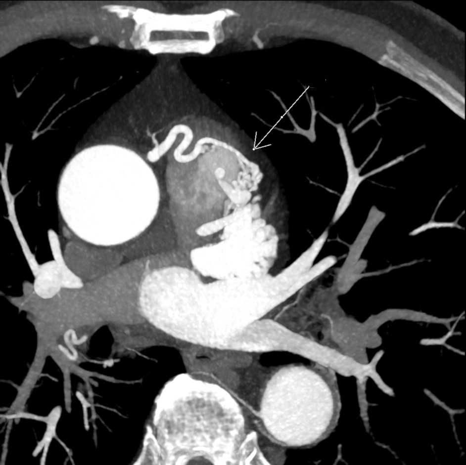
- Coronary arteriovenous fistula (white arrow) on axial arterial phase MIP CT. The MIP image nicely shows diffusion of the resulting contrast blush within the pulmonary artery.
- This findings are noticed incidentally at CT cardiac imaging for coronary stents visualisation.
- In this case surgical correction was not indicated due to asymptomatic clinical presentation and elderly age.
- CT follow-up was recommended.
- Case courtesy of Dr Irina Haidzel, Radiopaedia.org, rID: 58931
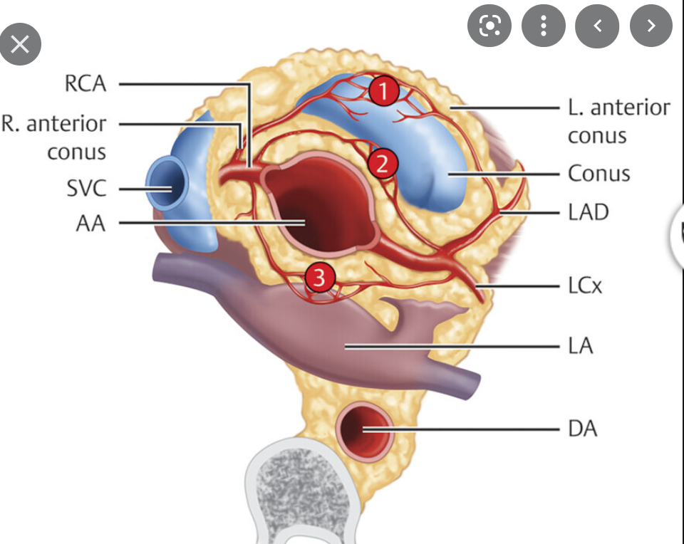
What is 3 on this diagram?
Variant anatomy
Kugel artery: collateral that connects the SA nodal artery and the AV nodal artery (anastomotic artery magnum)
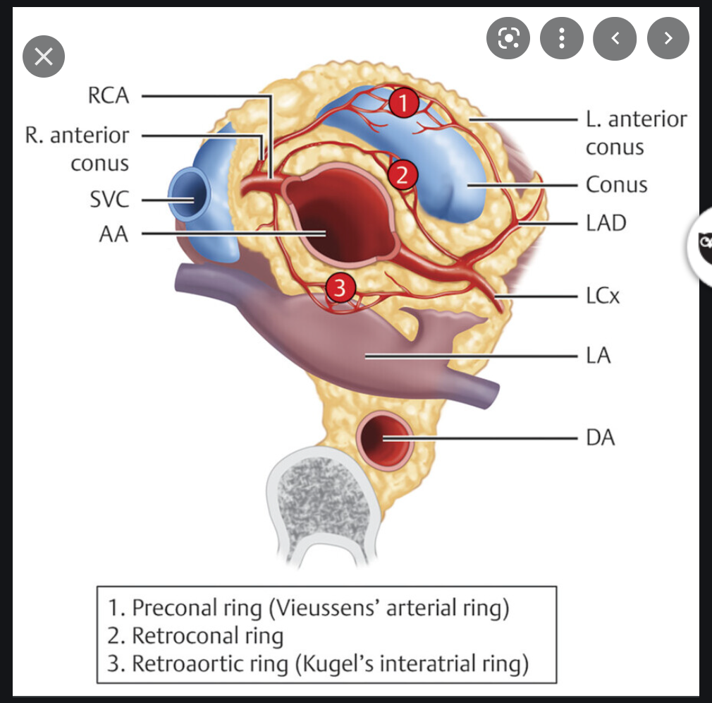
What is labeled ‘1’ on this diagram?

Vieussens ring: collateral branches from right conus artery to LAD.

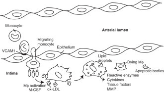
What are the 3 stages of Atherosclerotic CAD?
Atherosclerotic CAD ( Fig. 2.78 )
Now recognized as an inflammatory condition with established cascade of events. Three stages:
- Intimal fatty streaks (nonobstructive, clinically silent)
- Development of active inflammation with monocyte recruitment, macrophages (foam cells), fibrous plaques during adulthood (narrowing of lumen: angina)
- Late occlusive disease: calcifications, hemorrhage (angina, AMI)
What are the 7 main risk factors for coronary artery disease?
- Strong correlation
- Elevated CRP, LDL
- Family members with atherosclerotic disease
- Smoking
- HTN
- Hyperlipidemia
- Diabetes
- Male
- Weaker correlation
- Obesity
- Stress
- Sedentary life
What is the treatment for coronary artery disease?
Treatment
- Reversal of risk factors (diet, smoking cessation)
- Medication (statins)
- Transluminal coronary angioplasty, coronary stents
- Surgery
- Saphenous vein aortocoronary bypass
- Left internal mammary coronary bypass
What is the annual mortality of:
- One-vessel disease:
- Two-vessel disease:
- Three-vessel disease:
What two factors double mortality?
Annual Mortality
- One-vessel disease: 2%–3%
- Two-vessel disease: 3%–7%
- Three-vessel disease: 6%–11%
- Low EF, doubles mortality
- Abnormal wall motion, doubles mortality
What are the radiographic features of Coronary Artery disease?
Radiographic Features
- Plain radiograph
- Calcification of coronary arteries are the most reliable plain radiograph sign of CAD (90% specificity in symptomatic patients), but calcified coronary arteries are not necessarily stenotic.
- LV aneurysm is the second most reliable plain radiograph sign of CAD.
- It develops in 20% of MIs.
- Location
- Anteroapical wall: 70%
- Inferior wall: 20%
- Posterior wall: 10%
- CHF causing:
- Pulmonary edema
- Least reliable sign of CAD
- Case courtesy of Dr Maxime St-Amant, Radiopaedia.org, rID: 20697
- CT-scan shows an aneurysm which seem to originate from the proximal right coronary artery. The aneurysm collar is larger than it would be expected in a pseudoaneurysm. Coronary angiography could be done to further confirm the diagnosis
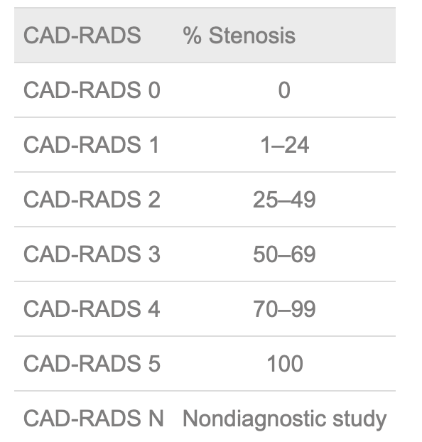
Stenosis of ___ is considered significant in all coronary arteries except ___ in which threshold is ___.
Stenosis of >70% (CAD-RADS ≥4) is considered significant in all coronary arteries except left main, in which threshold is 50%.
Stenosis occurs primarily in which arteries?
Coronary angiography
Stenosis occurs primarily in:
Proximal portions of major arteries
LAD > RCA > LCx
Collaterals develop if____ of the coronary diameter is obstructed;
two types of anastomosis:
Collaterals develop if >90% of the coronary diameter is obstructed; two types of anastomosis:
- Connections between branches of the same coronary artery (homocoronary)
- Connections between the branches of the three major coronary arteries (intercoronary)
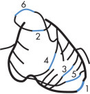
What are the 6 common pathways of intercoronary anastomoses?
Common pathways of intercoronary anastomoses ( Fig. 2.79 ) in descending order of frequency are:
- Surface of apex
- Surface of pulmonary conus
- Between anterior and posterior septal branches
- In the AV groove: LCx and distal RCA
- On the surface of the RV wall
- On the atrial wall around SA node
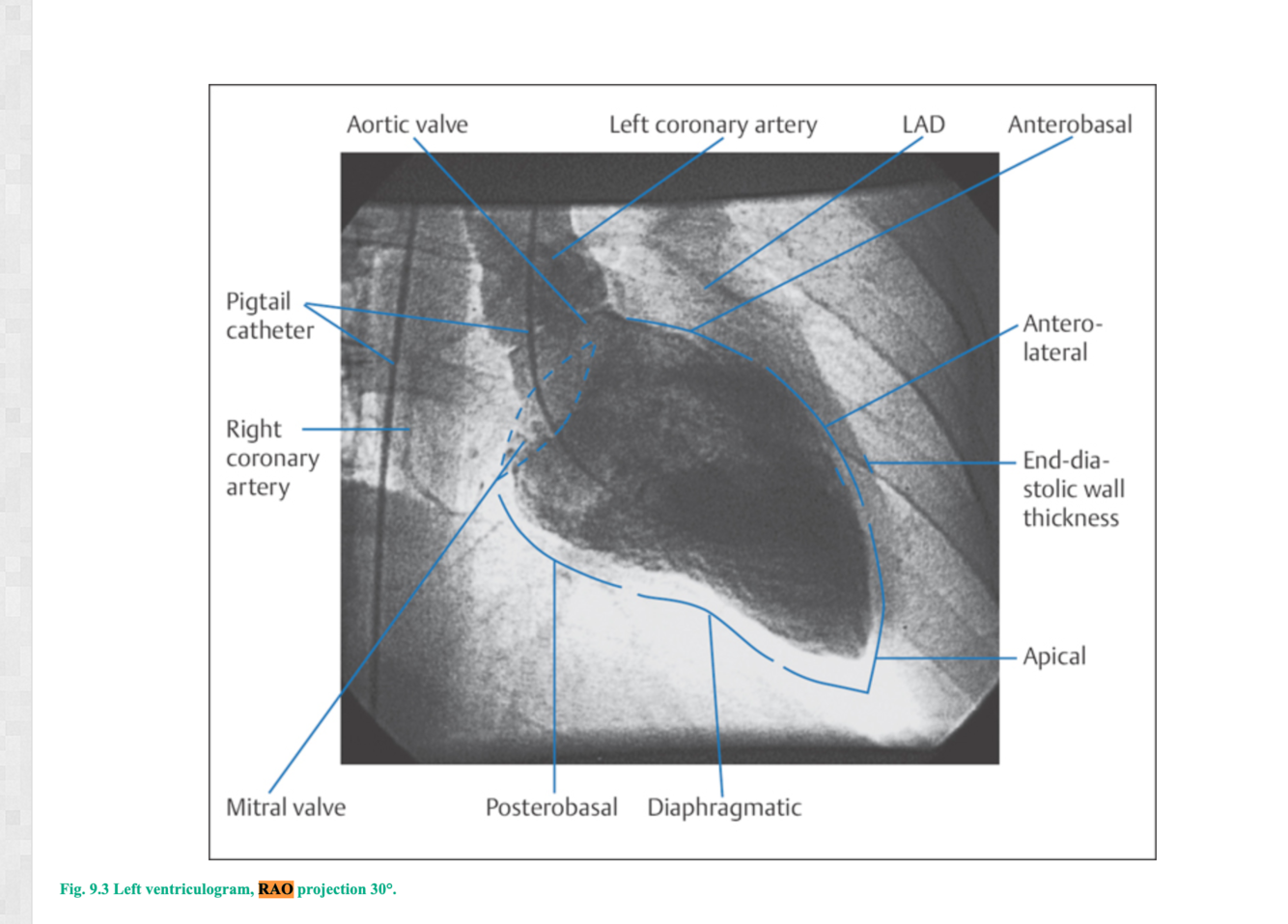
In left ventriculography, which view is the most helpful?
What can you evaluate?
Left ventriculography
- RAO view most helpful
- Evaluate
- LV function,
- valvular insufficiency,
- shunts,
- mural thrombus
- Evaluate
What is CAD RADS
what are the different scores?
- GRADING OF STENOSIS ON CORONARY COMPUTED TOMOGRAPHIC ANGIOGRAPHY (CAD-RADS)
- CAD-RADS
- Coronary Artery Disease—Reporting and Data System.
- MODIFIERS:
- If more than one modifier is present, the slash symbol (“/”) should follow each modifier in the following order:
- first, modifier N (nondiagnostic);
- second, modifier S (stent);
- third, modifier G (graft);
- fourth, modifier V (vulnerability).
- If more than one modifier is present, the slash symbol (“/”) should follow each modifier in the following order:
Presentation
Anterior ischaemia on ECG.
Patient Data
Age: 25 years
Gender: Female
Kawasaki Disease (Mucocutaneous Lymph Node Syndrome)
- Idiopathic acute febrile multisystem disease in children.
- Most cases are self-limited and without complications.
- Mortality from AMI: 3%.
- Treatment is with aspirin and gamma globulin.
- Clinical Findings
- Fever and cervical lymphadenopathy
- Desquamating rash on palms/soles
- Vasculitis of coronary arteries
Case courtesy of Assoc Prof Craig Hacking, Radiopaedia.org, rID: 35218
25F hx of Kawasaki and AMI at 8months old.
Right Coronary Artery (RCA): 2 fusiform aneurysms without thrombosis- 1) proximal segment 15 mm long and 8 mm in diameter with mural calcification and 2) mid segment 7 mm long and 6.5 mm in diameter at the origin of a small AM branch.
What are the rad features of Kawasaki Disease?
- Radiographic Features
- Spectrum of coronary disease
- Aneurysm:
- present in 25%
- (most are multiple when present)
- usually in proximal segments and detectable by US
- Cardiac CTA can visualize the coronary arteries beyond their proximal portions, identify giant aneurysms (>8 mm) and evaluate for complications such as thrombosis.
- Stenoses
- Occlusion
- Rupture
- Aneurysm:
- Transient gallbladder hydrops
- Spectrum of coronary disease
What sign is this?
What is the anatomy of the structure associated with this sign?
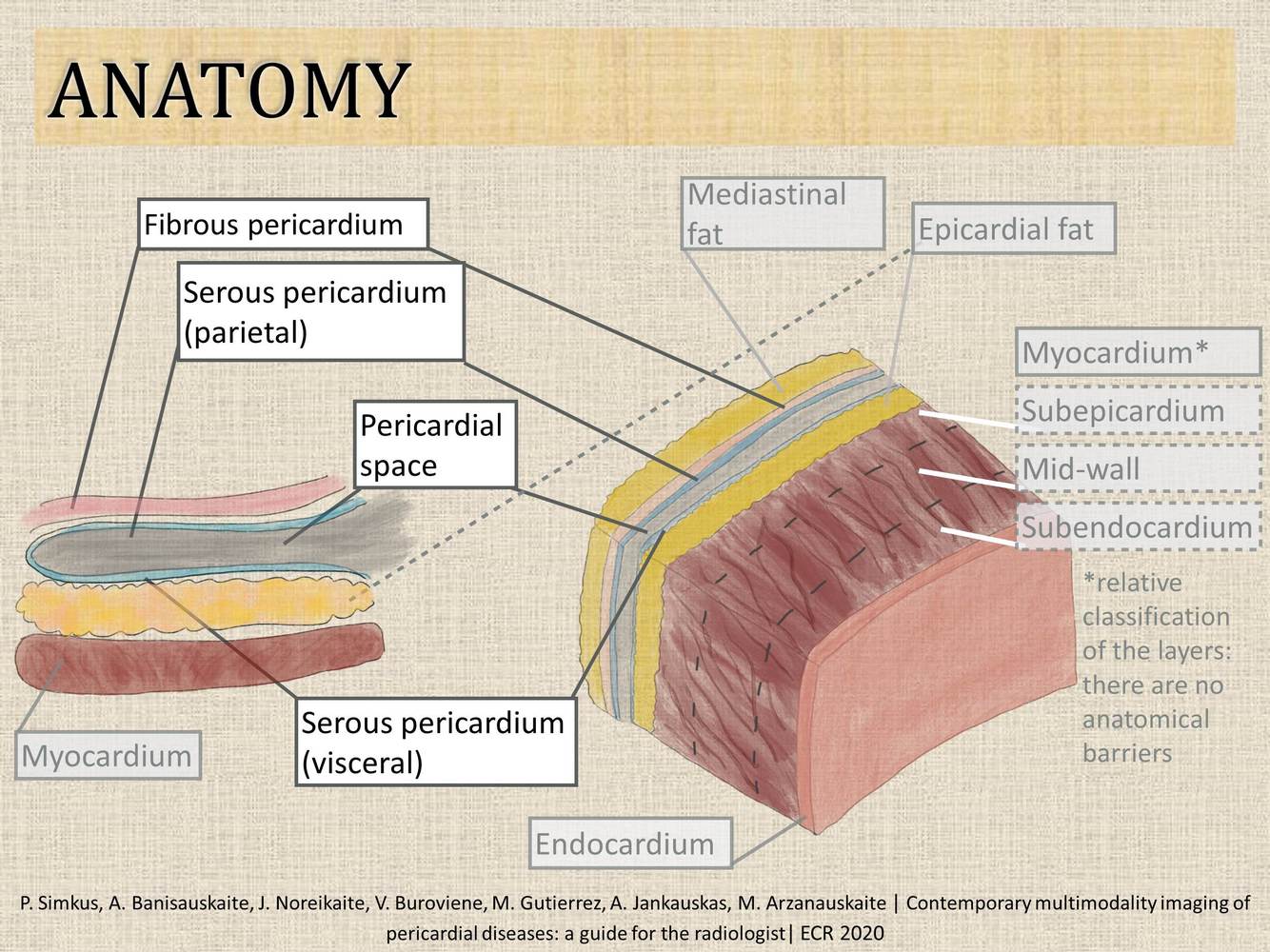
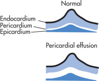
Pericardium Normal Anatomy
- Pericardium consists of two layers:
- External fibrous pericardium
- Internal serous epicardium
- The normal pericardial cavity has 10 to 50 mL of clear serous fluid.
- Normal structures:
- Fat stripe:
- fat on surface of heart beneath pericardium seen on lateral CXR
- Superior pericardial recess (commonly seen by CT or MRI)
- Fat stripe:
Congenital Absence of the Pericardium
- Types:
- May be total or partial.
- Partial absence is more common, occurs mainly on the left and is usually asymptomatic.
- Large defects may cause cardiac strangulation.
- Small defects are usually asymptomatic.
- Radiographic Features
- Total absence of the pericardium
- Mimics the appearance of the large silhouette seen in pericardial effusions
- Partial absence of the pericardium
- Heart is shifted and rotated into left pleural cavity
- PA view looks like an RAO view
- Heart is separated from the sternum on cross-table lateral view
- Left hilar mass: herniated left atrial appendage and pulmonary trunk
- Total absence of the pericardium
- Cardiac CT and MRI
- Total or partial absence of pericardium
- Exaggerated levoposition of heart
- Excess lung tissue between aorta and PA
- Radiographic features
- Plain radiograph
- Chest radiographic findings are usually subtle and non-specific. Features may include:
- apparent elevation of the cardiac apex
- right cardiac border might be indistinct due to leftward displacement and rotation of the heart
- prominent pulmonary artery segment (PA): both the medial and the lateral borders of the main PA might be seen more clearly as a result of the lack of pericardial reflection between the aorta and the PA
- lucency caused by the interposition of the lung between the aorta and main pulmonary artery segment
- the cardiophrenic space is increased on the frontal chest radiograph
- appearances may form the Snoopy sign, which is said to be pathognomonic for pericardial agenesis 18,19
- Case Discussion
- Plain radiograph
Pericardial agenesis (partial) is a rare cardiac defect. Radiologically, it presents with the following :
levoposition of the heart
prominent pulmonary artery
air interface in the aorto-pulmonary window or between the base of the heart and the diapgragm
CT-scan is better than MRI for this diagnosis, since it has better spatial resolution. The pericardial defect will be seen. A complete pericardial defect is a benign condition, while patients with partial agenesis may have cardiac herniation through the defect. They should be managed surgically.
* **Case courtesy of Dr Maxime St-Amant, Radiopaedia.org, rID: 20695**
What is the dx?
What is the underlying pathology?
% that are asymptomatic

- Pericardial Cysts
- Pericardial cysts represent congenital malformations (persistent coelom).
- 90% unilocular, 10% multilocular
- 75% are asymptomatic; occur at all ages
- If there is communication with pericardial cavity, the entity is termed pericardial diverticulum.
- Radiographic Features
- Well-defined, rounded soft tissue density on plain radiograph
- Most common location: cardiophrenic angles
- Other locations: anterior and middle mediastinum
- CT is helpful in establishing diagnosis.
- Case courtesy of Dr Henry Knipe, Radiopaedia.org, rID: 26335
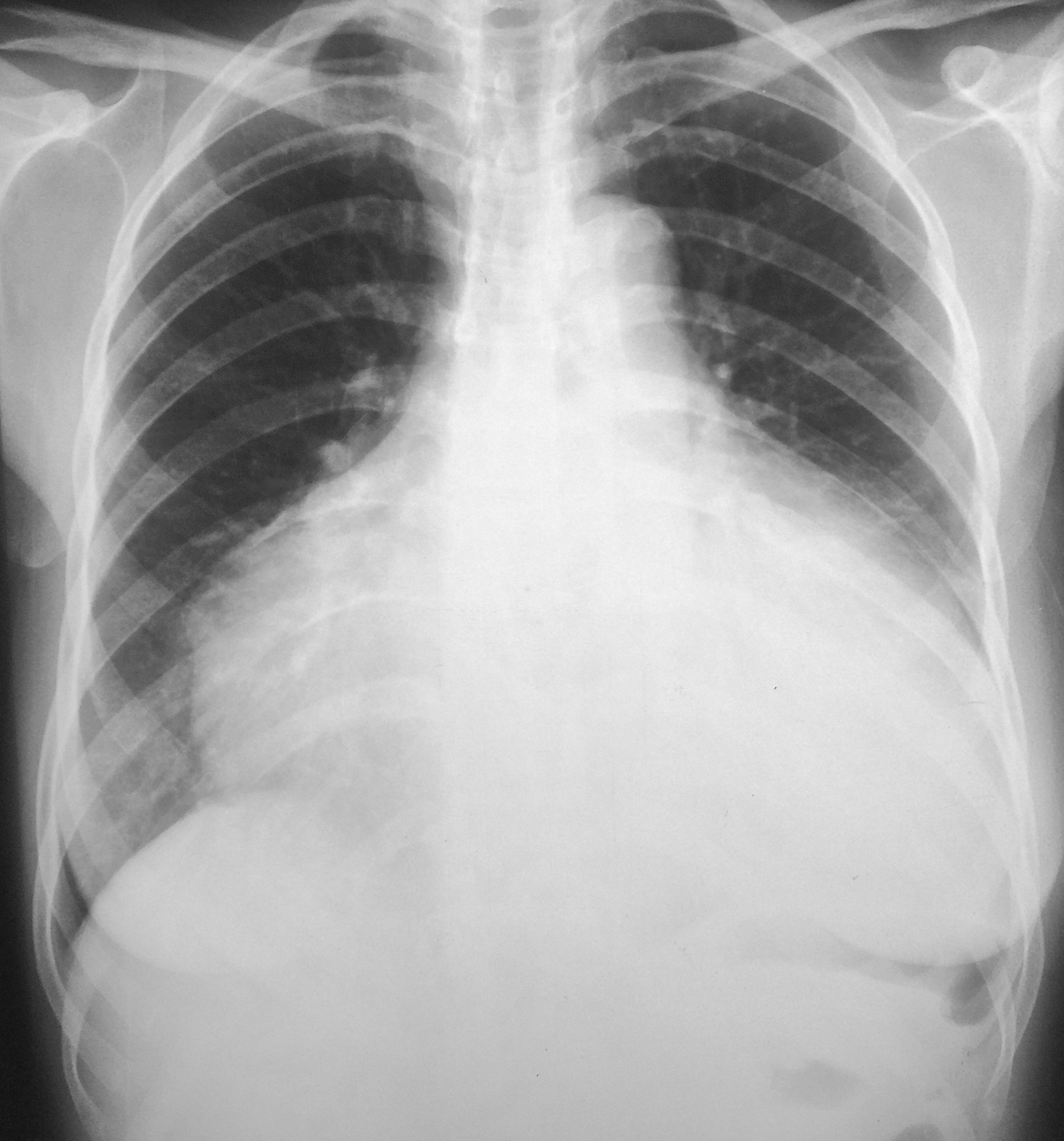
- Large well-defined mass projects in the right mid-to-lower zones obscures the right heart border.
Case Discussion
The patient underwent excision of this mass via a VATS procedure. Histology demonstrated a pericardial (mesothelial) cyst.
What are the 6 broad categories of causes of Pericardial effusion?
16 causes
- Tumor
- Metastases (melanoma, breast, lung)
- Inflammatory/idiopathic
- Rheumatic heart disease
- Collagen vascular disease
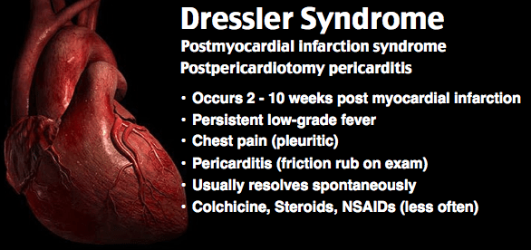
- Dressler syndrome
- Postpericardiotomy syndrome
- Drug hypersensitivity
- Infectious
- Viral
- Pyogenic
- Tuberculosis (TB)
- Metabolic
- Uremia
- Myxedema
- Trauma
- Hemopericardium
- Postoperative (frequently after pacemaker implantation and EP ablations)
- Vascular
- Acute MI
- Aortic dissection
- Ventricular rupture
- Case courtesy of Dr Vincent Tatco, Radiopaedia.org, rID: 43323
What is the dx?
What volume of fluid is required to be detectable?
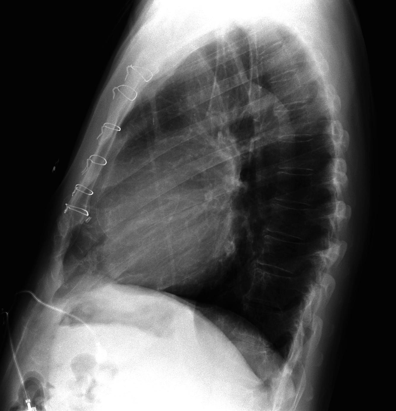
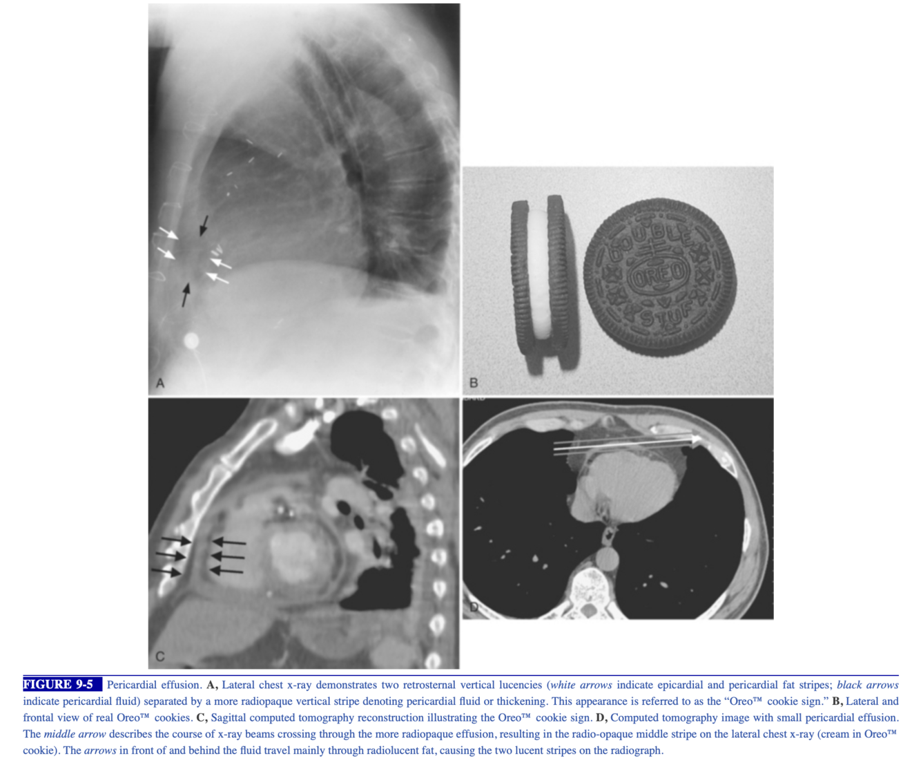
what sign is this?
Radiographic Features ( Fig. 2.80 )
- Plain radiograph
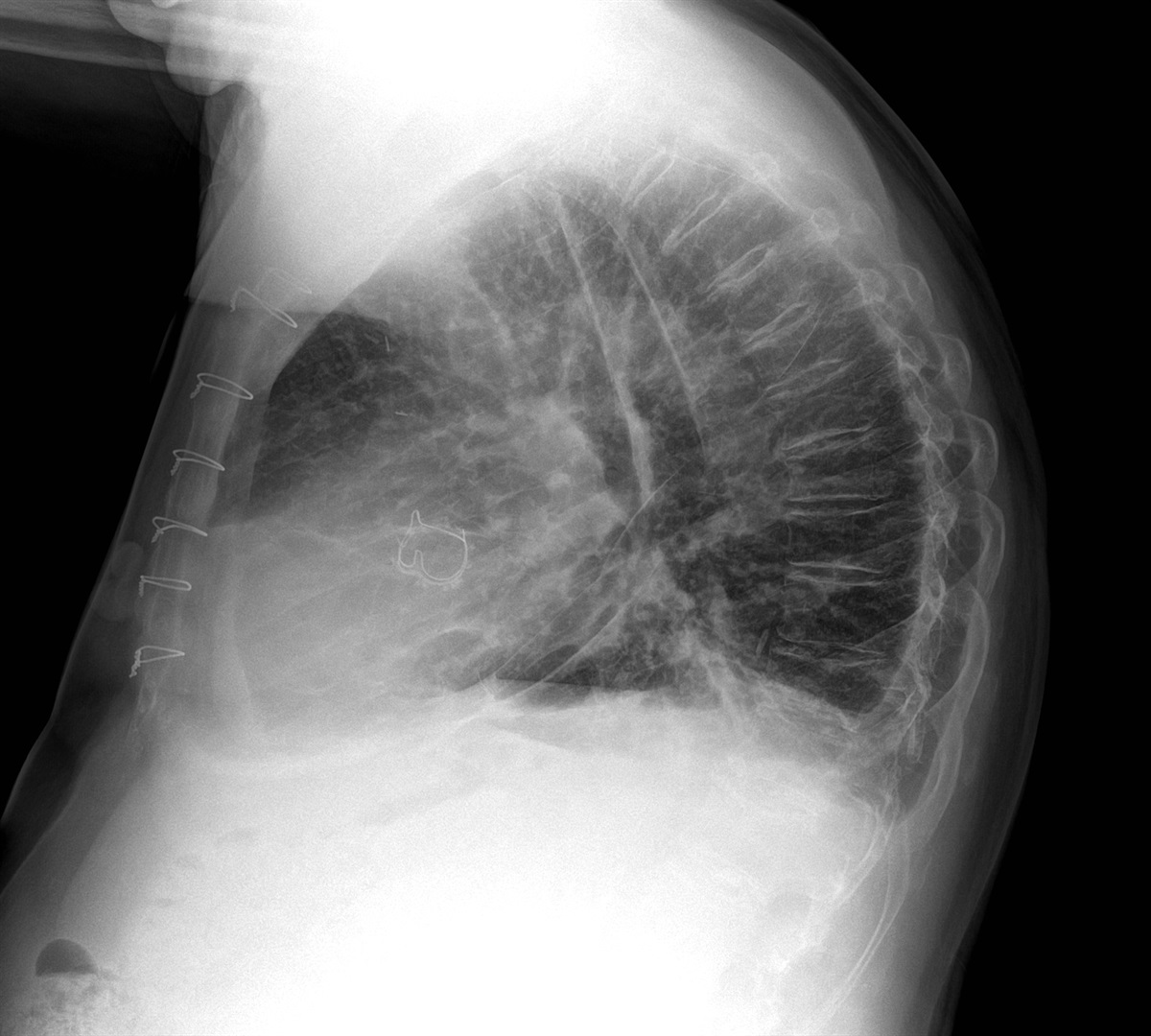
- >250 mL is necessary to be detectable.
- Oreo cookie sign on lateral view:
- subpericardial fat stripe measures >10 mm (a stripe 1–5 mm can be normal).
- Symmetrical enlargement of cardiac silhouette (water-bottle sign)
- Postsurgical loculated pericardial effusion may mimic an LV aneurysm.
- US
- Study of choice
- Echo-free space between epicardium and pericardium
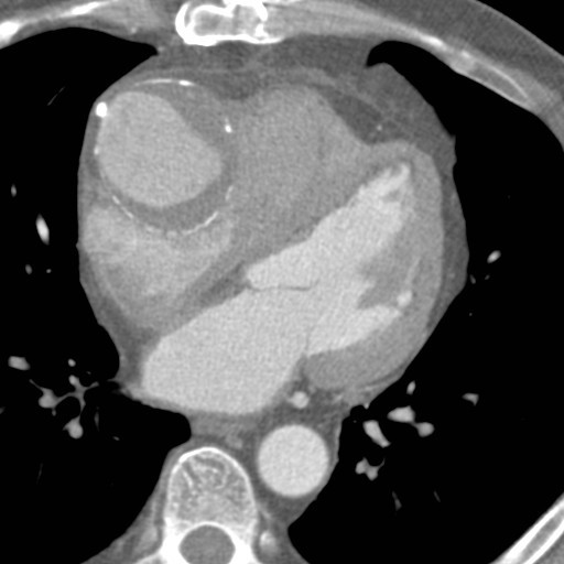
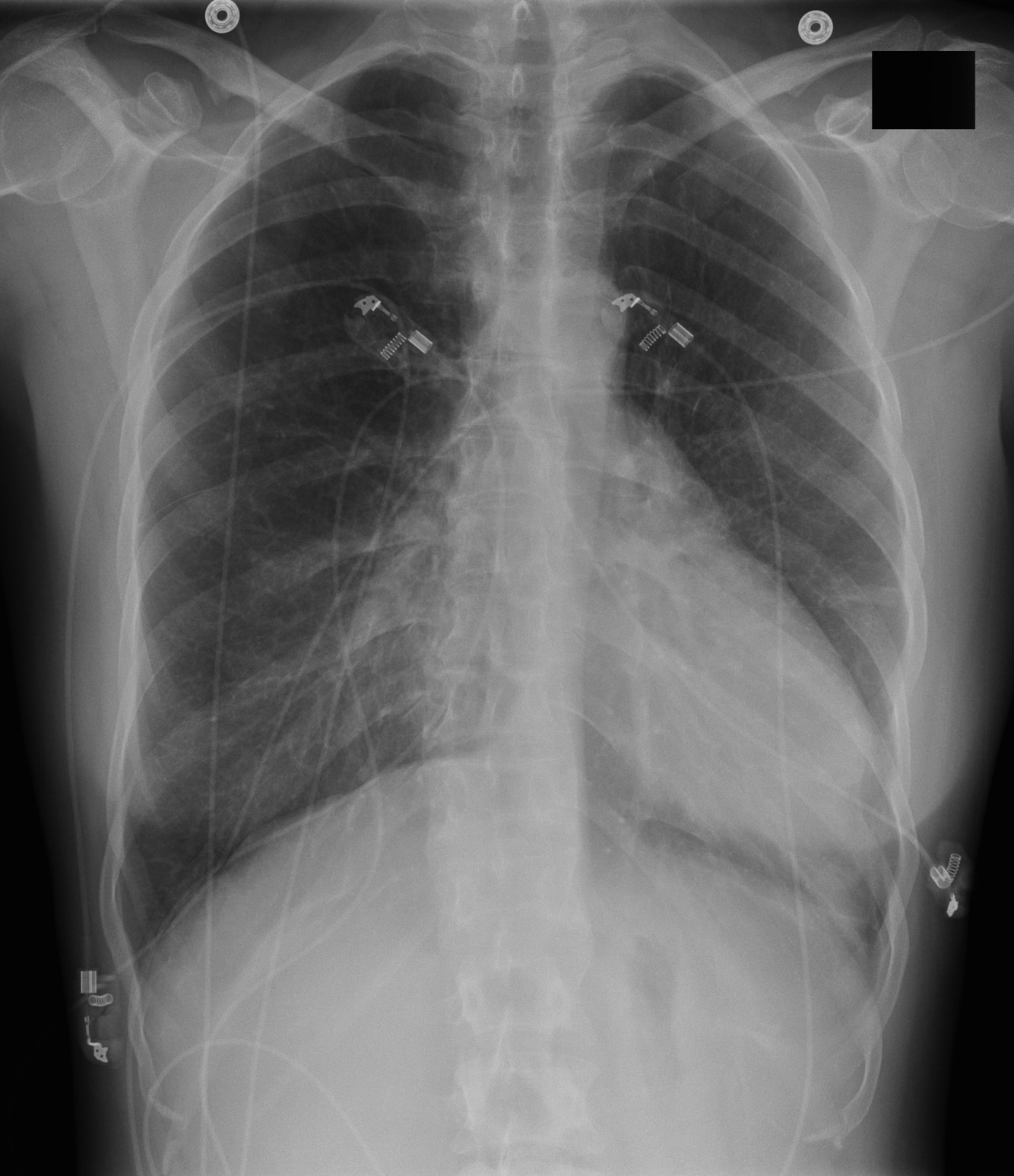
67-year-old male with history of prosthetic aortic valve stenosis, now with dyspnea on exertion and lower extremity swelling
Vertical retrosternal opaque stripes representative of possible pericardial effusion (“oreo cookie sign”).
https://www.auntminnie.com/index.aspx?sec=olce&sub=mlib&pag=cpages&ce_id=12564&pno=1


-
CHEST IMAGING 1100
-
CHEST IMAGING 2100
-
CHEST IMAGING 343
-
CHEST IMAGING 4 (TUMOURS)81
-
CHEST IMAGING 5 PATHOLOGY2
-
CARDIAC IMAGING 1100
-
CARDIAC IMAGING 2100
-
CARDIAC IMAGING 3100
-
CARDIAC IMAGING 443
-
GIT 1101
-
GIT 2100
-
GIT 3100
-
GIT 4102
-
Hepatobilary155
-
Biliary System76
-
Pancreas66
-
Spleen24
-
Adrenal Glands70
-
GENITOURINARY IMAGING 197
-
GENITOURINARY IMAGING 2100
-
GENITOURINARY IMAGING 3100
-
GENITOURINARY IMAGING 4100
-
GENITOURINARY IMAGING 545
-
RETROPERITONEUM33
-
Male Pelvis10
-
GIT PATHOLOGY31
-
Skeletal Dysplasias18
-
MSK104
-
MSK Crack the Core70
-
MSK 295
-
MSK 3100
-
Neuro100
-
Neuro 222
-
NEURO 375
-
Head and Neck 1100
-
Head and Neck 2100
-
Head and Neck 3100
-
Head and Neck 4100
-
Head and Neck 555
-
DDX Head and Neck35
-
Vascular10
-
IR30
-
BREAST IMAGING52
-
OBSTETRICS17
-
GYNAECOLOGY40
-
PAEDIATRICS 196
-
PAEDIATRICS 297
-
PAEDIATRICS 395
-
Nuclear Medicine 134
-
PET CT16
-
Syndromes94
-
HAEMATOLOGY6
-
PATHOLOGY 141
-
Crack the core WHen I Say you say...489
-
Physics31
-
crack the core exam case companion18
-
EPONYMOUS Diseases/signs22
-
What the F&^# is that word?10
-
Radiology Signs25
-
Mnemonics36
-
GIT Pathology1
-
NEURO MRI PHYSICS14
-
GREAT CHEST XRAY CASES1
-
THIS PATIENT IS TYPICAL OF X CONDITION2
