What has similar pattern as ATN but occurs later in the posttransplant period.
Cyclosporine toxicity has similar pattern as ATN but occurs later in the posttransplant period.
What are the imaging features of chronic Cystitis?
What is this condition?
Imaging Features
- Cystitis cystica:
- serous fluid–filled cysts;
- multiple smooth round filling defects
- Cystitis glandularis:
- mucin-secreting glandular hypertrophy:
- multiple cyst like filling defects along mucosa
- Same findings as in acute cystitis
https://www.liebertpub.com/doi/10.1089/cren.2017.0010
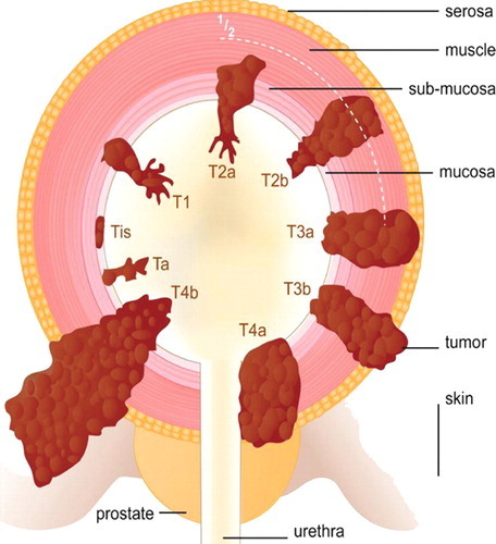
Describe the staging of bladder cancer
Staging
T1: mucosal and submucosal tumors
T2: superficial muscle layer is involved
T3a: deep muscular wall involved
T3b: perivesicular fat involved
T4: other organs invaded
N: the presence and distribution of malignant adenopathy affects the prognosis.
https://pubs.rsna.org/doi/10.1148/rg.322115125
Malacoplakia
Rare inflammatory condition that most commonly affects the bladder.
Yellow-brown subepithelial plaques consist of mononuclear histiocytes that contain Michaelis-Gutmann bodies.
On IVP, multiple mural filling defects with flat or convex border are seen, giving a cobblestone appearance.
Obstruction is a rare complication.
Mimics Malignancy.
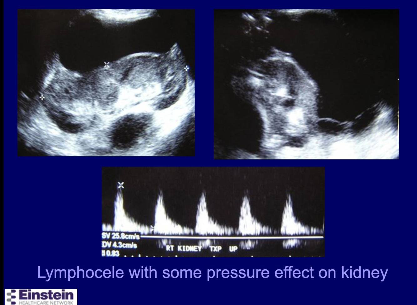
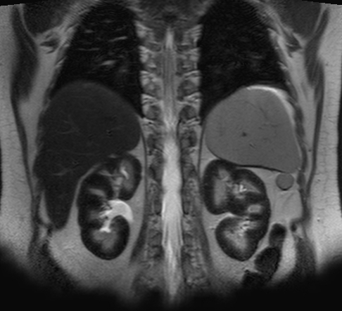
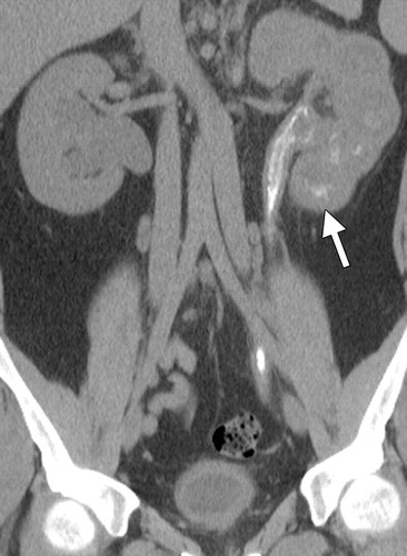
How often do perirenal fluid collections occur in renal transplants?
What % persist?
Perirenal Fluid Collections
Perirenal fluid collections occur in 40% of transplants.
The collections persist in 15%.
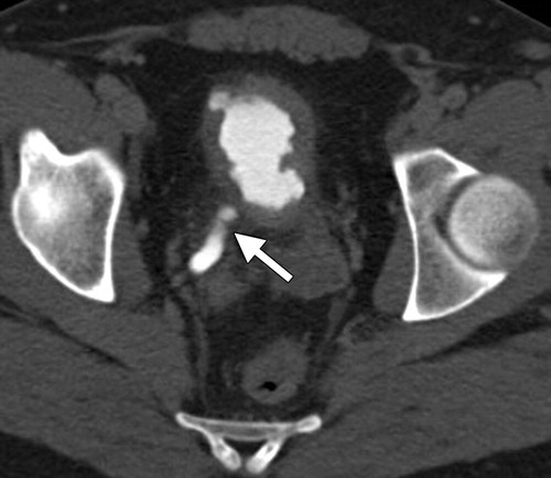
Lymphocele with pressure effect on Tx Kidney
What are 4 causes of this condition?

Vesicovaginal fistula:
- surgery,
- catheters,
- cancer,
- radiation
Case courtesy of Dr Ian Bickle, Radiopaedia.org, rID: 48793
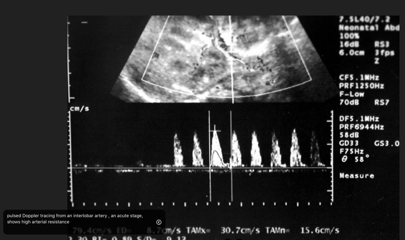
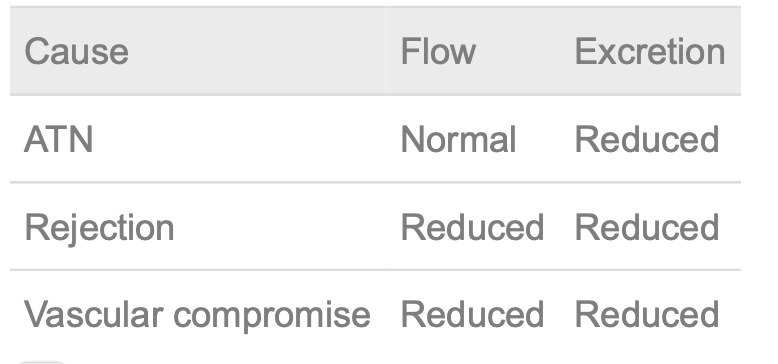
What are the imaging features of ATN?
on US
on MAG3
on Urogram
Imaging Features
- Smooth large kidneys
- Normal renal perfusion (MAG 3 angiography)
- Diminished or absent opacification after IV contrast administration
- Persistent dense nephrogram at late time points, 75%
- Variable US features:
- Increased cortical echogenicity with normal corticomedullary junction
- Increased echogenicity of pyramids
Striated Nephrogram:
Case courtesy of Dr David Cuete, Radiopaedia.org, rID: 28077
Radiographic features
Imaging demonstrates preserved renal parenchyma perfusion, but with minimal or absent excretion into the urinary collecting system.
Fluoroscopy / CT urography
Imaging with iodinated contrast typically demonstrates an immediate or mildly delayed nephrogram, but without excretion into the collecting system. Delayed 24 hour imaging would also demonstrate persistent nephrogram or striated nephrogram due to stasis of contrast within the renal tubules 3,4.
Ultrasound
Ultrasound is usually performed in this setting to assess the renal parenchyma and exclude other causes of obstruction. In acute tubular necrosis, the kidneys usually have a normal appearance on ultrasound, but may be enlarged and increased echogenicity 5.
What are 3 causes of Bladder adenocarcinoma?
Adenocarcinoma, 2%
- Bladder exstrophy
- Urachal remnant
- Cystitis glandularis; 10% pass mucus in urine
What are 5 causes of bladder outlet obstruction in children?
- Posterior urethral valves (most common in males)
- Ectopic ureterocele (most common in females)
- Bladder neck obstruction
- Urethral stricture
- Prune-belly syndrome
What are the signs of renal vein thrombosis in the kidneys on USS?
Early and late phases
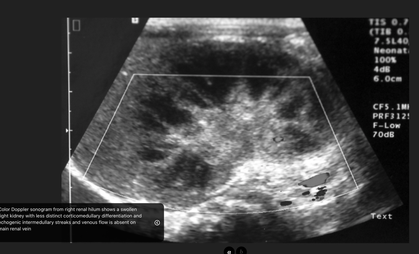
Kidneys
- Renal enlargement
- US:
- Early
- hypoechoic cortex (early edema);
- hyperechoic cortex after 10 days
- (fibrosis, cellular infiltrates
- preserved corticomedullary differentiationm (CMD);
- late phase (several weeks):
- decreased size,
- hyperechoic kidney with loss of CMD
- Early
https://www.eurorad.org/case/4596
What are the imaging features of this?
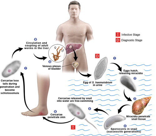
Schistosomiasis
Imaging Features
- Extensive calcifications in bladder wall and ureter (hallmark)
- Inflammatory pseudopolyps: “bilharziomas”
- Ureteral strictures, fistulas
- SCC (suspect when previously identified calcifications have changed in appearance)
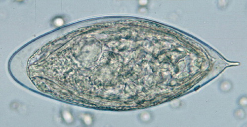
Figure 2 Photomicrograph (original magnification, ×200; hematoxylin-eosin stain) shows a fresh ovum from S haematobium, floating in a human urine specimen. Note the terminal spine at one end of the ovum.
Adult schistosomes do not usually cause an inflammatory reaction in the venous system. In fact, their presence there is associated with increased protection of the host against reinfection by cercariae. In general, dead eggs and dead flukes cause a more severe inflammatory reaction than living ones do (22).
Pathologic changes in the urinary tract due to schistosomiasis are far more common in chronic infections than in acute ones. Such changes result from the deposition of eggs (not adult flukes) in and around vessels, which leads to chronic inflammatory lesions and induces an immune response with granuloma formation and associated fibrotic changes (23).
https://pubs.rsna.org/doi/10.1148/rg.324115162
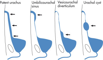
What are the four types of
Congenital urachal anomalies?
- Patent urachus
- Umbilical–urachal sinus
- Vesicourachal diverticulum
- Urachal Cyst
The majority of patients with urachal abnormalities (except those with a patent urachus) are asymptomatic.
However, these patients may become symptomatic if these abnormalities are associated with infection.
What are the 7 causes of Acute Tubular Necrosis?
2 categories
which category is more common?
- Renal ischemia, 60%
- Surgery, transplant, other causes
- Pregnancy related
- Nephrotoxins, 40%
- Radiographic contrast material, now controversial. Risk is probably real in patients with GFR ≤30
- Aminoglycosides
- Antineoplastic agents
- Hemoglobin, myoglobin
- Chemicals: organic solvents, HgCl 2
What are the imaging features of Leukoplakia?
What is the significance of Leukoplakia?
- Imaging Features
- Mucosal thickening
- Filling defect
- Treatment and prognosis
- Leukoplakia is considered a premalignant condition.
- There is an association with bladder neoplasia in 25% of cases.
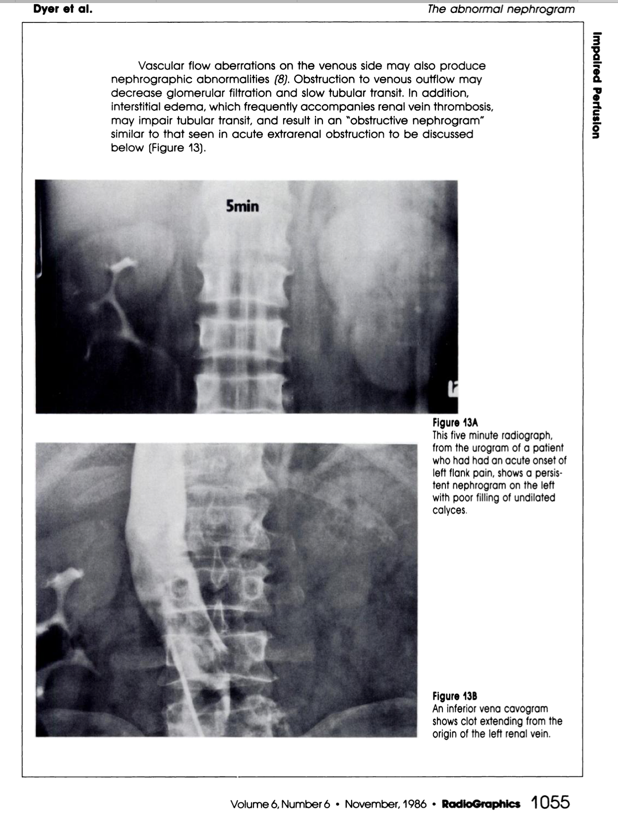
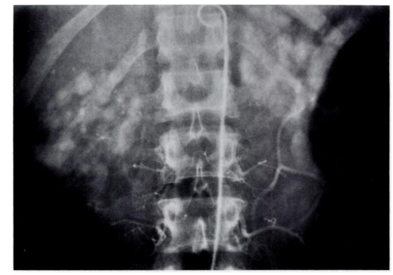
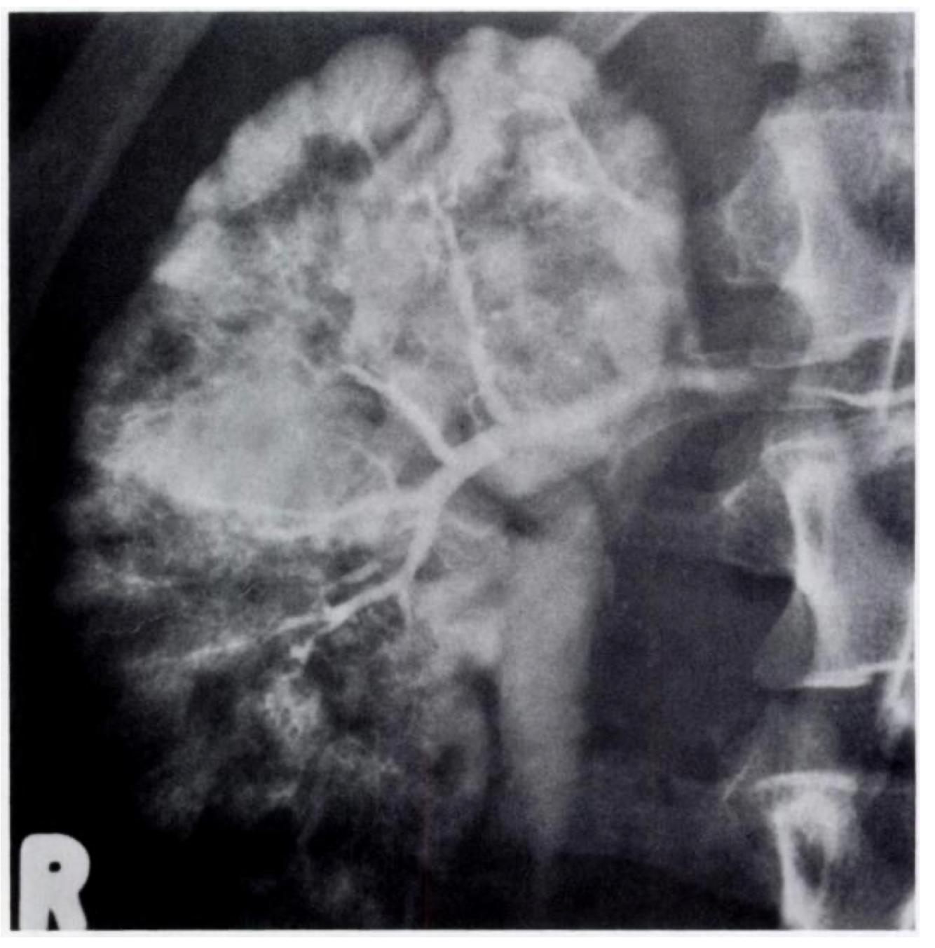
What are the signs of RVT on IVP?
What sign is this?
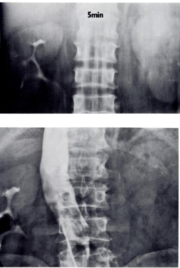
Delayed nephrogram
IVP:
- little opacification,
- prolonged nephrogram,
- striated nephrogram (stasis in collecting tubules);
- intrarenal collecting system is stretched and compressed by edema
https://pubs.rsna.org/doi/pdf/10.1148/radiographics.6.6.3685518
What is Bladder Leukoplakia a/w?
What is the underlying pathological process
Leukoplakia
Squamous metaplasia of transitional cell epithelium (keratinization).
Associated with:
- chronic infection (80%) and
- calculi (40%).
Bladder > renal pelvis > ureter
r. Premalignant? Clinical findings include hematuria in 30% and passage of desquamated keratinized epithelial layers.
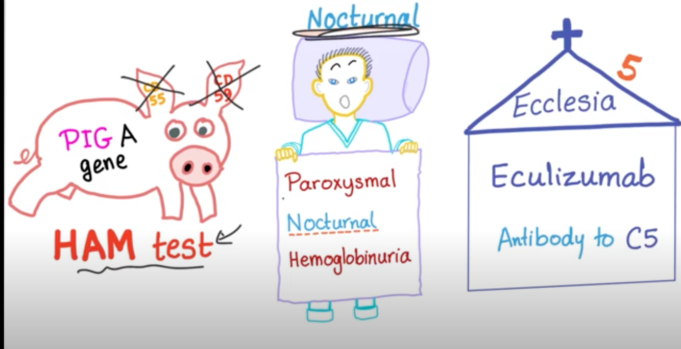
What are the findings of FLOW and Excretion with the following?
(NUCLEAR SCANS, INTRAVENOUS PYELOGRAM)
Paroxysmal Nocturnal Hemoglobinuria
Rare acquired hemolytic disorder. The renal cortex appears hypointense on T2/T2* because of hemosiderin deposition.
Case courtesy of Dr Hani Makky Al Salam, Radiopaedia.org, rID: 14460
Radiopedia:
- Renal haemosiderosis results from accumulation of haemosiderin in the kidneys.
- It is usually considered a benign and incidental radiologic finding and rarely results in clinically apparent renal dysfunction.
Pathology
- Renal haemosiderosis is a known complication of the following conditions:
- chronic intravascular haemolytic states such as haemolytic anaemias like sickle cell anaemia and thalassaemia 1,3
- paroxysmal nocturnal haemoglobinuria (PNH)
- mechanical haemolysis from prosthetic cardiac valve
https://www.youtube.com/watch?v=6OwlmIMU7L4
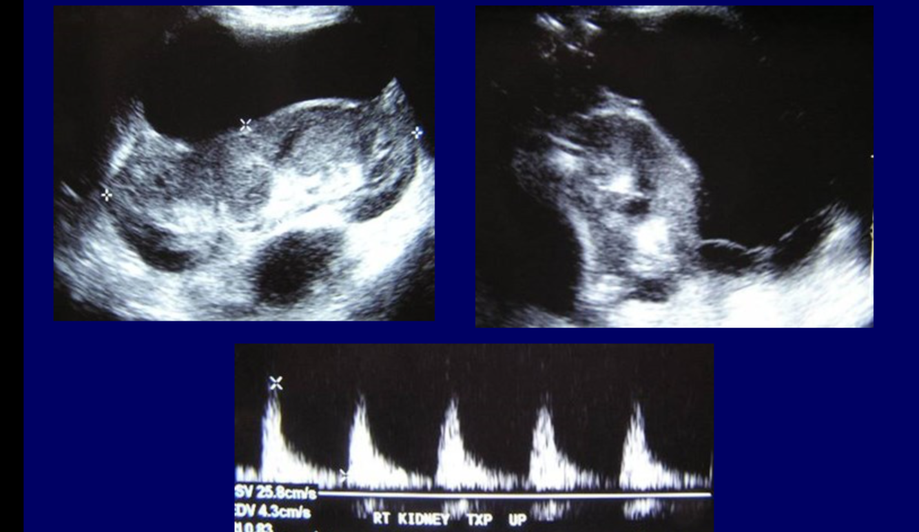
What are 6 vascular complications RE renal Transplant?
For each what are the causes/treatment options?
Vascular Complications
- RVT:
- most occur in first 3 days after transplantation.
- Renal artery occlusion or stenosis.
- Anastomotic stenosis is treated with angioplasty with up to 87% success rate.
- Infarction
- Pseudoaneurysm of anastomosis:
- surgical treatment
- AV fistula:
- usually from renal biopsy;
- if symptomatic, embolization is performed.
- Ureterovesical anastomosis obstruction may result from
- edema,
- stricture,
- ischemia,
- rejection,
- extrinsic compression, or
- compromised position of kidney.
What rarely occurs after 1 month post renal transplant?
ATN rarely occurs beyond 1 month after transplant.
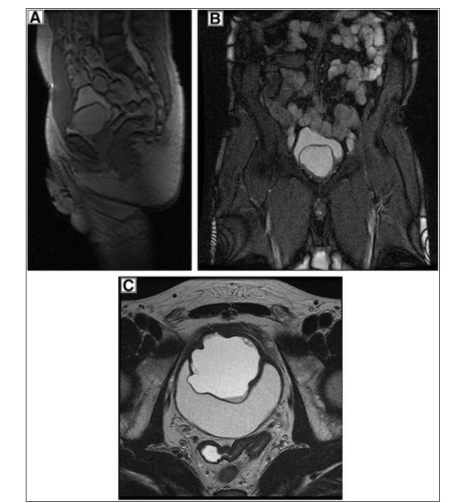
What are the imaging features of bladder cancer?
What is the preferred modality for local staging?
Imaging Features
- Mass in bladder wall
- MRI is now preferred for local staging
- Obstructive uropathy because of involvement of ureteric orifices
SIGN
What sign is seen here?
Tuberculosis
- Chronic interstitial cystitis that usually ends in fibrosis.
- Typically coexists with renal TB.
- Imaging Features
- Cystitis cystica or glandularis often coexist, causing filling defects in bladder.
- THIMBLE. BLADDER Small, contracted thick-walled bladder
- Mural calcification (less common)
https: //www.researchgate.net/figure/Thimble-bladder-Axial-image-A-and-curved-MPR-in-the-coronal-plane-B-of-the_fig5_315832565
https: //pubs.rsna.org/doi/10.1148/rg.323115004
SPOTTED NEPHROGRAM
Figure 5A
A late arterial phase from an aorto- gram shows bilateral spotted neph- rognams in a patient with polyartenitis nodosa.
Figure 5B
This is a late phase from a selective renal arteniogram in the same patient; it demonstrates occlusion of multiple peripheral vessels with cortical irregu- lanity.
https://pubs.rsna.org/doi/pdf/10.1148/radiographics.6.6.3685518
What is the only renal process with normal renal flow but reduced excretion.
ATN is the only renal process with normal renal flow but reduced excretion.


-
CHEST IMAGING 1100
-
CHEST IMAGING 2100
-
CHEST IMAGING 343
-
CHEST IMAGING 4 (TUMOURS)81
-
CHEST IMAGING 5 PATHOLOGY2
-
CARDIAC IMAGING 1100
-
CARDIAC IMAGING 2100
-
CARDIAC IMAGING 3100
-
CARDIAC IMAGING 443
-
GIT 1101
-
GIT 2100
-
GIT 3100
-
GIT 4102
-
Hepatobilary155
-
Biliary System76
-
Pancreas66
-
Spleen24
-
Adrenal Glands70
-
GENITOURINARY IMAGING 197
-
GENITOURINARY IMAGING 2100
-
GENITOURINARY IMAGING 3100
-
GENITOURINARY IMAGING 4100
-
GENITOURINARY IMAGING 545
-
RETROPERITONEUM33
-
Male Pelvis10
-
GIT PATHOLOGY31
-
Skeletal Dysplasias18
-
MSK104
-
MSK Crack the Core70
-
MSK 295
-
MSK 3100
-
Neuro100
-
Neuro 222
-
NEURO 375
-
Head and Neck 1100
-
Head and Neck 2100
-
Head and Neck 3100
-
Head and Neck 4100
-
Head and Neck 555
-
DDX Head and Neck35
-
Vascular10
-
IR30
-
BREAST IMAGING52
-
OBSTETRICS17
-
GYNAECOLOGY40
-
PAEDIATRICS 196
-
PAEDIATRICS 297
-
PAEDIATRICS 395
-
Nuclear Medicine 134
-
PET CT16
-
Syndromes94
-
HAEMATOLOGY6
-
PATHOLOGY 141
-
Crack the core WHen I Say you say...489
-
Physics31
-
crack the core exam case companion18
-
EPONYMOUS Diseases/signs22
-
What the F&^# is that word?10
-
Radiology Signs25
-
Mnemonics36
-
GIT Pathology1
-
NEURO MRI PHYSICS14
-
GREAT CHEST XRAY CASES1
-
THIS PATIENT IS TYPICAL OF X CONDITION2
